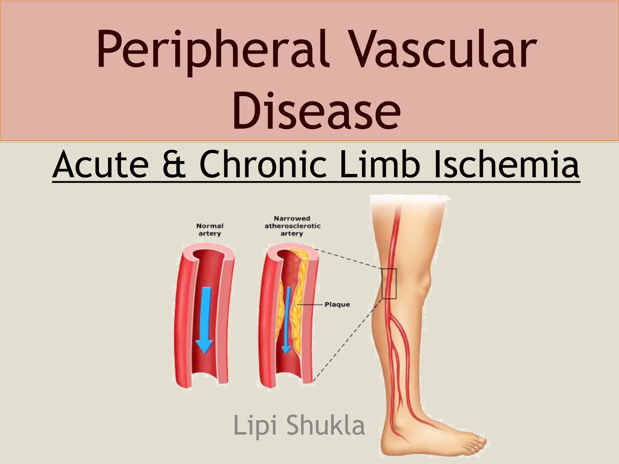
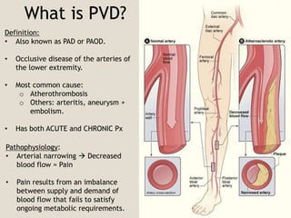
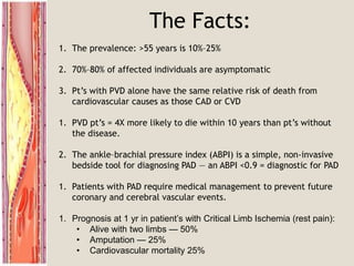
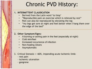
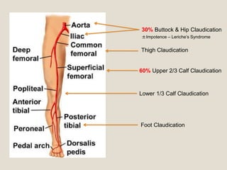
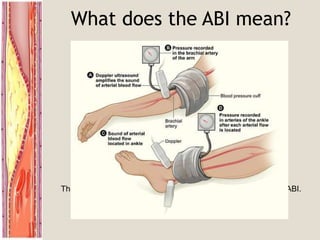
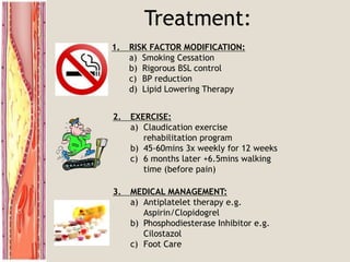
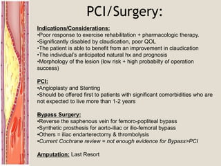
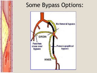
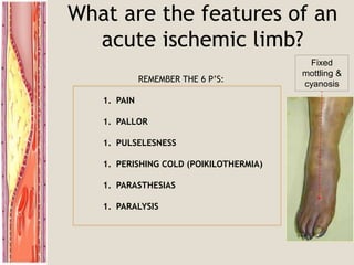
Peripheral Vascular
Disease
Acute & Chronic Limb Ischemia
Lipi Shukla
What is PVD?
Definition:
• Also known as PAD or PAOD.
• Occlusive disease of the arteries of
the lower extremity.
• Most common cause:
o Atherothrombosis
o Others: arteritis, aneurysm +
embolism.
• Has both ACUTE and CHRONIC Px
Pathophysiology:
• Arterial narrowing  Decreased
blood flow = Pain
• Pain results from an imbalance
between supply and demand of
blood flow that fails to satisfy
ongoing metabolic requirements.
The Facts:
1. The prevalence: >55 years is 10%–25%
2. 70%–80% of affected individuals are asymptomatic
3. Pt’s with PVD alone have the same relative risk of death from
cardiovascular causes as those CAD or CVD
1. PVD pt’s = 4X more likely to die within 10 years than pt’s without
the disease.
2. The ankle–brachial pressure index (ABPI) is a simple, non-invasive
bedside tool for diagnosing PAD — an ABPI <0.9 = diagnostic for PAD
1. Patients with PAD require medical management to prevent future
coronary and cerebral vascular events.
1. Prognosis at 1 yr in patient’s with Critical Limb Ischemia (rest pain):
• Alive with two limbs — 50%
• Amputation — 25%
• Cardiovascular mortality 25%
Risk Factors:
Typical Patient:
• Smoker (2.5-3x)
• Diabetic (3-4x)
• Hypertension
• Hx of Hypercholesterolemia/AF/IHD/CVA
• Age ≥ 70 years.
• Age 50 - 69 years with a history of smoking or diabetes.
• Age 40 - 49 with diabetes and at least one other risk factor for
atherosclerosis.
• Leg symptoms suggestive of claudication with exertion or
ischemic pain at rest.
• Abnormal lower extremity pulse examination.
• Known atherosclerosis at other sites (eg, coronary, carotid, or
renal artery disease).
Chronic PVD History:
3. Critical Stenosis = >60%, impending acute ischemic limb:
- rest pain
- ischemic ulceration
- gangrene
2. Other Symptom/Signs:
• A burning or aching pain in the feet (especially at night)
• Cold skin/feet
• Increased occurrence of infection
• Non-healing Ulcers
• Asymptomatic
1. INTERMITTENT CLAUDICATION
• Derived from the Latin word ‘to limp’
• “Reproducible pain on exercise which is relieved by rest”
• Pain can also be reproduced by elevating the leg
• “my legs get sore at night and feel better when I hang them over
the edge of the bed”
Thigh Claudication
60% Upper 2/3 Calf Claudication
Lower 1/3 Calf Claudication
Foot Claudication
30% Buttock & Hip Claudication
±Impotence – Leriche’s Syndrome
DDx of Leg Pain
1. Vascular
a) DVT (as for risk factors)
b) PVD (claudication)
2. Neurospinal
a) Disc Disease
b) Spinal Stenosis (Pseudoclaudication)
3. Neuropathic
a) Diabetes
b) Chronic EtOH abuse
4. Musculoskeletal
a) OA (variation with weather + time of day)
b) Chronic compartment syndrome
Physical Examination:
Examination: What do to:
Inspection
Expose the skin
and look for:
• Thick Shiny Skin
• Hair Loss
• Brittle Nails
• Colour Changes (pallor)
• Ulcers
• Muscle Wasting
Palpation • Temperature (cool, bilateral/unilateral)
• Pulses: ?Regular, ?AAA
• Capillary Refill
• Sensation/Movement
Auscultation • Femoral Bruits
Ankle Brachial
Index (ABI)
= Systolic BP in ankle
Systolic BP in brachial artery
Buerger’s Test • Elevate the leg to 45° - and look for pallor
• Place the leg in a dependent position 90°& look
for a red flushed foot before returning to normal
• Pallor at <20° = severe PAD.
Pictures:
What does the ABI mean?
ABI Clinical Correlation
>0.9 Normal Limb
0.5-0.9 Intermittent Claudication
<0.4 Rest Pain
<0.15 Gangrene
CAUTION:
Patient’s with Diabetes + Renal Failure:
They have calcified arterial walls which can falsely elevate their ABI.
Investigations:
NON INVASIVE:
Duplex Ultrasound
 normal is triphasic  biphasic  monophasic  absent
BLOOD TESTS:
1. FBE/EUC/Homocysteine Levels
2. Coagulation Studies
3. Fasting Lipids and Fasting Glucose
4. HBA1C
WHEN TO IMAGE:
1. To image = to intervene
2. Pt’s with disabling symptoms where revascularisation is considered
3. To accurately depict anatomy of stenosis and plan for PCI or Surgery
4. Sometimes in pt’s with discrepancy in hx and clinical findings
ANGIOGRAPHY:
Non-invasive:
• CT Angiogram
• MR Angiogram
Invasive:
• Digital Subtraction Angiography
 Gold Standard
 Intervention at the same time
Tardus et parvus = small amplitude + slow rising pulse
CT Angiography Digital Subtraction Angiography
Value of angiography
Localizes the obstruction
Visualize the arterial tree & distal
run-off
Can diagnose an embolus:
Sharp cutoff, reversed meniscus or clot
silhouette
Treatment:
1. RISK FACTOR MODIFICATION:
a) Smoking Cessation
b) Rigorous BSL control
c) BP reduction
d) Lipid Lowering Therapy
3. MEDICAL MANAGEMENT:
a) Antiplatelet therapy e.g.
Aspirin/Clopidogrel
b) Phosphodiesterase Inhibitor e.g.
Cilostazol
c) Foot Care
2. EXERCISE:
a) Claudication exercise
rehabilitation program
b) 45-60mins 3x weekly for 12 weeks
c) 6 months later +6.5mins walking
time (before pain)
PCI/Surgery:
Indications/Considerations:
•Poor response to exercise rehabilitation + pharmacologic therapy.
•Significantly disabled by claudication, poor QOL
•The patient is able to benefit from an improvement in claudication
•The individual’s anticipated natural hx and prognosis
•Morphology of the lesion (low risk + high probabilty of operation
success)
PCI:
•Angioplasty and Stenting
•Should be offered first to patients with significant comorbidities who are
not expected to live more than 1-2 years
Bypass Surgery:
•Reverse the saphenous vein for femoro-popliteal bypass
•Synthetic prosthesis for aorto-iliac or ilio-femoral bypass
•Others = iliac endarterectomy & thrombolysis
•Current Cochrane review = not enough evidence for Bypass>PCI
Amputation: Last Resort
Some Bypass Options:
Mr. X presents with an acutely
painful leg:
You have had a busy day in the ED and the next
patient to see is:
Mr. X – a 60 yr old gentleman with a very painful leg.
He tells you that he woke up this morning with an
excruciating pain in his left leg and has never felt
this pain before.
? Embolism (AF/Recent Infarct/Anuerysm)
? Thrombosis of native vessel or graft
?Trauma
MUST RULE OUT ACUTE LIMB ISCHEMIA
What are the features of an
acute ischemic limb?
REMEMBER THE 6 P’S:
1. PAIN
1. PALLOR
1. PULSELESNESS
1. PERISHING COLD (POIKILOTHERMIA)
1. PARASTHESIAS
1. PARALYSIS
Fixed
mottling &
cyanosis
History & Exam Findings
Further Hx:
• Smokes 20cigs/day for 30 years
• 4 months of ‘leg cramps’ in BOTH legs
• 2-3 weeks of intermittent chest palpitations
• Has not seen a Dr. in the last month
Examination:
• Inspection:
o LLL: below the knee is pale/cool
• Palpation:
o Irregularly irregular pulse
o LLL Capillary return is sluggish
o No pulses palpable below L femoral artery
o All pulses palpable but appear reduced in R leg
o Normal Sensation + Movement bilaterally
Impression?
60yo male with a L Acute Ischemic limb on the background of heavy
smoking, untreated AF and symptomatic PVD.
What will you do now?
1. CALL THE VASCULAR REGISTRAR
2. ORDER INVESTIGATIONS
a) FBE
b) EUC
c) Coagulation Studies
d) Group and Hold
e) 12 Lead ECG
f) Chest XR
3. INITATE ACUTE MANAGEMENT:
a) Analgesia
b) Commence IV heparin
c) Call Radiology for Angiography if limb still viable
d) Discuss with registrar:
i) Thrombotic cause  ?cathetar induced thrombolysis
ii) Embolic cause  ?embolectomy
iii) All other measures not possible  Bypass/Amputation
Simple measures to improve
existing perfusion:
• Keep the foot dependant
• Avoid pressure over the heel
• Avoid extremes of temperature
(cold induces vasospasm)
• Maximum tissue oxygenation
(oxygen inhalation)
• Correct hypotension
Mr. X’s Complication
- Angiogram is done in radiology
- Shows acute thrombosis of L popliteal artery
- Cathetar induced urokinase and heparin infusion is started
…. 3-4 hours later
-Severe calf pain in the reperfused limb
-All pulses are present
-Leg is swollen, tense and +++ tender
REPERFUSION INJURY!
-Restored blood flow can lead to unwanted local + systemic effects
1) Washout =
oMetabolic Acidosis
oHyperkalemia
oARF (myoglobinuria)
oNon-cardiac APO
2) Compartment Syndrome =
oMay need fasciotomy
Learning Outcomes
1. Risk factors for PVD
2. Recognise signs and symptoms of chronic ischemia of the lower limbs
3. Differential diagnosis for leg pain
4. Examine a chronic ischemic limb
5. Understand medical/surgical of management of PVD
6. Recognise an acute ischemic limb
7. Know it is important to call the vascular registrar ASAP
8. Know what investigations to order in the ED
9. Be aware of the manifestations of reperfusion injury
Questions?
References:
Uptodate Articles:
- Clinical features, diagnosis & natural history of lower extremity PAD
- Treatment of chronic critical limb ischemia
- Indications for surgery in the patient with lower extremity claudication
• Norgren L, Hiatt WR, Dormandy JA, et al. Inter-Society Consensus for the
Management of Peripheral Arterial Disease (TASC II). J Vasc Surg 2007; 45 Suppl S:S5
• McDaniel MD, Cronenwett JL. Basic data related to the natural history of
intermittent claudication. Ann Vasc Surg 1989; 3:273.
• Lane DA, Lip GYH. Treatment of hypertension in peripheral arterial disease.
Cochrane Database of Systematic Reviews 2009, Issue 4. Art. No.: CD003075. DOI:
10.1002/14651858.CD003075.pub2
• Murabito JM, Evans JC, Nieto K, et al. Prevalence and clinical correlates of
peripheral arterial disease in the Framingham Offspring Study. Am Heart J 2002;
143:961
• Peripheral arterial disease: prognostic significance and prevention of
atherothrombotic complicationsPaul E Norman, John W Eikelboom and Graeme J
HankeyMJA 2004; 181 (3): 150-154
• http://www.imagingpathways.health.wa.gov.au/includes/dipmenu/limb_is/summar
y.html

Peripheral-Vascular-Disease-Surgical-Presentation.ppt

  • 1.
    Peripheral Vascular Disease Acute &Chronic Limb Ischemia Lipi Shukla
  • 2.
    What is PVD? Definition: •Also known as PAD or PAOD. • Occlusive disease of the arteries of the lower extremity. • Most common cause: o Atherothrombosis o Others: arteritis, aneurysm + embolism. • Has both ACUTE and CHRONIC Px Pathophysiology: • Arterial narrowing  Decreased blood flow = Pain • Pain results from an imbalance between supply and demand of blood flow that fails to satisfy ongoing metabolic requirements.
  • 3.
    The Facts: 1. Theprevalence: >55 years is 10%–25% 2. 70%–80% of affected individuals are asymptomatic 3. Pt’s with PVD alone have the same relative risk of death from cardiovascular causes as those CAD or CVD 1. PVD pt’s = 4X more likely to die within 10 years than pt’s without the disease. 2. The ankle–brachial pressure index (ABPI) is a simple, non-invasive bedside tool for diagnosing PAD — an ABPI <0.9 = diagnostic for PAD 1. Patients with PAD require medical management to prevent future coronary and cerebral vascular events. 1. Prognosis at 1 yr in patient’s with Critical Limb Ischemia (rest pain): • Alive with two limbs — 50% • Amputation — 25% • Cardiovascular mortality 25%
  • 4.
    Risk Factors: Typical Patient: •Smoker (2.5-3x) • Diabetic (3-4x) • Hypertension • Hx of Hypercholesterolemia/AF/IHD/CVA • Age ≥ 70 years. • Age 50 - 69 years with a history of smoking or diabetes. • Age 40 - 49 with diabetes and at least one other risk factor for atherosclerosis. • Leg symptoms suggestive of claudication with exertion or ischemic pain at rest. • Abnormal lower extremity pulse examination. • Known atherosclerosis at other sites (eg, coronary, carotid, or renal artery disease).
  • 5.
    Chronic PVD History: 3.Critical Stenosis = >60%, impending acute ischemic limb: - rest pain - ischemic ulceration - gangrene 2. Other Symptom/Signs: • A burning or aching pain in the feet (especially at night) • Cold skin/feet • Increased occurrence of infection • Non-healing Ulcers • Asymptomatic 1. INTERMITTENT CLAUDICATION • Derived from the Latin word ‘to limp’ • “Reproducible pain on exercise which is relieved by rest” • Pain can also be reproduced by elevating the leg • “my legs get sore at night and feel better when I hang them over the edge of the bed”
  • 6.
    Thigh Claudication 60% Upper2/3 Calf Claudication Lower 1/3 Calf Claudication Foot Claudication 30% Buttock & Hip Claudication ±Impotence – Leriche’s Syndrome
  • 7.
    DDx of LegPain 1. Vascular a) DVT (as for risk factors) b) PVD (claudication) 2. Neurospinal a) Disc Disease b) Spinal Stenosis (Pseudoclaudication) 3. Neuropathic a) Diabetes b) Chronic EtOH abuse 4. Musculoskeletal a) OA (variation with weather + time of day) b) Chronic compartment syndrome
  • 8.
    Physical Examination: Examination: Whatdo to: Inspection Expose the skin and look for: • Thick Shiny Skin • Hair Loss • Brittle Nails • Colour Changes (pallor) • Ulcers • Muscle Wasting Palpation • Temperature (cool, bilateral/unilateral) • Pulses: ?Regular, ?AAA • Capillary Refill • Sensation/Movement Auscultation • Femoral Bruits Ankle Brachial Index (ABI) = Systolic BP in ankle Systolic BP in brachial artery Buerger’s Test • Elevate the leg to 45° - and look for pallor • Place the leg in a dependent position 90°& look for a red flushed foot before returning to normal • Pallor at <20° = severe PAD.
  • 9.
  • 10.
    What does theABI mean? ABI Clinical Correlation >0.9 Normal Limb 0.5-0.9 Intermittent Claudication <0.4 Rest Pain <0.15 Gangrene CAUTION: Patient’s with Diabetes + Renal Failure: They have calcified arterial walls which can falsely elevate their ABI.
  • 11.
    Investigations: NON INVASIVE: Duplex Ultrasound normal is triphasic  biphasic  monophasic  absent BLOOD TESTS: 1. FBE/EUC/Homocysteine Levels 2. Coagulation Studies 3. Fasting Lipids and Fasting Glucose 4. HBA1C WHEN TO IMAGE: 1. To image = to intervene 2. Pt’s with disabling symptoms where revascularisation is considered 3. To accurately depict anatomy of stenosis and plan for PCI or Surgery 4. Sometimes in pt’s with discrepancy in hx and clinical findings
  • 12.
    ANGIOGRAPHY: Non-invasive: • CT Angiogram •MR Angiogram Invasive: • Digital Subtraction Angiography  Gold Standard  Intervention at the same time
  • 14.
    Tardus et parvus= small amplitude + slow rising pulse
  • 15.
    CT Angiography DigitalSubtraction Angiography Value of angiography Localizes the obstruction Visualize the arterial tree & distal run-off Can diagnose an embolus: Sharp cutoff, reversed meniscus or clot silhouette
  • 16.
    Treatment: 1. RISK FACTORMODIFICATION: a) Smoking Cessation b) Rigorous BSL control c) BP reduction d) Lipid Lowering Therapy 3. MEDICAL MANAGEMENT: a) Antiplatelet therapy e.g. Aspirin/Clopidogrel b) Phosphodiesterase Inhibitor e.g. Cilostazol c) Foot Care 2. EXERCISE: a) Claudication exercise rehabilitation program b) 45-60mins 3x weekly for 12 weeks c) 6 months later +6.5mins walking time (before pain)
  • 17.
    PCI/Surgery: Indications/Considerations: •Poor response toexercise rehabilitation + pharmacologic therapy. •Significantly disabled by claudication, poor QOL •The patient is able to benefit from an improvement in claudication •The individual’s anticipated natural hx and prognosis •Morphology of the lesion (low risk + high probabilty of operation success) PCI: •Angioplasty and Stenting •Should be offered first to patients with significant comorbidities who are not expected to live more than 1-2 years Bypass Surgery: •Reverse the saphenous vein for femoro-popliteal bypass •Synthetic prosthesis for aorto-iliac or ilio-femoral bypass •Others = iliac endarterectomy & thrombolysis •Current Cochrane review = not enough evidence for Bypass>PCI Amputation: Last Resort
  • 18.
  • 19.
    Mr. X presentswith an acutely painful leg: You have had a busy day in the ED and the next patient to see is: Mr. X – a 60 yr old gentleman with a very painful leg. He tells you that he woke up this morning with an excruciating pain in his left leg and has never felt this pain before. ? Embolism (AF/Recent Infarct/Anuerysm) ? Thrombosis of native vessel or graft ?Trauma MUST RULE OUT ACUTE LIMB ISCHEMIA
  • 20.
    What are thefeatures of an acute ischemic limb? REMEMBER THE 6 P’S: 1. PAIN 1. PALLOR 1. PULSELESNESS 1. PERISHING COLD (POIKILOTHERMIA) 1. PARASTHESIAS 1. PARALYSIS Fixed mottling & cyanosis
  • 21.
    History & ExamFindings Further Hx: • Smokes 20cigs/day for 30 years • 4 months of ‘leg cramps’ in BOTH legs • 2-3 weeks of intermittent chest palpitations • Has not seen a Dr. in the last month Examination: • Inspection: o LLL: below the knee is pale/cool • Palpation: o Irregularly irregular pulse o LLL Capillary return is sluggish o No pulses palpable below L femoral artery o All pulses palpable but appear reduced in R leg o Normal Sensation + Movement bilaterally Impression? 60yo male with a L Acute Ischemic limb on the background of heavy smoking, untreated AF and symptomatic PVD.
  • 22.
    What will youdo now? 1. CALL THE VASCULAR REGISTRAR 2. ORDER INVESTIGATIONS a) FBE b) EUC c) Coagulation Studies d) Group and Hold e) 12 Lead ECG f) Chest XR 3. INITATE ACUTE MANAGEMENT: a) Analgesia b) Commence IV heparin c) Call Radiology for Angiography if limb still viable d) Discuss with registrar: i) Thrombotic cause  ?cathetar induced thrombolysis ii) Embolic cause  ?embolectomy iii) All other measures not possible  Bypass/Amputation Simple measures to improve existing perfusion: • Keep the foot dependant • Avoid pressure over the heel • Avoid extremes of temperature (cold induces vasospasm) • Maximum tissue oxygenation (oxygen inhalation) • Correct hypotension
  • 23.
    Mr. X’s Complication -Angiogram is done in radiology - Shows acute thrombosis of L popliteal artery - Cathetar induced urokinase and heparin infusion is started …. 3-4 hours later -Severe calf pain in the reperfused limb -All pulses are present -Leg is swollen, tense and +++ tender REPERFUSION INJURY! -Restored blood flow can lead to unwanted local + systemic effects 1) Washout = oMetabolic Acidosis oHyperkalemia oARF (myoglobinuria) oNon-cardiac APO 2) Compartment Syndrome = oMay need fasciotomy
  • 24.
    Learning Outcomes 1. Riskfactors for PVD 2. Recognise signs and symptoms of chronic ischemia of the lower limbs 3. Differential diagnosis for leg pain 4. Examine a chronic ischemic limb 5. Understand medical/surgical of management of PVD 6. Recognise an acute ischemic limb 7. Know it is important to call the vascular registrar ASAP 8. Know what investigations to order in the ED 9. Be aware of the manifestations of reperfusion injury
  • 25.
  • 26.
    References: Uptodate Articles: - Clinicalfeatures, diagnosis & natural history of lower extremity PAD - Treatment of chronic critical limb ischemia - Indications for surgery in the patient with lower extremity claudication • Norgren L, Hiatt WR, Dormandy JA, et al. Inter-Society Consensus for the Management of Peripheral Arterial Disease (TASC II). J Vasc Surg 2007; 45 Suppl S:S5 • McDaniel MD, Cronenwett JL. Basic data related to the natural history of intermittent claudication. Ann Vasc Surg 1989; 3:273. • Lane DA, Lip GYH. Treatment of hypertension in peripheral arterial disease. Cochrane Database of Systematic Reviews 2009, Issue 4. Art. No.: CD003075. DOI: 10.1002/14651858.CD003075.pub2 • Murabito JM, Evans JC, Nieto K, et al. Prevalence and clinical correlates of peripheral arterial disease in the Framingham Offspring Study. Am Heart J 2002; 143:961 • Peripheral arterial disease: prognostic significance and prevention of atherothrombotic complicationsPaul E Norman, John W Eikelboom and Graeme J HankeyMJA 2004; 181 (3): 150-154 • http://www.imagingpathways.health.wa.gov.au/includes/dipmenu/limb_is/summar y.html

Editor's Notes

  • #4 There are currently insufficient data to recommend routine population screening for asymptomatic PAD using the ABPI. - In recent years, it has become evident that PAD is an important predictor of substantial coronary and cerebral vascular risk Patients with symptomatic PAD have a 15-year accrued survival rate of about 22%, compared with a survival rate of 78% in patients without symptoms of PAD. Patients with critical leg ischaemia, who have the lowest ABPI values, have an annual mortality of 25%
  • #5 Patients at risk — Based in part upon the above observations, the 2005 American College of Cardiology/American Heart Association (ACC/AHA) guidelines on PAD, which were produced in collaboration with major vascular medicine, vascular surgery, and interventional radiology societies, identified the following groups at risk for lower extremity PAD Risk Factors: Atherosclerosis (same as RF’s for CAD and CVD) Smoking (2.5-3x) Diabetes 3-4x Hypertension, increased age >50, male and family history RARE: homocysteinuria
  • #6 Asymptomatic — 20 to 50 percent ** Unfortunately, however, a PAD diagnosis can be missed since nearly 50% of patients are asymptomatic or have atypical symptoms.  Thus, a high index of suspicion is necessary in patients presenting with potential risk factors. Classic claudication — 10 to 35 percent Critical limb ischemia — 1 to 2 percent Cramp or tingling which recurs on walking the same distance
  • #7 Buttock and hip — aortoiliac disease Thigh — aortoiliac or common femoral artery Upper two-thirds of the calf — superficial femoral artery Lower one-third of the calf — popliteal artery Foot claudication — tibial or peroneal artery
  • #8 Osteoarthritis of the hip or knee joints — Osteoarthritis can be distinguished clinically from aortoiliac disease because osteoarthritic pain may not disappear promptly after exercise, may be associated with weather changes, and may vary in intensity from day to day (usually worse in the morning or upon wakening) Neurogenic claudication — Neurogenic claudication, also called pseudoclaudication, describes a pain syndrome due to lumbar neurospinal canal compression, which is usually due to osteophytic narrowing of the neurospinal canal. The clinical presentation often helps to distinguish vasculogenic (ie, true) claudication from pseudoclaudication. Unlike true claudication, which occurs with walking and is relieved by stopping, pseudoclaudication causes pain with erect posture (lumbar lordosis) and is relieved by sitting or lying down. Patients with pseudoclaudication may also find symptomatic relief by leaning forward and straightening the spine (usually done with pushing a shopping cart or leaning against a wall). (See "Lumbar spinal stenosis: Pathophysiology, clinical features, and diagnosis".)
  • #9 Auscultate for femoral (1/2 way between the ASIS and pubic symphysis)
  • #10 ULCER associated with claudication + signs of ischaemia occur on dorsum of foot + anterior skin ↓ pulses, cold to touch, hairless skin Painful, punched out edge
  • #11 Take the highest measurement in both limbs low ABI is also predictive of an increased risk of all-cause and cardiovascular mortality [39,40] and of the development of coronary artery calcification 95% sensitive in detecting angiogram positive disease and around 99% specific in identifying supposedly healthy subjects
  • #12 Peripheral artery wave forms: arterial form is triphasic – consists of forward flow in systolic peak, reversal of flow in early diastole and forward flow in late diastole. This becomes impaired (eliminated reverse flow due to stenosis of vessel and becomes biphasic  decreased systolic peak and increase of flow in diastole) The decision to image is a decision to intervene if a suitable lesion is identified and is only applicable to a minority of patients with intermittent claudication, and then only after risk factors have been addressed and medical management followed. There is also a role for imaging in the small group of patients in whom there is a discrepancy between the history and objective clinical signs. The purpose of imaging is to assess the anatomical location, morphology and extent of disease in order to determine suitability for intervention and occasionally to differentiate atherosclerotic PAD from other causes such as neurogenic claudication and entrapment.
  • #13 Imaging is largely reserved for patients with disabling symptoms in whom revascularisation is planned. In these patients, accurate depiction of the vascular anatomy is critical for clinical decision making as the distribution and severity of disease are key factors determining whether revascularisation should be by endovascular techniques or open surgery. IV-DSA uses a computer technique which compares an x-ray image of a region of the body before and after radiopaque iodine based dye has been injected intravenously into the body. Tissues and blood vessels on the first image are digitally subtracted from the second image, leaving a clear picture of the artery which can then be studied independently and in isolation from the rest of the body.
  • #17 HBA1C as close to 6.0 as possible (Selective B-1 blockade ok Anti-hypertensive medications may worsen the PAD symptoms by reducing blood flow and supply of oxygen to the limbs, and may have long-term effects on disease progression). controversial due to the presumed peripheral haemodynamic consequences of beta blockers, leading to worsening symptoms of intermittent claudication. There is currently no evidence that beta blockers adversely affect walking distance in people with intermittent claudication. However, due to the lack of large published trials beta blockers should be used with caution if clinically indicated. Aim LDL 2.6mmol/L with PAD Aim LDL <1.8mmol/L with ATH in other vessels Improved endothelial dysfunction via increases in nitric oxide synthase and prostacyclin [40]. (See "Endothelial dysfunction".)Reduced local inflammation that is induced by muscle ischemia by decreasing free radicals [41].Increased exercise pain tolerance [38].Induction of vascular angiogenesis [42].Improved muscle metabolism by favorable effects on muscle carnitine metabolism and other pathways [43].Reductions in blood viscosity and red cell aggregation BEWARE HF with cilostazol (inhibits platelet aggregation and acts as an arterial vasodilator) Two compared ACE inhibitors against placebo. In the HOPE study there was a significant reduction in the number of cardiovascular events in 168 patients receiving ramipril (OR 0.72, 95% confidence interval 0.58 to 0.91). In the second trial using perindopril in a small numbers of patients, there was a marginal increase in claudication distance but no change in ankle brachial pressure index (ABPI) and a reduction in maximum walking distance.The third trial in patients undergoing angioplasty suggested that the calcium antagonist verapamil reduced restenosis, although this was not reflected in the maintenance of a high ABPI. Another small study demonstrated no significant difference in arterial intima-media thickness with men receiving the thiazide diuretic hydrochlorathiazide compared to those receiving the alpha-adrenoreceptor blocker doxazosin.
  • #18 (ie has , such as angina, heart failure, chronic obstructive pulmonary disease, or orthopedic problems). Most procedures are performed for severe claudication but approximately 30 to 40 percent are done for limb salvage In patients with unilateral disease with acceptable aortic inflow, procedures include iliac endarterectomy, aortoiliac or iliofemoral bypass.These procedures should be performed in conjunction with femoral-femoral bypass in bilateral iliac artery occlusive disease if the patient is not a suitable candidate for bilateral aortofemoral bypass grafting.Axillofemoral bypass should not be used for the treatment of intermittent claudication except in very limited settings, such as chronic infra-aortic occlusion associated with severe symptoms in a patient who is not a candidate for aortobifemoral bypass.For infrainguinal (outflow) disease [2]:Bypasses to the popliteal artery above or below the knee should be constructed with an autogenous vein from the ipsilateral or contralateral leg or arms, if possible.It is reasonable to use a synthetic graft to the popliteal artery below the knee only if no autogenous vein is available.The evidence is less well established for femorotibial artery bypasses with autologous vein, an approach that may be considered in rare instances.The efficacy of synthetic grafts to the popliteal artery above the knee is not well established because of reduced patency rates.Femorotibial bypasses with synthetic grafts should not be performed.
  • #23 5000U bolus with 1000U/hr infusion afterwards Heparin protocol in hospital A cathetar inserted directly into the throbus and agents like urokinase/streptokinase or tPA used.
  • #24 inflam. mediators/myoglobin/ K+ and free radicals