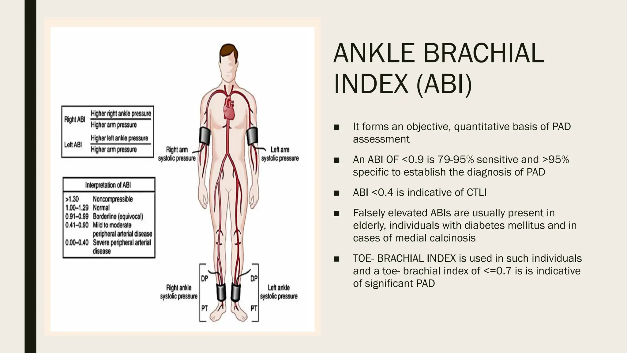
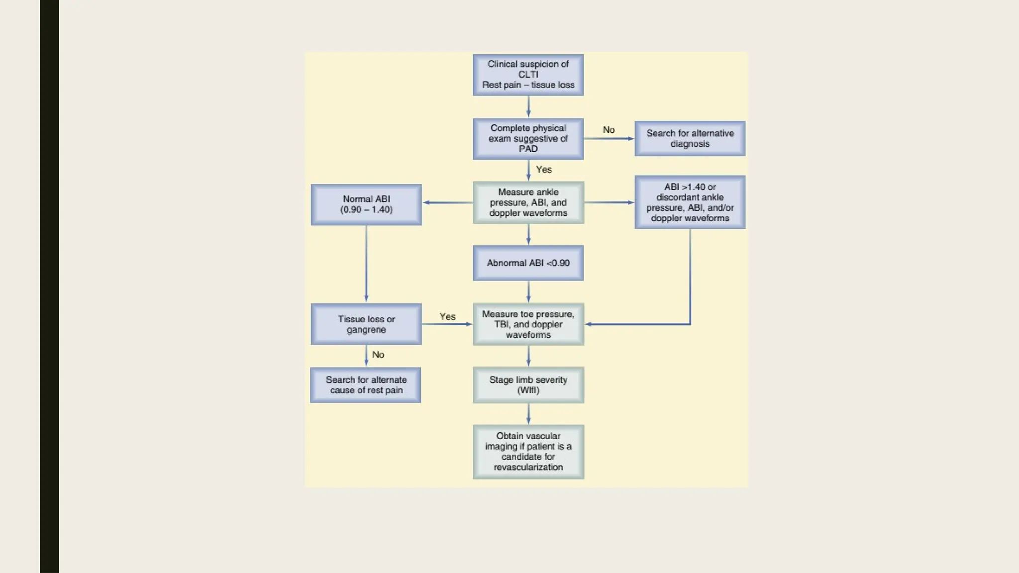
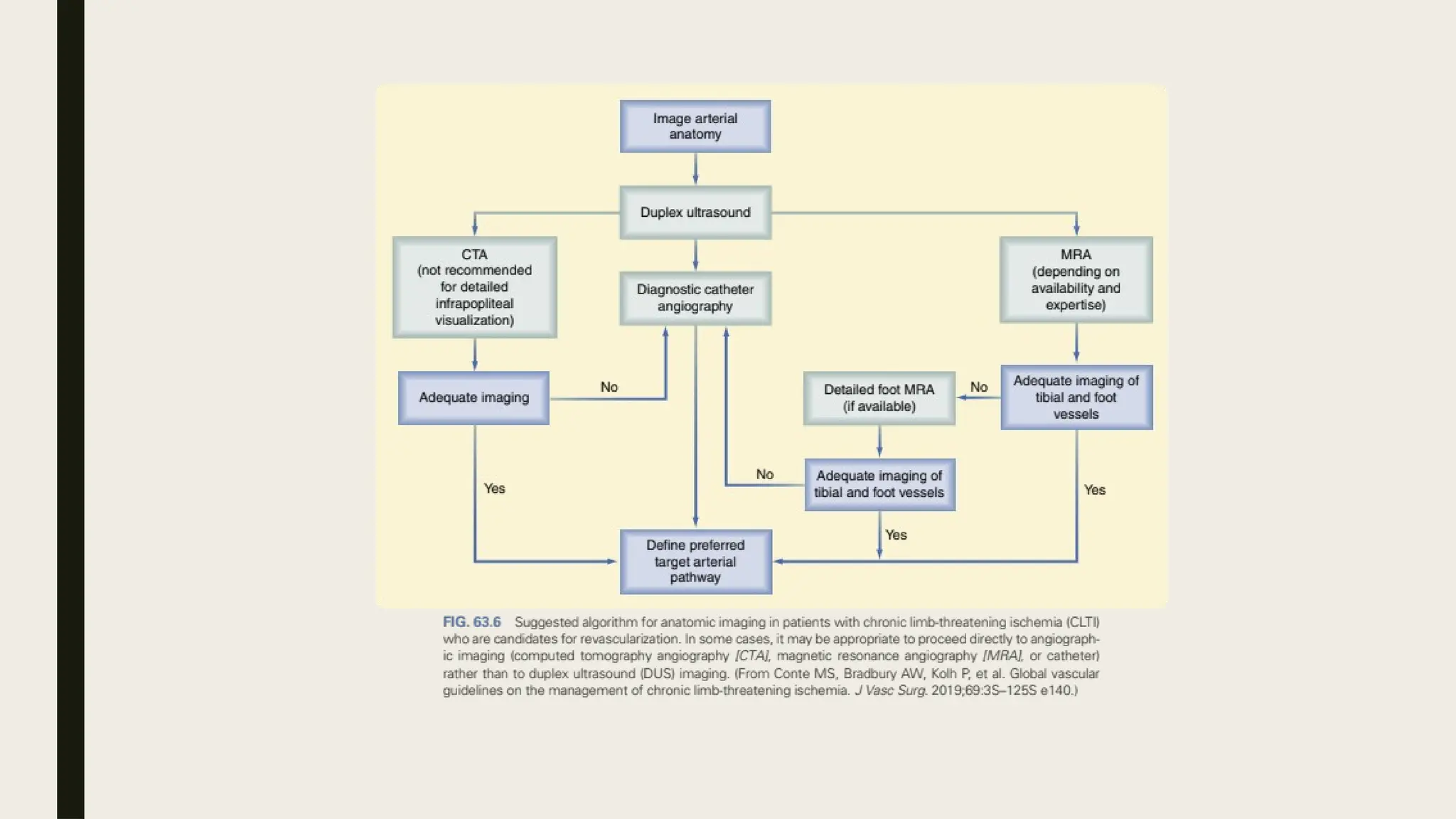
PERIPHERAL
ARTERIAL DISEASE
Presented by- Raksha R Thobbi- JR1- General Surgery
Under the guidance of- Dr Shalu Gupta ma’am
Moderators- Dr Bhupen Songra sir
Dr Garima Agarwal ma'am
Dr Sumit Choudhary sir
Dr Pahun Garg sir
DEFINITION
■ PAD or PVD is a slow and progressive disorder of the blood vessels.
■ Narrowing, blockage or spasms in a blood vessels can cause PAD
■ It can affect any blood vessels outside of the heart including arteries, veins or
lymphatic vessels. Organs supplied by these vessels such as the brains or legs, may
not get enough blood flow for healthy function, legs and feet being most often
affected
■ CRITICAL LIMB ISCHEMIA is defined as a clinical syndrome by the presence of PAD
in combination with rest pain, gangrene, lower limb ulceration for more than 2
weeks
SYMPTOMS
■ About half the people diagnosed with PVD have no symptoms. For those symptoms,
the most common first symptom is painful leg cramping that occurs with exercise
and is relieved by rest; known as intermittent claudication
■ During rest, the muscles need less blood flow, so the pain disappears. It may occur
in one or both legs. This depends on the location of the clogged or narrow artery.
■ Changes in the skin, including decreased skin temperature, or thin, brittle, shiny
skin on the legs and feet
■ Weak pulses in the legs and the feet
■ Gangrene (dead tissue due to lack of blood flow)
■ Hair loss on the legs
■ Impotence
■ Wounds that won't heal over pressure points, such as heels or ankles
■ Numbness, weakness, or heaviness in muscles
■ Pain (described as burning or aching) at rest, commonly in the toes and at night
while lying flat
■ Paleness when the legs are elevated
■ Reddish-blue discoloration of the extremities
■ Restricted mobility
■ Severe pain when the artery is very narrow or blocked
■ Thickened, opaque toenails
RISK FACTORS
■ MODIFIABLE FACTORS
1. Age (higher risk over age 50)
2. History of heart disease
3. Male gender
4. Postmenopausal women
5. Family history of high cholesterol,
high blood pressure, or peripheral
vascular disease
■ NON-MODIFIABLE FACTORS
1. Coronary artery disease
2. Diabetes
3. High cholesterol
4. High blood pressure
5. Overweight
6. Physical inactivity
7. Smoking or use of tobacco products
8. Those who smoke or have diabetes
have the highest risk of complications
from PVD. This is because these risk
factors cause impaired blood flow.
COMPLICATIONS
■ Complications of PVD most often occur because of decreased or absent blood flow.
■ Amputation (loss of a limb)
■ Poor wound healing
■ Restricted mobility due to pain or discomfort
■ Severe pain in the affected extremity
■ Stroke (three times more likely in people with PVD)
EVALUATION OF PATIENT
■ Patient should be evaluated for pertinent symptoms and associated disability
■ Past medical history including operative history
■ Assessment of all major cardiovascular risk factors (smoking, diabetes,
hypertension, hyperlipidemia, obesity, sedentary lifestyle)
■ Palpation of all accessible peripheral pulses
■ Auscultation of neck, abdomen, groin for bruits; Auscultation of heart and lungs
■ Palpation of abdomen, femoral and popliteal regions for aneurysm
■ evaluation of extremities- temperature changes, signs muscle atrophy, distal hair
loss, ulcers
• All patients with foot ulcers should be tested for neuropathy to detect
loss of protective sensation
PROBE TO BONE TEST
If bone is visualised or palpable,
osteomyelitis is to be considered
SEMMES WEINSTEIN
MONOFILAMENT TEST
ANKLE BRACHIAL
INDEX (ABI)
■ It forms an objective, quantitative basis of PAD
assessment
■ An ABI OF <0.9 is 79-95% sensitive and >95%
specific to establish the diagnosis of PAD
■ ABI <0.4 is indicative of CTLI
■ Falsely elevated ABIs are usually present in
elderly, individuals with diabetes mellitus and in
cases of medial calcinosis
■ TOE- BRACHIAL INDEX is used in such individuals
and a toe- brachial index of <=0.7 is is indicative
of significant PAD
INVESTIGATIONS
■ DOPPLER to find out the site of block—hand held Doppler can be used (Doppler:
Christian Johann Doppler, Austrian physicist). It is combination of B mode
ultrasound and Doppler study. Difference in transmitted beam of the ultrasound
and reflected beam is called as Doppler shift which is assessed and converted into
audible signals. It is used to study the site, extent, severity of block, and also
about collaterals. Audible sound is heard with normal flow, and sound is
important. Turbulence is heard when there is stenosed partially blocked artery.
Audible sound will be absent if there is complete block. Using Doppler probe
blood pressure at various levels can be assessed. Pulse wave tracing along the
artery is also important. Arterial diameter, blood flow rate, velocity of flowing
blood, assessment of stenosed segment is properly done using Doppler.
■ Treadmill test/ECG/ECHOCARDIPOGRAPHY to assess cardiac/ coronary status.
■ USGto see aneurysm/aorta and its anatomical changes/other vessels in the
abdomen/other organs.
■ PLETHYSMOGRAPHY: It measures the blood flow in limbs. Water filled volume recorder; air filled
volume recorder; mercury in silastic gauze is used after occluding the venous outflow. It is a
noninvasive method. Segmental plethysmog- raphy using occlusion cuffs of 65 mmHg pressure
is placed at thigh, calf and ankle levels and then quantitative measure of pulsation is done.
■ BROWN’S VASOMOTOR INDEX: Specific nerve of the ischaemic limb is anaesthetised like
posterior tibial nerve or ulnar nerve (local anaesthetia or spinal anaesthesia is given to
anaesthetise entire limb). If the ischaemic disease is at vasospasm stage (like in TAO), nerve
block will relieve the sympathetic vasospasm and skin temperature rises. It is compared to
mouth temperature of the patient. Rise in skin temperature—rise in mouth temperature) divided
by rise in mouth temperature is called as Brown’s vasomotor index. If it is more than 3.5, it is
due to vasospasm. If less than 3.5, sympathectomy is not beneficial.
■ TRANSCUTANEOUS OXIMETRY: By placing polarographic electrodes over the skin over thigh, leg
and foot of oxygen tension (tcPO2) can be measured which is reflection of underlying tissue
perfusion. Normal tcPO2 in the foot is 50-60 mmHg. Level less than 40 mmHg shows
inadequate wound healing. Level below 10 mmHg suggests critical ischaemia with complete
failure of wound healing.
• DIGITAL SUBTRACTION ANGIOGRAPHY
(DSA): Here vessel (artery) is delineated
in a better way by
eliminating other tissues through
computer system. AV fistulas,
haemangiomas, lesion in circle of Willis,
vascular tumours, other vascular
anomalies are well made out.
Dye is injected either to an artery or
vein. Injecting into a vein is technically
easier but larger dose of dye is required.
Injecting into an artery is technically
difficult but small dose of dye is
sufficient.
Advantages: Only vascular system is
visualised; other stems are eliminated by
computer subtraction. Small lesion, its
location and details are better observed
with greater clarity.
CT ANGIOGRAM/MR ANGIOGRAM
UPPER LIMB ISCHEMIA
■ Types- acute and chronic
■ Acute- causes- embolism; trauma; iatrogenic; post AV fistula steal syndrome; aortic
dissection
■ Chronic- causes- arteritis; atherosclerosis; TAO of upper limb; fibromuscular
dysplasia; APLA; hypercoagulable states; PCV; cold agglutinins
■ Symptoms- upper limb claudication, weakness, wasting of arm, forearm and hand
muscles; ischemic changes in skin, tapering of finger tips, brittle nails
RAYNAUD’S PHENOMENON
■ Episodic vasospasm/ arteriolar spasm
■ Sequence of clinical features
1. Local syncope: vasospasm- white cold palm with tingling and numbness
2. Local asphyxia: accumulation of deoxygenated blood- cyanosis and burning
sensation
3. Local recovery: relief of spasm- flushing of digits and palm with pain
4. Local gangrene: persistent spasm beyond ischemia time- ulceration/gangrene
■ Coffman criteria- episodic attacks od well demarcated reversible self limiting colour
changes for 1-20 minutes- on exposure to cold- symmetric- lasting for 2 years
■ Types of Raynaud’s-
1. Primary: due to increased sensitivity of alpha 2 receptors to norepinephrine, 5HT
and TXA2; decreased to NO and endothelin 1
2. Secondary- secondary to underlying vasculitis
■ Clinical manifestations: bilateral, young females; stage of syncope, asphyxia and
recovery; normal pulses
■ Treatment:
■ Avoid stimulants- exposure to cold, machinery
■ Low dose aspirin; CCB; endothelin inhibitors; prostacyclin analogues; cervical
sympathectomy
THORACIC OUTLET SYNDROME
■ Neurovascular disorder resulting from
compression of the brachial plexus and/or
subclavian vessels in the interval between
the neck and axilla.
• interscalene triangle-proximal space
contents brachial plexus trunks, subclavian
artery, subclavian vein does not pass
through interscalene triangle, runs
beneath anterior scalene muscle prior to
entering the costoclavicular space
• costoclavicular space- middle space
separated from the interscalene triangle
by the first rib borders; contents- brachial
plexus divisions subclavian artery and vein
• retropectoralis minor space distal space;
also known as the thoraco-coraco-pectoral
space or subcoracoid space; contents-
brachial plexus cords;axillary artery and
vein
■ HISTORY:
1. Neurogenic symptoms: non radicular pain, numbness, paresthesias, weakness
2. Vascular: cyanotic swelling of limb with distended veins; unilateral episodic Raynaud
type symptoms associated with cyanosis and erythema
3. Muscle atrophy: atrophy of abductor pollicis brevis, hypothenar muscles, interossei-
GILLIATT-SUMNER HAND
■ CAUSES:
1. Soft tissue abnormalities- scalene muscle abnormalities; anomalous costoclavicular
ligament (paget schroetter syndrome) ; soft tissue tumours like Pancoast tumours;
2. Osseous- cervical rib; prominent c7 process; abnormal clavicle
3. Vacular- embolic events; aneurysm formation; CLTI
■ SPECIFIC TESTS FOR THROACIC OUTLET OBSTRUCTION:
1. SUPRACLAVICULAR PRESSURE test- compression at interscalene triangle
2. ADSON’s test
3. Costoclavicular manuever
4. Wright’s test
5. Roos test/ elevated arm stress test
6. Cyriax release test
BUERGER’S DISEASE(TAO)
■ Segmental, progressive, occlusive, inflammation of small and medium vessels
■ Seen in young males (20-40 years) with h/o smoking
■ Carboxyhaemoglobin formed causes vasospasm and initmal hyperplasia leading to
thrombosis and obliteration of vessels leading to panarteritis
■ Classification of TAO:
1. Type 1- upper limb TAO
2. Type 2- involving legs/feet- crural/infrapopliteal
3. Type 3- femoropopliteal
4. Type 4- Aortoiliofemoral
5. Type 5- generalised
■ SHIANOYA’S CRITERIA:
1. Tobacco use; only in males
2. Disease onset before 45 years of age
3. Distal extremity involved first without embolic or atherosclerotic features
4. Absence of T2DM and hyperlipidemia
5. With or without thrombophlebitis
■ CLINICAL FEATURES- intermittent claudication in foot and calf, recurrent migratory
superficial thrombophlebitis, absent/feeble distal pulses
■ On angiogram- corkscrew appearance of vessel due to dilation of vasovasrum;
inverted tree/ spider leg collaterals
■ TREATMENT: quit smoking
■ Vasodilators
■ Pentoxiphylline
■ Low dose aspirin
■ Prostacyclins, ticlopidine
■ Clopidogrel with statins
■ PDE inhibitors- cilastazole
■ Xanthine nicotinate supplementation
■ Surgical management- omentoplasty; profundoflasty; lumbar sympathectomy;
amputation, ilizarvo method for bone neogenesis
• Dysfunction of the endothelium is
the earliest hallmark of vessel
injury (Ross hypothesis of
atherosclerosis) and it can be
detected before histologic changes
associated with atherosclerosis
are evident. .
• After initial epithelial dysfunction,
the endothelium increases the
expression of leukocyte adhesion
molecules such as vascular cell
adhesion molecule-1, intracellular
adhesion molecule-1, and P- and
E-selectin, which increases the
adherence of macrophages and
other leukocytes.
• Permeability of the endothelium
also increases and permits entry
of leukocytes and lipoproteins into
the subendothelial space.
■ Chemokines and cytokines such as monocyte chemotactic protein-1 and interleukin-8
further enhance the recruitment of leukocytes and smooth muscle cells (SMCs) into the
subendothelial space.
■ There are three stages of plaque, with the earliest stage termed the fatty streak. Fatty
streaks are focal, yellow, usually linear streaks that can be seen on the luminal surface of
arteries and are evident in most individuals after three years of age.
■ These streaks are microscopically macrophages (foam cells) full of lipid that accumulate
in the intima. They often occur at branch points. Atherosclerotic plaque also tends to
develop at branch points.
■ The intermediate stage is a fibrofatty lesion characterized by increased deposition of
layers of matrix around layered macrophages, T lymphocytes, and SMCs. The most
advanced stage is the complicated or fibrous plaque. .
■ Rupture of the cap characterizes an unstable plaque, which exposes the vessel lumen to
lipid and cellular debris, leading to the thrombotic complications associated with
atherosclerotic plaque
CLASSIFICATION OF PAD
LIFESTYLE MODIFICATIONS
■ Tobacco abuse is a frequent comorbid condition for patients with PAD and
specifically those with CLTI. The extent of cigarette smoking has been
shown to correlate with PAD severity. Tobacco abuse leads to higher MACEs
in patients with PAD and also contributes to PAD disease progression.
■ Exercise has been shown to have clear benefits for patients with PAD and
intermittent claudication and should be the attempted prior to
revascularization in these patients.
■ Exercise has been shown to improve walking distances in claudicants by
increasing calf blood flow, improving endothelial function, reducing local
inflammation, and inducing angiogenesis.
■ The general recommendations are for these patients to perform a
minimum of 45 to 60 minutes of exercise, 3 times per week for 12 weeks,
typically walking on a treadmill.
MEDICAL MANAGEMENT
■ Antithrombotic therapy is strongly recommended for all patients with PAD to reduce major
adverse cardiac events (MACEs), defined as a composite of nonfatal stroke, nonfatal
myocardial infarction, and cardiovascular death as a mainstay of this therapy is low-dose
aspirin.
■ Recent data suggests that further benefit might result from the use of alternative
antiplatelets agents such as clopidogrel or ticlopidine for lowering MACEs
■ A meta-analysis performed comparing single-agent antithrombotic use in PAD patients
suggested that clopidogrel monotherapy was most effective for lowering MACEs. Currently,
there is no clear ben- e t for dual antiplatelet therapy (DAPT) or systemic anticoagulation in
patients with PAD to lower MACEs, though there are several ongoing clinical trials to evaluate
this issue further.
■ Lipid-lowering therapy is essential in patients with PAD and has been demonstrated to
decrease MACEs. Additionally, there appears to be a direct anti-inflammatory effect in PAD
patients, which has been postulated to lead to atherosclerotic plaque stability and reduce
vascular events. It has been well established that high-intensity statin therapy decreases
MACEs in patients with PAD. Specifically, this includes high intensity rosuvastatin (20–40
mg/day) or simvastatin (40–80 mg/day).
■ Control of hypertension has been shown to decrease MACEs in patients with PAD.
Data suggests that targeting systolic blood pressure (SBP) less than 140 mm Hg
and diastolic blood pressure (DBP) less than 90 mm achieves optimal reduction in
MACEs in patients with PAD.
■ Specific categories of antihypertensives have not clearly been demonstrated to be
optimal in PAD patients, with angiotensin-converting enzyme (ACE) inhibitors,
calcium channel blockers, beta blockers, and diuretics all being effective to lower
MACEs.
■ Diabetes mellitus is a significant risk factor and contributor to the development of
atherosclerosis and PAD. The extent and severity of disease correlates with blood
glucose control. Therefore, glycemic control should be a focus of care in patients
with PAD specific goal is for patients is to maintain a Hemoglobin A1c level of less
than 7%. There has been a noted advantage for using metformin as the primary
hypoglycemic agent for patients with Type II diabetes and CLTI. Adjunctive
medications as well as insulin should be considered to achieve this HbA1c target.
ENDOVASCULAR THERAPY
■ Access should be obtained in every case using a combination of anatomic landmarks (based on
palpable bony or fluoroscopic landmarks), pulse palpation (if present), and ultrasound guidance.
■ retrograde common femoral artery access is most commonly used for aortic and common iliac
artery interventions, with up and over retrograde common femoral artery access for contralateral
external iliac and superficial femoral/above-knee popliteal interventions.
■ Antegrade common or proximal superficial femoral artery accesses are preferred by many when
treating infrageniculate disease, and because of the characteristics of proximal and distal caps for
longer or calcified occlusive lesions, retrograde access of a tibial or pedal vessel is often used
either alone or in combination with antegrade access in treating more complex CLTI cases.
■ Once the lesion(s) are identified, they must be crossed with a suitable wire to allow treatment.
■ Once the culprit arterial lesion has been successfully crossed, balloon angioplasty can be
performed. Balloon angioplasty fractures the plaque and may cause focal dissection, so
appropriate sizing is important.
■ Stents are reserved by some for residual stenosis, dissection or other complications of plain
balloon angioplasty. However, for iliac lesions, data would suggest that primary stenting is superior
to angioplasty (percutaneous transluminal angioplasty) alone
OPEN REVASCULARISATION
PROCEDURES
■ CT angiography can be considered for
assessment of inflow disease, although its use in
the lower extremity vessels, especially in the CLTI
cohort, is limited by the frequency of small, long
segment calcified vessels in patients with CLTI.
■ Magnetic resonance angiography and duplex
ultrasonography can also be successfully used for
preoperative planning.
■ Similarly, detailed assessment must be performed
to identify the most suitable distal target artery.
We are proponents of conventional digital
subtraction arteriography to identify the ideal
distal target.
■ Assessment of the inflow must be performed prior to performing a bypass.
■ In patients with a normal palpable ipsilateral femoral pulse and triphasic common
femoral Doppler arterial waveforms, intervention on the inflow is unlikely to be required.
■ In patients without a palpable femoral pulse or an abnormal femoral Doppler waveform
(especially when bilateral), additional preoperative imaging should be considered.
■ If disease exists in the ipsilateral aortoiliac segment or common femoral artery, these
should be treated either concurrently or prior to performing the infrainguinal bypass.
Both endovascular and open surgical options exist to optimize inflow, including
aortofemoral bypass, iliac balloon angioplasty with or without stenting, and common
femoral endarterectomy with or without profundaplasty.
■ The general principle in selecting a distal target vessel is to choose the most proximal
vessel distal to hemodynamically significant disease that has continuous runoff to the
foot through at least one tibial vessel.
■ Conduit selection is perhaps the
most critical factor underlying
successful infrainguinal bypass.
Autologous vein conduits include
ipsilateral and contralateral great
saphenous vein (GSV), short
saphenous vein (SSV), femoral
vein, arm (basilic and cephalic)
vein, endarterectomized superficial
femoral artery, cryopreserved vein,
and radial artery.
■ Prosthetic conduits include Dacron,
heparin-bonded Dacron, human
umbilical vein,
polytetrafluoroethylene (PTFE) with
and without covalently bonded
heparin, and expanded PTFE
bonded with heparin.
AORTO FEMORAL BYPASS
■ The typical reconstruction performed is
an aorto bifemoral bypass with the
outflow target either the common
femoral arteries or the profunda femoris
arteries bilaterally.
■ The preferred proximal aortic
anastomosis by many vascular surgeons
is end-to-end, as it is thus easier to
cover/protect the prosthetic from the
adjacent duodenum and although
unproven, it has been felt by some to be
hemodynamically superior.
■ There are certain settings, however, in
which an end-to-side proximal
anastomosis may be considered or even
be mandatory such as hypogastric
vessels.
COMPLICATIONS
■ Major complications following infrainguinal bypass include wound problems, graft occlusion, graft
infection, bleeding, and death.
■ The PREVENT III trial demonstrated the following complication rates associated with infrainguinal
vein bypass procedures: death (2.7%), myocardial infraction (4.7%), major amputation (1.8%),
graft occlusion (5.2%), major wound complication (4.8%), and graft hemorrhage (0.4%).
■ Late complications include lymphedema, infection, graft aneurysm, and graft stenosis or
occlusion.
■ Early graft occlusion is typically associated with technical or judgment error and should be
remedied as soon as possible.
■ Intermediate and late graft occlusion occurs due to a number of underlying causes, including
intimal hyperplasia (with a peak incidence in the first 18 postoperative months), anastomotic
aneurysm, and recurrent atherosclerotic disease. These should generally only be treated for high-
grade restenosis or whenever thepatient has return of symptoms or a nonhealing wound.
■ For vein grafts, structured serial duplex graft surveillance has been shown to reduce intermediate
and late bypass graft occlusion.
POST PROCEDURE SURVEILLANCE
■ The Society for Vascular Surgery guideline
suggests continued clinical follow up at 3 months
and then subsequently at 6-month intervals.
■ A general guideline following open surgical
revascularization includes early postoperative
assessment within 4 weeks of intervention and
then at 3-, 6, and 12-month intervals following the
operation. Thereafter, surveillance can be
continued every 6 to 12 months.
■ Vein graft surveillance is generally recommended
every 3 to 6 months for the first 2 years, and then
annually thereafter.
BIBLIOGRAPHY
■ Sabiston- textbook of surgery
■ SRB’s manual of surgery
■ Bailey and love’s short practice of surgery
■ Society of vascular surgeons' guidelines
THANK YOU

Peripheral arteial diaease presentation slideshow

  • 1.
    PERIPHERAL ARTERIAL DISEASE Presented by-Raksha R Thobbi- JR1- General Surgery Under the guidance of- Dr Shalu Gupta ma’am Moderators- Dr Bhupen Songra sir Dr Garima Agarwal ma'am Dr Sumit Choudhary sir Dr Pahun Garg sir
  • 2.
    DEFINITION ■ PAD orPVD is a slow and progressive disorder of the blood vessels. ■ Narrowing, blockage or spasms in a blood vessels can cause PAD ■ It can affect any blood vessels outside of the heart including arteries, veins or lymphatic vessels. Organs supplied by these vessels such as the brains or legs, may not get enough blood flow for healthy function, legs and feet being most often affected ■ CRITICAL LIMB ISCHEMIA is defined as a clinical syndrome by the presence of PAD in combination with rest pain, gangrene, lower limb ulceration for more than 2 weeks
  • 3.
    SYMPTOMS ■ About halfthe people diagnosed with PVD have no symptoms. For those symptoms, the most common first symptom is painful leg cramping that occurs with exercise and is relieved by rest; known as intermittent claudication ■ During rest, the muscles need less blood flow, so the pain disappears. It may occur in one or both legs. This depends on the location of the clogged or narrow artery. ■ Changes in the skin, including decreased skin temperature, or thin, brittle, shiny skin on the legs and feet ■ Weak pulses in the legs and the feet ■ Gangrene (dead tissue due to lack of blood flow) ■ Hair loss on the legs ■ Impotence
  • 4.
    ■ Wounds thatwon't heal over pressure points, such as heels or ankles ■ Numbness, weakness, or heaviness in muscles ■ Pain (described as burning or aching) at rest, commonly in the toes and at night while lying flat ■ Paleness when the legs are elevated ■ Reddish-blue discoloration of the extremities ■ Restricted mobility ■ Severe pain when the artery is very narrow or blocked ■ Thickened, opaque toenails
  • 5.
    RISK FACTORS ■ MODIFIABLEFACTORS 1. Age (higher risk over age 50) 2. History of heart disease 3. Male gender 4. Postmenopausal women 5. Family history of high cholesterol, high blood pressure, or peripheral vascular disease ■ NON-MODIFIABLE FACTORS 1. Coronary artery disease 2. Diabetes 3. High cholesterol 4. High blood pressure 5. Overweight 6. Physical inactivity 7. Smoking or use of tobacco products 8. Those who smoke or have diabetes have the highest risk of complications from PVD. This is because these risk factors cause impaired blood flow.
  • 6.
    COMPLICATIONS ■ Complications ofPVD most often occur because of decreased or absent blood flow. ■ Amputation (loss of a limb) ■ Poor wound healing ■ Restricted mobility due to pain or discomfort ■ Severe pain in the affected extremity ■ Stroke (three times more likely in people with PVD)
  • 7.
    EVALUATION OF PATIENT ■Patient should be evaluated for pertinent symptoms and associated disability ■ Past medical history including operative history ■ Assessment of all major cardiovascular risk factors (smoking, diabetes, hypertension, hyperlipidemia, obesity, sedentary lifestyle) ■ Palpation of all accessible peripheral pulses ■ Auscultation of neck, abdomen, groin for bruits; Auscultation of heart and lungs ■ Palpation of abdomen, femoral and popliteal regions for aneurysm ■ evaluation of extremities- temperature changes, signs muscle atrophy, distal hair loss, ulcers
  • 9.
    • All patientswith foot ulcers should be tested for neuropathy to detect loss of protective sensation PROBE TO BONE TEST If bone is visualised or palpable, osteomyelitis is to be considered SEMMES WEINSTEIN MONOFILAMENT TEST
  • 10.
    ANKLE BRACHIAL INDEX (ABI) ■It forms an objective, quantitative basis of PAD assessment ■ An ABI OF <0.9 is 79-95% sensitive and >95% specific to establish the diagnosis of PAD ■ ABI <0.4 is indicative of CTLI ■ Falsely elevated ABIs are usually present in elderly, individuals with diabetes mellitus and in cases of medial calcinosis ■ TOE- BRACHIAL INDEX is used in such individuals and a toe- brachial index of <=0.7 is is indicative of significant PAD
  • 11.
    INVESTIGATIONS ■ DOPPLER tofind out the site of block—hand held Doppler can be used (Doppler: Christian Johann Doppler, Austrian physicist). It is combination of B mode ultrasound and Doppler study. Difference in transmitted beam of the ultrasound and reflected beam is called as Doppler shift which is assessed and converted into audible signals. It is used to study the site, extent, severity of block, and also about collaterals. Audible sound is heard with normal flow, and sound is important. Turbulence is heard when there is stenosed partially blocked artery. Audible sound will be absent if there is complete block. Using Doppler probe blood pressure at various levels can be assessed. Pulse wave tracing along the artery is also important. Arterial diameter, blood flow rate, velocity of flowing blood, assessment of stenosed segment is properly done using Doppler. ■ Treadmill test/ECG/ECHOCARDIPOGRAPHY to assess cardiac/ coronary status. ■ USGto see aneurysm/aorta and its anatomical changes/other vessels in the abdomen/other organs.
  • 12.
    ■ PLETHYSMOGRAPHY: Itmeasures the blood flow in limbs. Water filled volume recorder; air filled volume recorder; mercury in silastic gauze is used after occluding the venous outflow. It is a noninvasive method. Segmental plethysmog- raphy using occlusion cuffs of 65 mmHg pressure is placed at thigh, calf and ankle levels and then quantitative measure of pulsation is done. ■ BROWN’S VASOMOTOR INDEX: Specific nerve of the ischaemic limb is anaesthetised like posterior tibial nerve or ulnar nerve (local anaesthetia or spinal anaesthesia is given to anaesthetise entire limb). If the ischaemic disease is at vasospasm stage (like in TAO), nerve block will relieve the sympathetic vasospasm and skin temperature rises. It is compared to mouth temperature of the patient. Rise in skin temperature—rise in mouth temperature) divided by rise in mouth temperature is called as Brown’s vasomotor index. If it is more than 3.5, it is due to vasospasm. If less than 3.5, sympathectomy is not beneficial. ■ TRANSCUTANEOUS OXIMETRY: By placing polarographic electrodes over the skin over thigh, leg and foot of oxygen tension (tcPO2) can be measured which is reflection of underlying tissue perfusion. Normal tcPO2 in the foot is 50-60 mmHg. Level less than 40 mmHg shows inadequate wound healing. Level below 10 mmHg suggests critical ischaemia with complete failure of wound healing.
  • 13.
    • DIGITAL SUBTRACTIONANGIOGRAPHY (DSA): Here vessel (artery) is delineated in a better way by eliminating other tissues through computer system. AV fistulas, haemangiomas, lesion in circle of Willis, vascular tumours, other vascular anomalies are well made out. Dye is injected either to an artery or vein. Injecting into a vein is technically easier but larger dose of dye is required. Injecting into an artery is technically difficult but small dose of dye is sufficient. Advantages: Only vascular system is visualised; other stems are eliminated by computer subtraction. Small lesion, its location and details are better observed with greater clarity.
  • 14.
  • 15.
    UPPER LIMB ISCHEMIA ■Types- acute and chronic ■ Acute- causes- embolism; trauma; iatrogenic; post AV fistula steal syndrome; aortic dissection ■ Chronic- causes- arteritis; atherosclerosis; TAO of upper limb; fibromuscular dysplasia; APLA; hypercoagulable states; PCV; cold agglutinins ■ Symptoms- upper limb claudication, weakness, wasting of arm, forearm and hand muscles; ischemic changes in skin, tapering of finger tips, brittle nails
  • 16.
    RAYNAUD’S PHENOMENON ■ Episodicvasospasm/ arteriolar spasm ■ Sequence of clinical features 1. Local syncope: vasospasm- white cold palm with tingling and numbness 2. Local asphyxia: accumulation of deoxygenated blood- cyanosis and burning sensation 3. Local recovery: relief of spasm- flushing of digits and palm with pain 4. Local gangrene: persistent spasm beyond ischemia time- ulceration/gangrene ■ Coffman criteria- episodic attacks od well demarcated reversible self limiting colour changes for 1-20 minutes- on exposure to cold- symmetric- lasting for 2 years
  • 17.
    ■ Types ofRaynaud’s- 1. Primary: due to increased sensitivity of alpha 2 receptors to norepinephrine, 5HT and TXA2; decreased to NO and endothelin 1 2. Secondary- secondary to underlying vasculitis ■ Clinical manifestations: bilateral, young females; stage of syncope, asphyxia and recovery; normal pulses ■ Treatment: ■ Avoid stimulants- exposure to cold, machinery ■ Low dose aspirin; CCB; endothelin inhibitors; prostacyclin analogues; cervical sympathectomy
  • 18.
    THORACIC OUTLET SYNDROME ■Neurovascular disorder resulting from compression of the brachial plexus and/or subclavian vessels in the interval between the neck and axilla. • interscalene triangle-proximal space contents brachial plexus trunks, subclavian artery, subclavian vein does not pass through interscalene triangle, runs beneath anterior scalene muscle prior to entering the costoclavicular space • costoclavicular space- middle space separated from the interscalene triangle by the first rib borders; contents- brachial plexus divisions subclavian artery and vein • retropectoralis minor space distal space; also known as the thoraco-coraco-pectoral space or subcoracoid space; contents- brachial plexus cords;axillary artery and vein
  • 19.
    ■ HISTORY: 1. Neurogenicsymptoms: non radicular pain, numbness, paresthesias, weakness 2. Vascular: cyanotic swelling of limb with distended veins; unilateral episodic Raynaud type symptoms associated with cyanosis and erythema 3. Muscle atrophy: atrophy of abductor pollicis brevis, hypothenar muscles, interossei- GILLIATT-SUMNER HAND ■ CAUSES: 1. Soft tissue abnormalities- scalene muscle abnormalities; anomalous costoclavicular ligament (paget schroetter syndrome) ; soft tissue tumours like Pancoast tumours; 2. Osseous- cervical rib; prominent c7 process; abnormal clavicle 3. Vacular- embolic events; aneurysm formation; CLTI
  • 20.
    ■ SPECIFIC TESTSFOR THROACIC OUTLET OBSTRUCTION: 1. SUPRACLAVICULAR PRESSURE test- compression at interscalene triangle 2. ADSON’s test 3. Costoclavicular manuever 4. Wright’s test 5. Roos test/ elevated arm stress test 6. Cyriax release test
  • 21.
    BUERGER’S DISEASE(TAO) ■ Segmental,progressive, occlusive, inflammation of small and medium vessels ■ Seen in young males (20-40 years) with h/o smoking ■ Carboxyhaemoglobin formed causes vasospasm and initmal hyperplasia leading to thrombosis and obliteration of vessels leading to panarteritis ■ Classification of TAO: 1. Type 1- upper limb TAO 2. Type 2- involving legs/feet- crural/infrapopliteal 3. Type 3- femoropopliteal 4. Type 4- Aortoiliofemoral 5. Type 5- generalised
  • 22.
    ■ SHIANOYA’S CRITERIA: 1.Tobacco use; only in males 2. Disease onset before 45 years of age 3. Distal extremity involved first without embolic or atherosclerotic features 4. Absence of T2DM and hyperlipidemia 5. With or without thrombophlebitis ■ CLINICAL FEATURES- intermittent claudication in foot and calf, recurrent migratory superficial thrombophlebitis, absent/feeble distal pulses ■ On angiogram- corkscrew appearance of vessel due to dilation of vasovasrum; inverted tree/ spider leg collaterals
  • 23.
    ■ TREATMENT: quitsmoking ■ Vasodilators ■ Pentoxiphylline ■ Low dose aspirin ■ Prostacyclins, ticlopidine ■ Clopidogrel with statins ■ PDE inhibitors- cilastazole ■ Xanthine nicotinate supplementation ■ Surgical management- omentoplasty; profundoflasty; lumbar sympathectomy; amputation, ilizarvo method for bone neogenesis
  • 24.
    • Dysfunction ofthe endothelium is the earliest hallmark of vessel injury (Ross hypothesis of atherosclerosis) and it can be detected before histologic changes associated with atherosclerosis are evident. . • After initial epithelial dysfunction, the endothelium increases the expression of leukocyte adhesion molecules such as vascular cell adhesion molecule-1, intracellular adhesion molecule-1, and P- and E-selectin, which increases the adherence of macrophages and other leukocytes. • Permeability of the endothelium also increases and permits entry of leukocytes and lipoproteins into the subendothelial space.
  • 25.
    ■ Chemokines andcytokines such as monocyte chemotactic protein-1 and interleukin-8 further enhance the recruitment of leukocytes and smooth muscle cells (SMCs) into the subendothelial space. ■ There are three stages of plaque, with the earliest stage termed the fatty streak. Fatty streaks are focal, yellow, usually linear streaks that can be seen on the luminal surface of arteries and are evident in most individuals after three years of age. ■ These streaks are microscopically macrophages (foam cells) full of lipid that accumulate in the intima. They often occur at branch points. Atherosclerotic plaque also tends to develop at branch points. ■ The intermediate stage is a fibrofatty lesion characterized by increased deposition of layers of matrix around layered macrophages, T lymphocytes, and SMCs. The most advanced stage is the complicated or fibrous plaque. . ■ Rupture of the cap characterizes an unstable plaque, which exposes the vessel lumen to lipid and cellular debris, leading to the thrombotic complications associated with atherosclerotic plaque
  • 26.
  • 31.
    LIFESTYLE MODIFICATIONS ■ Tobaccoabuse is a frequent comorbid condition for patients with PAD and specifically those with CLTI. The extent of cigarette smoking has been shown to correlate with PAD severity. Tobacco abuse leads to higher MACEs in patients with PAD and also contributes to PAD disease progression. ■ Exercise has been shown to have clear benefits for patients with PAD and intermittent claudication and should be the attempted prior to revascularization in these patients. ■ Exercise has been shown to improve walking distances in claudicants by increasing calf blood flow, improving endothelial function, reducing local inflammation, and inducing angiogenesis. ■ The general recommendations are for these patients to perform a minimum of 45 to 60 minutes of exercise, 3 times per week for 12 weeks, typically walking on a treadmill.
  • 32.
    MEDICAL MANAGEMENT ■ Antithrombotictherapy is strongly recommended for all patients with PAD to reduce major adverse cardiac events (MACEs), defined as a composite of nonfatal stroke, nonfatal myocardial infarction, and cardiovascular death as a mainstay of this therapy is low-dose aspirin. ■ Recent data suggests that further benefit might result from the use of alternative antiplatelets agents such as clopidogrel or ticlopidine for lowering MACEs ■ A meta-analysis performed comparing single-agent antithrombotic use in PAD patients suggested that clopidogrel monotherapy was most effective for lowering MACEs. Currently, there is no clear ben- e t for dual antiplatelet therapy (DAPT) or systemic anticoagulation in patients with PAD to lower MACEs, though there are several ongoing clinical trials to evaluate this issue further. ■ Lipid-lowering therapy is essential in patients with PAD and has been demonstrated to decrease MACEs. Additionally, there appears to be a direct anti-inflammatory effect in PAD patients, which has been postulated to lead to atherosclerotic plaque stability and reduce vascular events. It has been well established that high-intensity statin therapy decreases MACEs in patients with PAD. Specifically, this includes high intensity rosuvastatin (20–40 mg/day) or simvastatin (40–80 mg/day).
  • 33.
    ■ Control ofhypertension has been shown to decrease MACEs in patients with PAD. Data suggests that targeting systolic blood pressure (SBP) less than 140 mm Hg and diastolic blood pressure (DBP) less than 90 mm achieves optimal reduction in MACEs in patients with PAD. ■ Specific categories of antihypertensives have not clearly been demonstrated to be optimal in PAD patients, with angiotensin-converting enzyme (ACE) inhibitors, calcium channel blockers, beta blockers, and diuretics all being effective to lower MACEs. ■ Diabetes mellitus is a significant risk factor and contributor to the development of atherosclerosis and PAD. The extent and severity of disease correlates with blood glucose control. Therefore, glycemic control should be a focus of care in patients with PAD specific goal is for patients is to maintain a Hemoglobin A1c level of less than 7%. There has been a noted advantage for using metformin as the primary hypoglycemic agent for patients with Type II diabetes and CLTI. Adjunctive medications as well as insulin should be considered to achieve this HbA1c target.
  • 35.
  • 38.
    ■ Access shouldbe obtained in every case using a combination of anatomic landmarks (based on palpable bony or fluoroscopic landmarks), pulse palpation (if present), and ultrasound guidance. ■ retrograde common femoral artery access is most commonly used for aortic and common iliac artery interventions, with up and over retrograde common femoral artery access for contralateral external iliac and superficial femoral/above-knee popliteal interventions. ■ Antegrade common or proximal superficial femoral artery accesses are preferred by many when treating infrageniculate disease, and because of the characteristics of proximal and distal caps for longer or calcified occlusive lesions, retrograde access of a tibial or pedal vessel is often used either alone or in combination with antegrade access in treating more complex CLTI cases. ■ Once the lesion(s) are identified, they must be crossed with a suitable wire to allow treatment. ■ Once the culprit arterial lesion has been successfully crossed, balloon angioplasty can be performed. Balloon angioplasty fractures the plaque and may cause focal dissection, so appropriate sizing is important. ■ Stents are reserved by some for residual stenosis, dissection or other complications of plain balloon angioplasty. However, for iliac lesions, data would suggest that primary stenting is superior to angioplasty (percutaneous transluminal angioplasty) alone
  • 39.
    OPEN REVASCULARISATION PROCEDURES ■ CTangiography can be considered for assessment of inflow disease, although its use in the lower extremity vessels, especially in the CLTI cohort, is limited by the frequency of small, long segment calcified vessels in patients with CLTI. ■ Magnetic resonance angiography and duplex ultrasonography can also be successfully used for preoperative planning. ■ Similarly, detailed assessment must be performed to identify the most suitable distal target artery. We are proponents of conventional digital subtraction arteriography to identify the ideal distal target.
  • 40.
    ■ Assessment ofthe inflow must be performed prior to performing a bypass. ■ In patients with a normal palpable ipsilateral femoral pulse and triphasic common femoral Doppler arterial waveforms, intervention on the inflow is unlikely to be required. ■ In patients without a palpable femoral pulse or an abnormal femoral Doppler waveform (especially when bilateral), additional preoperative imaging should be considered. ■ If disease exists in the ipsilateral aortoiliac segment or common femoral artery, these should be treated either concurrently or prior to performing the infrainguinal bypass. Both endovascular and open surgical options exist to optimize inflow, including aortofemoral bypass, iliac balloon angioplasty with or without stenting, and common femoral endarterectomy with or without profundaplasty. ■ The general principle in selecting a distal target vessel is to choose the most proximal vessel distal to hemodynamically significant disease that has continuous runoff to the foot through at least one tibial vessel.
  • 41.
    ■ Conduit selectionis perhaps the most critical factor underlying successful infrainguinal bypass. Autologous vein conduits include ipsilateral and contralateral great saphenous vein (GSV), short saphenous vein (SSV), femoral vein, arm (basilic and cephalic) vein, endarterectomized superficial femoral artery, cryopreserved vein, and radial artery. ■ Prosthetic conduits include Dacron, heparin-bonded Dacron, human umbilical vein, polytetrafluoroethylene (PTFE) with and without covalently bonded heparin, and expanded PTFE bonded with heparin.
  • 42.
    AORTO FEMORAL BYPASS ■The typical reconstruction performed is an aorto bifemoral bypass with the outflow target either the common femoral arteries or the profunda femoris arteries bilaterally. ■ The preferred proximal aortic anastomosis by many vascular surgeons is end-to-end, as it is thus easier to cover/protect the prosthetic from the adjacent duodenum and although unproven, it has been felt by some to be hemodynamically superior. ■ There are certain settings, however, in which an end-to-side proximal anastomosis may be considered or even be mandatory such as hypogastric vessels.
  • 43.
    COMPLICATIONS ■ Major complicationsfollowing infrainguinal bypass include wound problems, graft occlusion, graft infection, bleeding, and death. ■ The PREVENT III trial demonstrated the following complication rates associated with infrainguinal vein bypass procedures: death (2.7%), myocardial infraction (4.7%), major amputation (1.8%), graft occlusion (5.2%), major wound complication (4.8%), and graft hemorrhage (0.4%). ■ Late complications include lymphedema, infection, graft aneurysm, and graft stenosis or occlusion. ■ Early graft occlusion is typically associated with technical or judgment error and should be remedied as soon as possible. ■ Intermediate and late graft occlusion occurs due to a number of underlying causes, including intimal hyperplasia (with a peak incidence in the first 18 postoperative months), anastomotic aneurysm, and recurrent atherosclerotic disease. These should generally only be treated for high- grade restenosis or whenever thepatient has return of symptoms or a nonhealing wound. ■ For vein grafts, structured serial duplex graft surveillance has been shown to reduce intermediate and late bypass graft occlusion.
  • 44.
    POST PROCEDURE SURVEILLANCE ■The Society for Vascular Surgery guideline suggests continued clinical follow up at 3 months and then subsequently at 6-month intervals. ■ A general guideline following open surgical revascularization includes early postoperative assessment within 4 weeks of intervention and then at 3-, 6, and 12-month intervals following the operation. Thereafter, surveillance can be continued every 6 to 12 months. ■ Vein graft surveillance is generally recommended every 3 to 6 months for the first 2 years, and then annually thereafter.
  • 45.
    BIBLIOGRAPHY ■ Sabiston- textbookof surgery ■ SRB’s manual of surgery ■ Bailey and love’s short practice of surgery ■ Society of vascular surgeons' guidelines
  • 46.