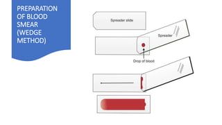
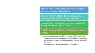
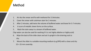
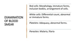
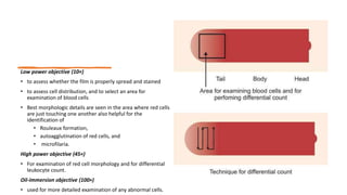
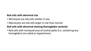
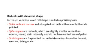
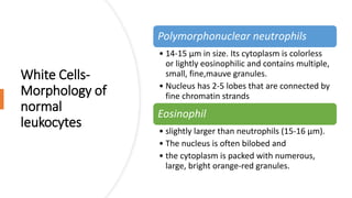
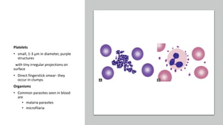
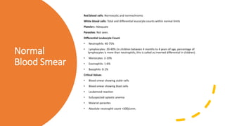
Blood Smear
DEFINITION
is a specimen for microscopic examination prepared by spreading a
drop of blood across a glass slide followed by staining with one of the
Romanowsky's stains
USES
1.the cause of anemia or thrombocytopenia
• identifying and typing of leukemia
• in diagnosing hemoparasitic infections (malaria, filaria, and
trypanosomiasis).
2. To monitor the effect of chemotherapy and radiotherapy
on bone marrow.
3. To provide direction for further investigations that will help in
arriving at the correct diagnosis (e.g. infections, drug toxicity, etc.).
PREPARATION
OF BLOOD
SMEAR
(WEDGE
METHOD)
Better blood cell morphology is obtained if smear is made
directly from a skin puncture.
If venous blood -prepared and stained within 2 hours of blood
collection
If collected in a syringe- the last drop of blood in the needle after
withdrawing (or first drop while dispensing) should be used
Aim of fixation is to prevent washing off of the smear from the
slide
If methanol is contaminated with water, sharpness of cell
morphology is lost and there is vacuolation of red cells.
Methanol should be acetone-free since acetone washes out
nuclear stain.
• 'spreader' slide—a glass slide with
absolutely smooth edges should be
selected
• 'spreader' slide should be narrower
• A well-spread blood smear
• (a) is tongue-shaped with a smooth
tail.
• (b) does not cover the entire area of
the slide .
• (c) has both thick and thin areas
with gradual transition, and
• (d) does not contain any lines or
holes
a thicker smear can be obtained by increasing the
angle and the speed of spreading
a thinner smear is obtained by decreasing the
'spreader' angle and the speed of spreading.
Heparin should not be used as an anticoagulant for
making blood films since it causes
platelet clumping and imparts a blue background to
the film.
• Recommended to stain blood films in reagent filled Coplin jars
• (uneven distribution of leukocytes (i.e. monocytes, neutrophils,
and abnormal cells are pushed towards the extreme tail end of
the smear)
• and distortion of red cell morphology at the edges.
Cleaning of
slides
New
slides
left overnight in a detergent solution,
washed in running tap water,
rinsed in distilled water, and wiped dry with a clean
cloth.
Before use, they are wiped with 95% methyl alcohol,
dried, and then kept covered to protect from dust.
Used
slides
soaked in a detergent solution at 60°C for 20 minutes,
washed in running tap water, rinsed in distilled water,
and then wiped dry.
Before use, they are wiped with 95% methyl alcohol,
dried, and then kept covered toprotect from dust.
STAINING
OF BLOOD
SMEAR
Leishman stain powder (0.6gram) is
mixed with water-free absolute
methyl alcohol (400 ml)
should be kept tightly stoppered in a
brown bottle and stored in a cool,
dark place at room temperature
stain should be kept for 3-5 days
before using since it improves the
quality of the stain.
Method
1. Air-dry the smear and fix with methanol for 2-3minutes.
2. Cover the smear with Leishman stain for 2 minutes.
3. After 2 minutes, add twice the volume of buffered water and leave for 5-7 minutes.
4. A scum of metallic sheen forms on the surface.
5. Wash the stain away in a stream of buffered water.
(Tap water can also be used for washing if it is not highly alkaline or highly acid)
6. Wipe the back of the slide clean and set it upright in the draining rack to
dry.
7. Mount the slide in a suitable mounting medium (e.g.DPX) with a clean and dry
25 × 25 mm coverslip.
A well-stained smear
• Red cells: pink-red or deep pink
• Polychromatic cells (Reticulocytes): Gray-
blue
• Neutrophils: Pale pink cytoplasm; mauve-
purple granules
• Eosinophils: Pale-pink cytoplasm; orange-
red granules
• Basophils: Blue cytoplasm; dark blue-violet
granules
• Monocytes: Gray-blue cytoplasm; fine
reddish (azurophil) granules
• Small lymphocytes: Dark blue cytoplasm
• Platelets: Purple
• Nuclei of all cells: Purple-violet
EXAMINATION
OF BLOOD
SMEAR
Red cells: Morphology, immature forms,
inclusion bodies, arrangement of cells.
White cells: Differential count, abnormal
or immature forms.
Platelets: Adequacy, abnormal forms.
Parasites: Malaria, filaria
Low power objective (10×)
• to assess whether the film is properly spread and stained
• to assess cell distribution, and to select an area for
examination of blood cells
• Best morphologic details are seen in the area where red cells
are just touching one another also helpful for the
identification of
• Rouleaux formation,
• autoagglutination of red cells, and
• microfilaria.
High power objective (45×)
• For examination of red cell morphology and for differential
leukocyte count.
Oil-immersion objective (100×)
• used for more detailed examination of any abnormal cells.
A well-stained smear is
• pink in color in thinner portion and purple-blue in thicker portion.
Excess blue coloration can be due to:
• (i) excessively thick smear,
• (ii) low concentration of eosin,
• (iii) impure dyes,
• (iv) too long staining time,
• (v) inadequate washing, or
• (vi) excessive alkaline pH of stain, buffer, or water.
Excess red coloration can be due to: (i) impure dyes or incorrect proportion of dyes,
• (ii) excessive acid pH of stain, buffer, or water (as the red cells take up more acid dye i.e. eosin),
• (iii) too short staining time, or
• (iv) excessive washing.
If there are granules of stain precipitate (masses of small black dots) on smear, stain needs to be filtered.
Red Cells
• Normal red cells are 7-8 μm in size.
• round with smooth contours, and stain deep pink
at the periphery and paler in the center.
• Area of central pallor is about 1/3rd the diameter
of the red cell..
• Normal red cells are described as normocytic (of
normal size) and normochromic (with normal
staining intensity i.e. hemoglobin content).
Morphologic
abnormalities of
red cells
Red cells with
abnormal size
Red cells with
abnormal
staining
Red cells with
abnormal
shape
Red cell
inclusions
Immature red
cells
Abnormal red
cell
arrangement
Red cells with abnormal size
• Microcytes are red cells smaller in size
• Macrocytes are red cells larger in size than normal
Red cells with abnormal staining (hemoglobin content):
• Red cells with increased area of central pallor (i.e. containing less
hemoglobin) are called as hypochromic.
Red cells with abnormal shape
Increased variation in red cell shape is called as poikilocytosis
• Sickle cells are narrow and elongated red cells with one or both ends
pointed
• Spherocytes are red cells, which are slightly smaller in size than
normal, round, stain intensely, and do not have central area of pallor
• Schistocytes are fragmented red cells-take various forms like helmet,
crescent, triangle, etc.
• Target cells are red cells with bull's eye
appearance. These red cells show a central
stained area and a peripheral stained rim with
unstained cytoplasm in Between
• Burr cells or echinocytes are small red cells with
regularly placed small projections on surface
• Acanthocytes are red cells with irregularly
spaced sharp projections of variable length on
surface
• Teardrop cells or dacryocytes have a tapering
droplike shape.
• Blister cells or hemi ghost cells are irregularly
contracted cells in which hemoglobin is
contracted and condensed away from the cell
membrane
• Bite cells result from removal of Heinz bodies by
the pitting action of the spleen
Variations in size and shape of red cells: (A) Microcytic hypochromic red cells in iron deficiency anemia; (B) Oval
macrocytes and a hypersegmented neutrophil in megaloblastic anemia; (C) Sickle cells in sickle cell anemia; (D)
Spherocytes
in hereditary spherocytosis; (E) Fragmented red cells or schistocytes in microangiopathic hemolytic anemia; (F)
Target cells
in hemoglobinopathy; (G) Burr cells in chronic renal failure; (H) Tear drop red cells in myelofibrosis; (I) Bite cells and
White Cells-
Morphology of
normal
leukocytes
Polymorphonuclear neutrophils
• 14-15 μm in size. Its cytoplasm is colorless
or lightly eosinophilic and contains multiple,
small, fine,mauve granules.
• Nucleus has 2-5 lobes that are connected by
fine chromatin strands
Eosinophil
• slightly larger than neutrophils (15-16 μm).
• The nucleus is often bilobed and
• the cytoplasm is packed with numerous,
large, bright orange-red granules.
Basophils are seen rarely on normal smears.
They are small (9-12 μm), round to oval cells, which contain very large,
coarse, deep purple Granules
Monocytes:the largest of the leukocytes (15-20 μm). It is irregular in shape,
with oval or clefted (kidney-shaped) nucleus and fine, delicate chromatin
Lymphocytes- small and large.
• small (7-8 μm).These cells have a high nuclear-cytoplasmic ratio with a thin
rim of deep blue cytoplasm. The nucleus is round or slightly clefted with
coarsely clumped chromatin.
• Large lymphocytes (10-15 μm) have a more abundant, pale blue cytoplasm.
Platelets
• small, 1-3 μm in diameter, purple
structures
with tiny irregular projections on
surface
• Direct fingerstick smear- they
occur in clumps.
Organisms
• Common parasites seen in blood
are
• malaria parasites
• microfilaria
Normal
Blood Smear
Red blood cells: Normocytic and normochromic
White blood cells: Total and differential leucocyte counts within normal limits
Platelets: Adequate
Parasites: Not seen.
Differential Leukocyte Count
• Neutrophils: 40-75%
• Lymphocytes: 20-40% (in children between 4 months to 4 years of age, percentage of
lymphocytes is more than neutrophils; this is called as inverted differential in children)
• Monocytes: 2-10%
• Eosinophils: 1-6%
• Basophils: 0-1%
Critical Values
• Blood smear showing sickle cells
• Blood smear showing blast cells
• Leukemoid reaction
• SuSuspected aplastic anemia
• Malarial parasites
• Absolute neutrophil count <500/cmm.

peripheral smear. DMLT.pptx

  • 1.
  • 2.
    DEFINITION is a specimenfor microscopic examination prepared by spreading a drop of blood across a glass slide followed by staining with one of the Romanowsky's stains
  • 3.
    USES 1.the cause ofanemia or thrombocytopenia • identifying and typing of leukemia • in diagnosing hemoparasitic infections (malaria, filaria, and trypanosomiasis). 2. To monitor the effect of chemotherapy and radiotherapy on bone marrow. 3. To provide direction for further investigations that will help in arriving at the correct diagnosis (e.g. infections, drug toxicity, etc.).
  • 4.
  • 5.
    Better blood cellmorphology is obtained if smear is made directly from a skin puncture. If venous blood -prepared and stained within 2 hours of blood collection If collected in a syringe- the last drop of blood in the needle after withdrawing (or first drop while dispensing) should be used Aim of fixation is to prevent washing off of the smear from the slide If methanol is contaminated with water, sharpness of cell morphology is lost and there is vacuolation of red cells. Methanol should be acetone-free since acetone washes out nuclear stain.
  • 6.
    • 'spreader' slide—aglass slide with absolutely smooth edges should be selected • 'spreader' slide should be narrower • A well-spread blood smear • (a) is tongue-shaped with a smooth tail. • (b) does not cover the entire area of the slide . • (c) has both thick and thin areas with gradual transition, and • (d) does not contain any lines or holes
  • 7.
    a thicker smearcan be obtained by increasing the angle and the speed of spreading a thinner smear is obtained by decreasing the 'spreader' angle and the speed of spreading. Heparin should not be used as an anticoagulant for making blood films since it causes platelet clumping and imparts a blue background to the film. • Recommended to stain blood films in reagent filled Coplin jars • (uneven distribution of leukocytes (i.e. monocytes, neutrophils, and abnormal cells are pushed towards the extreme tail end of the smear) • and distortion of red cell morphology at the edges.
  • 8.
    Cleaning of slides New slides left overnightin a detergent solution, washed in running tap water, rinsed in distilled water, and wiped dry with a clean cloth. Before use, they are wiped with 95% methyl alcohol, dried, and then kept covered to protect from dust. Used slides soaked in a detergent solution at 60°C for 20 minutes, washed in running tap water, rinsed in distilled water, and then wiped dry. Before use, they are wiped with 95% methyl alcohol, dried, and then kept covered toprotect from dust.
  • 9.
    STAINING OF BLOOD SMEAR Leishman stainpowder (0.6gram) is mixed with water-free absolute methyl alcohol (400 ml) should be kept tightly stoppered in a brown bottle and stored in a cool, dark place at room temperature stain should be kept for 3-5 days before using since it improves the quality of the stain.
  • 10.
    Method 1. Air-dry thesmear and fix with methanol for 2-3minutes. 2. Cover the smear with Leishman stain for 2 minutes. 3. After 2 minutes, add twice the volume of buffered water and leave for 5-7 minutes. 4. A scum of metallic sheen forms on the surface. 5. Wash the stain away in a stream of buffered water. (Tap water can also be used for washing if it is not highly alkaline or highly acid) 6. Wipe the back of the slide clean and set it upright in the draining rack to dry. 7. Mount the slide in a suitable mounting medium (e.g.DPX) with a clean and dry 25 × 25 mm coverslip.
  • 11.
    A well-stained smear •Red cells: pink-red or deep pink • Polychromatic cells (Reticulocytes): Gray- blue • Neutrophils: Pale pink cytoplasm; mauve- purple granules • Eosinophils: Pale-pink cytoplasm; orange- red granules • Basophils: Blue cytoplasm; dark blue-violet granules • Monocytes: Gray-blue cytoplasm; fine reddish (azurophil) granules • Small lymphocytes: Dark blue cytoplasm • Platelets: Purple • Nuclei of all cells: Purple-violet
  • 12.
    EXAMINATION OF BLOOD SMEAR Red cells:Morphology, immature forms, inclusion bodies, arrangement of cells. White cells: Differential count, abnormal or immature forms. Platelets: Adequacy, abnormal forms. Parasites: Malaria, filaria
  • 13.
    Low power objective(10×) • to assess whether the film is properly spread and stained • to assess cell distribution, and to select an area for examination of blood cells • Best morphologic details are seen in the area where red cells are just touching one another also helpful for the identification of • Rouleaux formation, • autoagglutination of red cells, and • microfilaria. High power objective (45×) • For examination of red cell morphology and for differential leukocyte count. Oil-immersion objective (100×) • used for more detailed examination of any abnormal cells.
  • 14.
    A well-stained smearis • pink in color in thinner portion and purple-blue in thicker portion. Excess blue coloration can be due to: • (i) excessively thick smear, • (ii) low concentration of eosin, • (iii) impure dyes, • (iv) too long staining time, • (v) inadequate washing, or • (vi) excessive alkaline pH of stain, buffer, or water. Excess red coloration can be due to: (i) impure dyes or incorrect proportion of dyes, • (ii) excessive acid pH of stain, buffer, or water (as the red cells take up more acid dye i.e. eosin), • (iii) too short staining time, or • (iv) excessive washing. If there are granules of stain precipitate (masses of small black dots) on smear, stain needs to be filtered.
  • 15.
    Red Cells • Normalred cells are 7-8 μm in size. • round with smooth contours, and stain deep pink at the periphery and paler in the center. • Area of central pallor is about 1/3rd the diameter of the red cell.. • Normal red cells are described as normocytic (of normal size) and normochromic (with normal staining intensity i.e. hemoglobin content).
  • 16.
    Morphologic abnormalities of red cells Redcells with abnormal size Red cells with abnormal staining Red cells with abnormal shape Red cell inclusions Immature red cells Abnormal red cell arrangement
  • 17.
    Red cells withabnormal size • Microcytes are red cells smaller in size • Macrocytes are red cells larger in size than normal Red cells with abnormal staining (hemoglobin content): • Red cells with increased area of central pallor (i.e. containing less hemoglobin) are called as hypochromic.
  • 18.
    Red cells withabnormal shape Increased variation in red cell shape is called as poikilocytosis • Sickle cells are narrow and elongated red cells with one or both ends pointed • Spherocytes are red cells, which are slightly smaller in size than normal, round, stain intensely, and do not have central area of pallor • Schistocytes are fragmented red cells-take various forms like helmet, crescent, triangle, etc.
  • 19.
    • Target cellsare red cells with bull's eye appearance. These red cells show a central stained area and a peripheral stained rim with unstained cytoplasm in Between • Burr cells or echinocytes are small red cells with regularly placed small projections on surface • Acanthocytes are red cells with irregularly spaced sharp projections of variable length on surface • Teardrop cells or dacryocytes have a tapering droplike shape. • Blister cells or hemi ghost cells are irregularly contracted cells in which hemoglobin is contracted and condensed away from the cell membrane • Bite cells result from removal of Heinz bodies by the pitting action of the spleen Variations in size and shape of red cells: (A) Microcytic hypochromic red cells in iron deficiency anemia; (B) Oval macrocytes and a hypersegmented neutrophil in megaloblastic anemia; (C) Sickle cells in sickle cell anemia; (D) Spherocytes in hereditary spherocytosis; (E) Fragmented red cells or schistocytes in microangiopathic hemolytic anemia; (F) Target cells in hemoglobinopathy; (G) Burr cells in chronic renal failure; (H) Tear drop red cells in myelofibrosis; (I) Bite cells and
  • 20.
    White Cells- Morphology of normal leukocytes Polymorphonuclearneutrophils • 14-15 μm in size. Its cytoplasm is colorless or lightly eosinophilic and contains multiple, small, fine,mauve granules. • Nucleus has 2-5 lobes that are connected by fine chromatin strands Eosinophil • slightly larger than neutrophils (15-16 μm). • The nucleus is often bilobed and • the cytoplasm is packed with numerous, large, bright orange-red granules.
  • 21.
    Basophils are seenrarely on normal smears. They are small (9-12 μm), round to oval cells, which contain very large, coarse, deep purple Granules Monocytes:the largest of the leukocytes (15-20 μm). It is irregular in shape, with oval or clefted (kidney-shaped) nucleus and fine, delicate chromatin Lymphocytes- small and large. • small (7-8 μm).These cells have a high nuclear-cytoplasmic ratio with a thin rim of deep blue cytoplasm. The nucleus is round or slightly clefted with coarsely clumped chromatin. • Large lymphocytes (10-15 μm) have a more abundant, pale blue cytoplasm.
  • 22.
    Platelets • small, 1-3μm in diameter, purple structures with tiny irregular projections on surface • Direct fingerstick smear- they occur in clumps. Organisms • Common parasites seen in blood are • malaria parasites • microfilaria
  • 23.
    Normal Blood Smear Red bloodcells: Normocytic and normochromic White blood cells: Total and differential leucocyte counts within normal limits Platelets: Adequate Parasites: Not seen. Differential Leukocyte Count • Neutrophils: 40-75% • Lymphocytes: 20-40% (in children between 4 months to 4 years of age, percentage of lymphocytes is more than neutrophils; this is called as inverted differential in children) • Monocytes: 2-10% • Eosinophils: 1-6% • Basophils: 0-1% Critical Values • Blood smear showing sickle cells • Blood smear showing blast cells • Leukemoid reaction • SuSuspected aplastic anemia • Malarial parasites • Absolute neutrophil count <500/cmm.