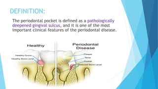
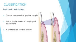
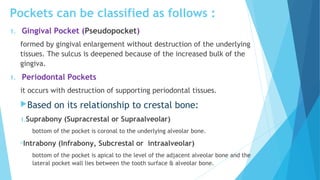
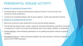
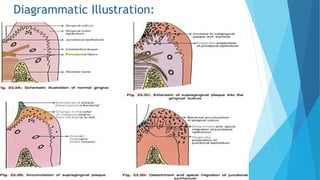
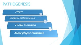
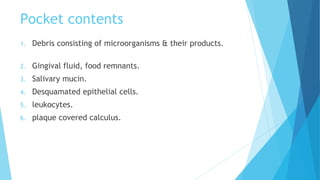
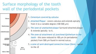
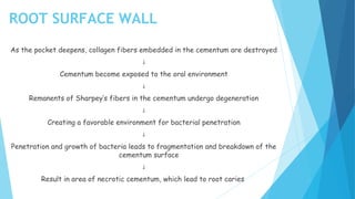
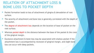
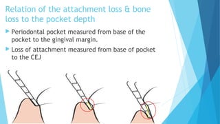
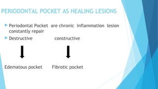
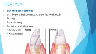
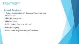
Periodontal pocket
Prepared by
Hana f.abdullah
OUTLINE
 Definition.
 Classification
 Pathogenesis
 Clinical features and histopathology
 Pocket Contents
 Morphology of pocket boundaries
 Measurement of periodontal pockets
 Treatment.
DEFINITION:
The periodontal pocket is defined as a pathologically
deepened gingival sulcus, and it is one of the most
important clinical features of the periodontal disease.
CLASSIFICATION
Based on its Morphology:
1. Coronal movement of gingival margin
2. Apical displacement of the gingival
attachment
3. A combination the two process
Pockets can be classified as follows :
1. Gingival Pocket (Pseudopocket)
formed by gingival enlargement without destruction of the underlying
tissues. The sulcus is deepened because of the increased bulk of the
gingiva.
1. Periodontal Pockets
it occurs with destruction of supporting periodontal tissues.
Based on its relationship to crestal bone:
1.Suprabony (Supracrestal or Supraalveolar)
bottom of the pocket is coronal to the underlying alveolar bone.
Intrabony (Infrabony, Subcrestal or intraalveolar)
bottom of the pocket is apical to the level of the adjacent alveolar bone and the
lateral pocket wall lies between the tooth surface & alveolar bone.
Suprabony pocket Infrabony pocket
1.Base of pocket is coronal to the level of
alveolar bone.
2.Horizontal pattern of bone destruction.
3.On facial and lingual surfaces , pdl
fibers beneath pocket follow their
normal oblique course.
4.Transeptal fibers are arranged
horizontally.
1.Base of pocket is apical to crest of
alveolar bone.
2.Vertical (angular) pattern of bone
destruction.
3.They follow angular pattern.
4. Transeptal fibers are arranged
obliquely.
A. Gingival Pocket B. Suprabony C. Intrabony Pocket
Based on number of surfaces involved:
1. Simple (involved one surface )
2. Compound (involved more than one surface )
3. Complex or Spiral – originating on one surface and twisting around
the tooth to involve one or more additional surfaces ( most
commonly found in furcation area)
I II III
Based on soft tissue wall of the pocket:
(1) Edematous Pocket.
(2) Fibrotic Pocket.
 
Based on the disease activity:
(1) Active Pocket.
(2) Inactive Pocket.
PERIODONTAL DISEASE ACTIVITY
1. PERIODS OF QUIESCENCE OR INACTIVITY
 Characterized by a reduced inflammatory response & little or no loss of bone and
connective tissue attachment.
 A build up of unattached plaque with its gram-negative, motile and anaerobic bacteria.
2. PERIODS OF EXACERBATION OR ACTIVITY
 Bone and connective tissue attachment are lost and the pocket deepens.
 This period may lasts for days, weeks, months & eventually followed by a period of remission
or quiescence in which G+ve bacteria proliferate and a more stable condition is established.
 Clinical features: shows bleeding spontaneous or on probing and greater amount of gingival
exudates.
 Histological Features : Pocket Epithelium appears thin and ulcerated, Infiltrate composed of
plasma cells & PMN leukocytes.
CLINICAL FEATURES HISTOPATHOLOGIC FEATURES
1. Bluish red,thickend marginal
gingiva
2. Gingival bleeding
3. Suppuration
4. Tooth mobility
5. Diastema formation
6. Symptoms-localised pain/pain
deep in the bone
1. 1.Circulatory stagnation.
2. Destruction of gingival fibers.
3. Atrophy of epithelium.
4. Edema and regeneration.
5. 2.Fibrotic changes .
6. 3.Increased vascularity, thinning
and degeneration of epithelium.
7. 4.Ulceration of inner aspect of
pocket wall.
8. 5.Suppuratiove inflammation of
inner wall.
PATHOGENESIS
 Presence of bacterial plaque on tooth surface
 Marginal gingiva become inflamed
 Gingiva sulcus deepens due to edematous enlargement of gingiva
 Gingiva pocket
 Anaerobic organisms tend to colonies the sub gingiva plaque
(Spirochetes and motile rods) (Due to an aerobic environment created in the pocket)
 Large number of PMN leukocytes and macrophages migrates to the gingiva tissue in
response to bacterial challenge
 Collagen fiber is loss
 Two mechanisms of collagen loss
 Lysosomal enzymes (Collagenase) released by PMN leukocytes
 Fibroblast phagocytoses collagen fibers by extending cytoplasmic process to the ligament
cementum interface Destruction of collagen fibers in gingival C.T. Collagen Matrix  
 coronal portion of the junctional epithelium get detached from the tooth
surface
 PMN cells migrates towards the coronal portion of junctional epithelium
 When volume of PMN leukocytes at the coronal portion of junctional
epithelium exceeds 60%, the epithelium cells separate from the tooth surface
 Pocket formation
 Plaque removal is difficult or impossible from deep pocket
 Favouring growth of pathogenic organism in that protected environment
 Further attachment loss
Diagrammatic Illustration:
PATHOGENESIS
Pocket contents
1. Debris consisting of microorganisms & their products.
2. Gingival fluid, food remnants.
3. Salivary mucin.
4. Desquamated epithelial cells.
5. leukocytes.
6. plaque covered calculus.
Surface morphology of the tooth
wall of the periodontal pockets
1. Cementum covered by calculus
2. Attached Plaque – covers calculus and extends apically
from it to a variable degree (100-500 µm)
3. The zone of unattached plaque Surround attached plaque
& extends apically to it.
4. The zone of attachment of Junctional Epithelium to the
tooth – this zone reduced to 100 µm (in periodontal
pocket) from 500 µm found in normal sulcus.
5. a zone of semi-destroyed connective tissue fibres – apical
to the JE
ROOT SURFACE WALL
As the pocket deepens, collagen fibers embedded in the cementum are destroyed
↓
Cementum become exposed to the oral environment
↓
Remanents of Sharpey’s fibers in the cementum undergo degeneration
↓
Creating a favorable environment for bacterial penetration
↓
Penetration and growth of bacteria leads to fragmentation and breakdown of the
cementum surface
↓
Result in area of necrotic cementum, which lead to root caries
RELATION OF ATTACHMENT LOSS &
BONE LOSS TO POCKET DEPTH
 Pocket formation leads to loss of attachment of gingiva & denudation of root
surface.
 The severity of attachment and bone loss is generally correlated with the depth of
the pocket.
 The degree of attachment loss depends on the location of base of pocket on the
root surface.
 Whereas pocket depth is the distance between the base of the pocket & the crest
of the gingival margin.
 Excessive attachment & bone loss may be associated with shallow pocket if the
attachment loss is accompanied by recession of gingival margin, and slight bone
loss can occur with deep pockets.
Relation of the attachment loss & bone
loss to the pocket depth
 Periodontal pocket measured from base of the
pocket to the gingival margin.
 Loss of attachment measured from base of pocket
to the CEJ
DIAGNOSIS/DETECTION OF POCKETS
 1.Careful exploration with a periodontal probe – accurate method.
 2.Radiograph: Pockets are not detected by radiographic examination
because pocket is a soft tissue change.
 Disadvantages of radiograph:
 Radiograph indicates areas of bone loss where pocket may be suspected,
they do not show pocket presence or depth.
 Radiograph show no difference before or after pocket elimination unless
bone has been modified
 Note: Gutta Percha points or Calibrated Silver points can be used with
radiograph to assist in determining the level of attachment of periodontal
pocket.
PERIODONTAL POCKET PROBING
The only reliable method of locating periodontal pockets and determining their extent is
careful probing along each tooth surface.
There are two different pocket depths -
1. Biologic or Histologic depth :- is the distance between the gingival margin and the base of
the pocket (the coronal end of the junctional epithelium.
2. Clinical or probing depth :- Is the distance from the gingival margin to which a probe
penetrates in to the pocket.
◉ According to several investigators - The probing force of 0.75 N or 25 gm have been
found to be well tolerated and accurate.
 In normal sulcus, the probe penetrates about one third to one half
the length of junctional epithelium
 In periodontal pocket with a short junctional epithelium the probe
penetrates beyond the apical end of junctional epithelium.
Vertical insertion of the probe (Left) may not detect interdental
craters, oblique positioning of the probe (Right) reaches the depth
of the crater.
Vertical Oblique
“Walking” the probe to explore the entire pocket
In the multirooted teeth the possibility of furcation involvement should be
carefully explored with specially designed probe (eg. Nabers probe)
PERIODONTAL POCKET AS HEALING LESIONS
 Periodontal Pocket are chronic inflammation lesion
constantly repair
 Destructive constructive
Edematous pocket Fibrotic pocket
TREATMENT
I. Non surgical treatment
1. oral hygiene instructions and their follow through.
2. Scaling.
3. Root planning.
4. Periodontal Medications
 Tetracycline
 Metranidazole
I. Surgical Treatment
 Pocket depth reduction through different surgical
procedures :
1. Gingival curettage.
2. Gingivectomy.
3. Periodontal flap procedures.
4. osseous surgery
5. Periodontal regenration preocedures.
TREATMENT
Periodontal pocket

Periodontal pocket

  • 1.
  • 2.
    OUTLINE  Definition.  Classification Pathogenesis  Clinical features and histopathology  Pocket Contents  Morphology of pocket boundaries  Measurement of periodontal pockets  Treatment.
  • 3.
    DEFINITION: The periodontal pocketis defined as a pathologically deepened gingival sulcus, and it is one of the most important clinical features of the periodontal disease.
  • 4.
    CLASSIFICATION Based on itsMorphology: 1. Coronal movement of gingival margin 2. Apical displacement of the gingival attachment 3. A combination the two process
  • 5.
    Pockets can beclassified as follows : 1. Gingival Pocket (Pseudopocket) formed by gingival enlargement without destruction of the underlying tissues. The sulcus is deepened because of the increased bulk of the gingiva. 1. Periodontal Pockets it occurs with destruction of supporting periodontal tissues. Based on its relationship to crestal bone: 1.Suprabony (Supracrestal or Supraalveolar) bottom of the pocket is coronal to the underlying alveolar bone. Intrabony (Infrabony, Subcrestal or intraalveolar) bottom of the pocket is apical to the level of the adjacent alveolar bone and the lateral pocket wall lies between the tooth surface & alveolar bone.
  • 6.
    Suprabony pocket Infrabonypocket 1.Base of pocket is coronal to the level of alveolar bone. 2.Horizontal pattern of bone destruction. 3.On facial and lingual surfaces , pdl fibers beneath pocket follow their normal oblique course. 4.Transeptal fibers are arranged horizontally. 1.Base of pocket is apical to crest of alveolar bone. 2.Vertical (angular) pattern of bone destruction. 3.They follow angular pattern. 4. Transeptal fibers are arranged obliquely.
  • 7.
    A. Gingival PocketB. Suprabony C. Intrabony Pocket
  • 8.
    Based on numberof surfaces involved: 1. Simple (involved one surface ) 2. Compound (involved more than one surface ) 3. Complex or Spiral – originating on one surface and twisting around the tooth to involve one or more additional surfaces ( most commonly found in furcation area) I II III
  • 9.
    Based on softtissue wall of the pocket: (1) Edematous Pocket. (2) Fibrotic Pocket.   Based on the disease activity: (1) Active Pocket. (2) Inactive Pocket.
  • 10.
    PERIODONTAL DISEASE ACTIVITY 1.PERIODS OF QUIESCENCE OR INACTIVITY  Characterized by a reduced inflammatory response & little or no loss of bone and connective tissue attachment.  A build up of unattached plaque with its gram-negative, motile and anaerobic bacteria. 2. PERIODS OF EXACERBATION OR ACTIVITY  Bone and connective tissue attachment are lost and the pocket deepens.  This period may lasts for days, weeks, months & eventually followed by a period of remission or quiescence in which G+ve bacteria proliferate and a more stable condition is established.  Clinical features: shows bleeding spontaneous or on probing and greater amount of gingival exudates.  Histological Features : Pocket Epithelium appears thin and ulcerated, Infiltrate composed of plasma cells & PMN leukocytes.
  • 11.
    CLINICAL FEATURES HISTOPATHOLOGICFEATURES 1. Bluish red,thickend marginal gingiva 2. Gingival bleeding 3. Suppuration 4. Tooth mobility 5. Diastema formation 6. Symptoms-localised pain/pain deep in the bone 1. 1.Circulatory stagnation. 2. Destruction of gingival fibers. 3. Atrophy of epithelium. 4. Edema and regeneration. 5. 2.Fibrotic changes . 6. 3.Increased vascularity, thinning and degeneration of epithelium. 7. 4.Ulceration of inner aspect of pocket wall. 8. 5.Suppuratiove inflammation of inner wall.
  • 12.
    PATHOGENESIS  Presence ofbacterial plaque on tooth surface  Marginal gingiva become inflamed  Gingiva sulcus deepens due to edematous enlargement of gingiva  Gingiva pocket  Anaerobic organisms tend to colonies the sub gingiva plaque (Spirochetes and motile rods) (Due to an aerobic environment created in the pocket)  Large number of PMN leukocytes and macrophages migrates to the gingiva tissue in response to bacterial challenge  Collagen fiber is loss  Two mechanisms of collagen loss  Lysosomal enzymes (Collagenase) released by PMN leukocytes  Fibroblast phagocytoses collagen fibers by extending cytoplasmic process to the ligament cementum interface Destruction of collagen fibers in gingival C.T. Collagen Matrix  
  • 13.
     coronal portionof the junctional epithelium get detached from the tooth surface  PMN cells migrates towards the coronal portion of junctional epithelium  When volume of PMN leukocytes at the coronal portion of junctional epithelium exceeds 60%, the epithelium cells separate from the tooth surface  Pocket formation  Plaque removal is difficult or impossible from deep pocket  Favouring growth of pathogenic organism in that protected environment  Further attachment loss
  • 14.
  • 15.
  • 16.
    Pocket contents 1. Debrisconsisting of microorganisms & their products. 2. Gingival fluid, food remnants. 3. Salivary mucin. 4. Desquamated epithelial cells. 5. leukocytes. 6. plaque covered calculus.
  • 17.
    Surface morphology ofthe tooth wall of the periodontal pockets 1. Cementum covered by calculus 2. Attached Plaque – covers calculus and extends apically from it to a variable degree (100-500 µm) 3. The zone of unattached plaque Surround attached plaque & extends apically to it. 4. The zone of attachment of Junctional Epithelium to the tooth – this zone reduced to 100 µm (in periodontal pocket) from 500 µm found in normal sulcus. 5. a zone of semi-destroyed connective tissue fibres – apical to the JE
  • 18.
    ROOT SURFACE WALL Asthe pocket deepens, collagen fibers embedded in the cementum are destroyed ↓ Cementum become exposed to the oral environment ↓ Remanents of Sharpey’s fibers in the cementum undergo degeneration ↓ Creating a favorable environment for bacterial penetration ↓ Penetration and growth of bacteria leads to fragmentation and breakdown of the cementum surface ↓ Result in area of necrotic cementum, which lead to root caries
  • 19.
    RELATION OF ATTACHMENTLOSS & BONE LOSS TO POCKET DEPTH  Pocket formation leads to loss of attachment of gingiva & denudation of root surface.  The severity of attachment and bone loss is generally correlated with the depth of the pocket.  The degree of attachment loss depends on the location of base of pocket on the root surface.  Whereas pocket depth is the distance between the base of the pocket & the crest of the gingival margin.  Excessive attachment & bone loss may be associated with shallow pocket if the attachment loss is accompanied by recession of gingival margin, and slight bone loss can occur with deep pockets.
  • 20.
    Relation of theattachment loss & bone loss to the pocket depth  Periodontal pocket measured from base of the pocket to the gingival margin.  Loss of attachment measured from base of pocket to the CEJ
  • 21.
    DIAGNOSIS/DETECTION OF POCKETS 1.Careful exploration with a periodontal probe – accurate method.  2.Radiograph: Pockets are not detected by radiographic examination because pocket is a soft tissue change.  Disadvantages of radiograph:  Radiograph indicates areas of bone loss where pocket may be suspected, they do not show pocket presence or depth.  Radiograph show no difference before or after pocket elimination unless bone has been modified  Note: Gutta Percha points or Calibrated Silver points can be used with radiograph to assist in determining the level of attachment of periodontal pocket.
  • 22.
    PERIODONTAL POCKET PROBING Theonly reliable method of locating periodontal pockets and determining their extent is careful probing along each tooth surface. There are two different pocket depths - 1. Biologic or Histologic depth :- is the distance between the gingival margin and the base of the pocket (the coronal end of the junctional epithelium. 2. Clinical or probing depth :- Is the distance from the gingival margin to which a probe penetrates in to the pocket. ◉ According to several investigators - The probing force of 0.75 N or 25 gm have been found to be well tolerated and accurate.
  • 23.
     In normalsulcus, the probe penetrates about one third to one half the length of junctional epithelium  In periodontal pocket with a short junctional epithelium the probe penetrates beyond the apical end of junctional epithelium.
  • 24.
    Vertical insertion ofthe probe (Left) may not detect interdental craters, oblique positioning of the probe (Right) reaches the depth of the crater. Vertical Oblique
  • 25.
    “Walking” the probeto explore the entire pocket In the multirooted teeth the possibility of furcation involvement should be carefully explored with specially designed probe (eg. Nabers probe)
  • 26.
    PERIODONTAL POCKET ASHEALING LESIONS  Periodontal Pocket are chronic inflammation lesion constantly repair  Destructive constructive Edematous pocket Fibrotic pocket
  • 27.
    TREATMENT I. Non surgicaltreatment 1. oral hygiene instructions and their follow through. 2. Scaling. 3. Root planning. 4. Periodontal Medications  Tetracycline  Metranidazole
  • 28.
    I. Surgical Treatment Pocket depth reduction through different surgical procedures : 1. Gingival curettage. 2. Gingivectomy. 3. Periodontal flap procedures. 4. osseous surgery 5. Periodontal regenration preocedures. TREATMENT