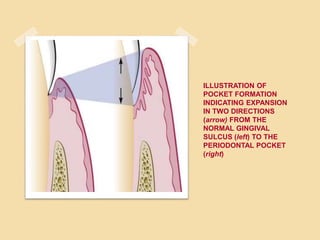
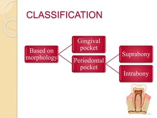
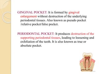
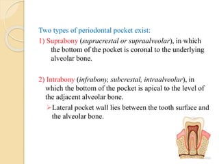
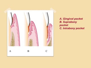
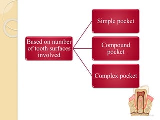
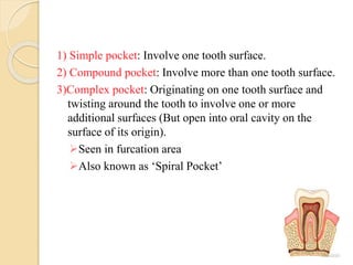
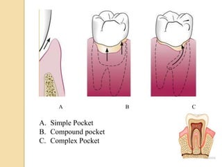
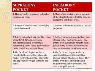
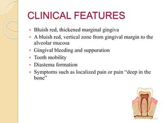
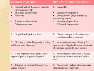
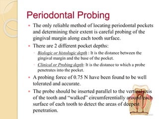
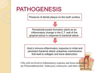
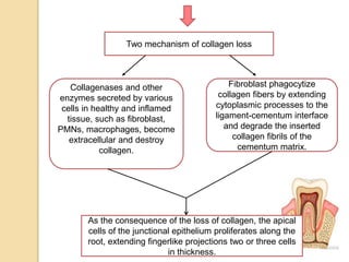
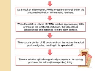
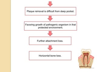
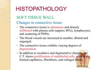
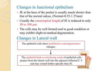
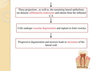
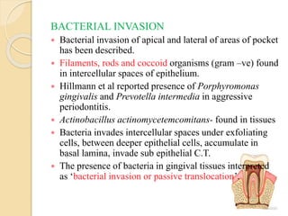
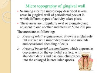
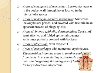
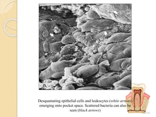
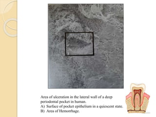
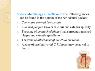
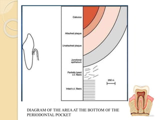
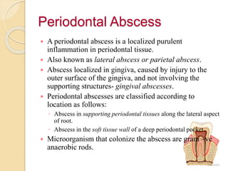
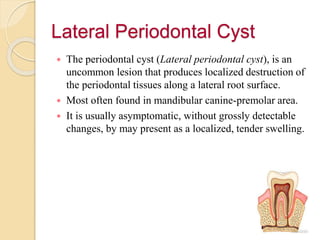
SlideShare a Scribd company logo
THE PERIODONTAL
POCKETS
Created by: HARPREET KAUR SIDHU
B.D.S. 3RD YEAR (2016-17)
ROLL NO.-35
OUTLINE
 Introduction
 Classification
 Clinical features
 The Periodontal Probing
 Pathogenesis
 Histopathology
◦ Bacterial invasion
◦ Mechanisms of Tissue destruction
◦ Micro topography of gingival wall
◦ Periodontal pockets as Healing lesion
◦ Pocket Content
◦ Root surface Walls
 Periodontal Disease Activity
 Site specificity
 Pulp changes associated with periodontal pockets
 Relationship of attachment loss and bone loss to pocket
depth
 Area between base of the pocket and alveolar bone
 Relationship of pocket to bone
 Periodontal Abscess
 Lateral Periodontal cyst
 Definition: The periodontal pocket is defined as a
pathologically deepened gingival sulcus.
 Cause: Deeping of gingival sulcus may occur by coronal
movement of gingival margin, apical displacement of
the gingival attachment, or combination of the two
processes.
 Normal: A probing depth of clinically normal gingival
sulcus in humans is 2-3 mm.
ILLUSTRATION OF
POCKET FORMATION
INDICATING EXPANSION
IN TWO DIRECTIONS
(arrow) FROM THE
NORMAL GINGIVAL
SULCUS (left) TO THE
PERIODONTAL POCKET
(right)
CLASSIFICATION
Based on
morphology
Gingival
pocket
Periodontal
pocket
Suprabony
Intrabony
GINGIVAL POCKET: It is formed by gingival
enlargement without destruction of the underlying
periodontal tissues. Also known as pseudo pocket
/relative pocket/false pocket.
PERIODONTAL POCKET: It produces destruction of the
supporting periodontal tissues, leading to loosening and
exfoliation of the teeth. It is also known as true or
absolute pocket.
Two types of periodontal pocket exist:
1) Suprabony (supracrestal or supraalveolar), in which
the bottom of the pocket is coronal to the underlying
alveolar bone.
2) Intrabony (infrabony, subcrestal, intraalveolar), in
which the bottom of the pocket is apical to the level of
the adjacent alveolar bone.
Lateral pocket wall lies between the tooth surface and
the alveolar bone.
A. Gingival pocket
B. Suprabony
pocket
C. Intrabony pocket
A B C
Based on number
of tooth surfaces
involved
Simple pocket
Compound
pocket
Complex pocket
1) Simple pocket: Involve one tooth surface.
2) Compound pocket: Involve more than one tooth surface.
3)Complex pocket: Originating on one tooth surface and
twisting around the tooth to involve one or more
additional surfaces (But open into oral cavity on the
surface of its origin).
Seen in furcation area
Also known as ‘Spiral Pocket’
A B C
A. Simple Pocket
B. Compound pocket
C. Complex Pocket
SUPRABONY
POCKET
INFRABONY
POCKET
1. Base of pocket is coronal to level of
the alveolar bone.
1. Base of the pocket is apical to crest
of the alveolar bone so that the bone is
adjacent to soft tissue wall.
2. Pattern of destruction of underlying
bone is horizontal.
2. Pattern of bone destruction is vertical
(angular).
3. Interproximally, transeptal fibers that
are restored during progressive
periodontal disease are arranged
horizontally in the space between base
of the pocket and alveolar bone.
3. Interproximally, transeptal fibers are
oblique rather than horizontal. They
extend from cementum beneath base of
pocket along alveolar bone and over
crest to cementum of adjacent tooth.
4. On facial and lingual surfaces,
periodontal ligament fibers beneath
pocket follow their normal horizontal-
oblique course between the tooth and
bone.
4. On facial and lingual surfaces,
periodontal ligament fibers follow
angular pattern of adjacent bone. They
extend from base of pocket along
alveolar bone and over crest to join
with outer periosteum.
CLINICAL FEATURES
 Bluish red, thickened marginal gingiva
 A bluish red, vertical zone from gingival margin to the
alveolar mucosa
 Gingival bleeding and suppuration
 Tooth mobility
 Diastema formation
 Symptoms such as localized pain or pain “deep in the
bone”
CLINICAL FEATURES HISTOPATHOLOGIC FEATURES
1. Gingival wall of the pocket presents
various degrees of:
• Bluish red discoloration
• Flaccidity
• A smooth, shiny surface
• Pitting on pressure.
1. Caused By:
• Circulatory stagnation
• Destruction of gingival fibers &
surrounding tissues.
• Atrophy of epithelium
• Edema & degeneration
2. Gingival wall pink and firm 2. Fibrotic changes predominate over
exudation and degeneration.
3. Bleeding is elicited by gently probing
soft tissue wall of pocket
3. Increased vascularity, thinning &
degeneration of epithelium and proximity
of engorged vessels to inner surface
4. When explored with a probe, inner
aspect of pocket is generally painful.
4. Pain on tactile stimulation is caused
by ulceration of inner aspect of pocket
wall.
5. Pus may be expressed by applying
digital pressure.
5. Pus occur in pockets with supurative
inflammation of inner wall.
Periodontal Probing
 The only reliable method of locating periodontal pockets
and determining their extent is careful probing of the
gingival margin along each tooth surface.
 There are 2 different pocket depths:
◦ Biologic or histologic depth : It is the distance between the
gingival margin and the base of the pocket.
◦ Clinical or Probing depth: It is the distance to which a probe
penetrates into the pocket.
 A probing force of 0.75 N have been found to be well
tolerated and accurate.
 The probe should be inserted parallel to the vertical axis
of the tooth and “walked” circumferentially around each
surface of each tooth to detect the areas of deepest
penetration.
PATHOGENESIS
Presence of dental plaque on the tooth surface.
Periodontal pocket formation starts as an
inflammatory change in the C.T. wall of the
gingival sulcus in response to bacterial attack.
Host’s immuno-inflammatory response to initial and
persistent bacterial attack unleashes mechanisms
that lead to collagen and bone destruction.
(The cells involved in inflammatory response and tissue destruction
are Polymorphonuclear leukocytes, monocytes, and other cells.)
Two mechanism of collagen loss
Collagenases and other
enzymes secreted by various
cells in healthy and inflamed
tissue, such as fibroblast,
PMNs, macrophages, become
extracellular and destroy
collagen.
Fibroblast phagocytize
collagen fibers by extending
cytoplasmic processes to the
ligament-cementum interface
and degrade the inserted
collagen fibrils of the
cementum matrix.
As the consequence of the loss of collagen, the apical
cells of the junctional epithelium proliferates along the
root, extending fingerlike projections two or three cells
in thickness.
As a result of inflammation, PMNs invade the coronal end of the
junctional epithelium in increasing numbers.
When the relative volume of PMNs reaches approximately 60%
or more of the junctional epithelium, the tissue loses
cohesiveness and detaches from the tooth surface.
Thus coronal portion of JE detaches from the root as the apical
portion migrates, resulting in its apical shift.
The oral sulcular epithelium gradually occupies an increasing
portion of the sulcus (then a pocket) lining.
Plaque removal is difficult from deep pocket.
Favoring growth of pathogenic organism in that
protected environment.
Further attachment loss.
Horizontal bone loss.
HISTOPATHOLOGY
SOFT TISSUE WALL
Changes in connective tissue
 The connective tissue is edematous and densely
infiltrated with plasma cells (approx 80%), lymphocytes,
and scattering of PMNs.
 The blood vessels are increased in number, dilated and
engorged.
 The connective tissue exhibits varying degrees of
degeneration.
 In addition to exudative and degenerative changes, the
C.T. shows proliferation of endothelial cells, with newly
formed capillaries, fibroblasts, and collagen fibers.
Changes in Junctional epithelium
 JE at the base of the pocket is usually much shorter than
that of the normal sulcus. (Normal-0.25-1.37mm)
 Usually the coronoapical length of JE is reduced to only
50 to 100 μm.
 The cells may be well formed and in good condition or
may exhibit slight-to-marked degeneration.
Changes in Lateral wall
The epithelial cells show proliferative and degenerative
changes.
The epithelial buds or interlacing cords of epithelial cells
project from the lateral wall into the adjacent inflamed C.T.
and may extend farther apically than JE.
These projections , as well as, the remaining lateral epithelium,
are densely infiltrated by leukocytes and edema from the inflamed
C.T.
Cells undergo vacuolar degeneration and rupture to form vesicles.
Progressive degeneration and necrosis leads to ulceration of the
lateral wall.
BACTERIAL INVASION
 Bacterial invasion of apical and lateral of areas of pocket
has been described.
 Filaments, rods and coccoid organisms (gram –ve) found
in intercellular spaces of epithelium.
 Hillmann et al reported presence of Porphyromonas
gingivalis and Prevotella intermedia in aggressive
periodontitis.
 Actinobacillus actinomycetemcomitans- found in tissues
 Bacteria invades intercellular spaces under exfoliating
cells, between deeper epithelial cells, accumulate in
basal lamina, invade sub epithelial C.T.
 The presence of bacteria in gingival tissues interpreted
as ‘bacterial invasion or passive translocation’.
Mechanism of tissue destruction
Inflammatory response triggered by bacterial plaque
Complex cascade events
Aimed at destroying and removing bacteria, necrotic cells,
and deleterious agents.
(It is a nonspecific process and host cells can damage or
destroy tissues by production of proteinases, cytokines,
and prostaglandins.)
Micro topography of gingival wall
 Scanning electron microscopy described several
areas in gingival wall of periodontal pocket in
which different types of activity takes place.
 These areas are irregularly oval or elongated and
adjacent to one another and measure 50 to 200 μm.
The areas are as following:
1) Areas of relative quiescence: Showing a relatively
flat surface with minor depression and mounds
and occasional shedding of cells
2) Areas of bacterial accumulation: which appears as
depressions on the epithelial surface, with
abundant debris and bacterial clumps penetrating
into the enlarged intercellular spaces.
3) Areas of emergence of leukocytes: Leukocytes appear
in the pocket wall through holes located in the
intercellular spaces.
4) Areas of leukocyte-bacteria interaction: Numerous
leukocytes are present and covered with bacteria in an
apparent process of phagocytosis.
5) Areas of intense epithelial desquamation: Consist of
semi attached and folded epithelial squames,
sometimes partially covered with bacteria.
6) Areas of ulceration: with exposed C.T.
7) Areas of hemorrhage: with numerous erythrocytes.
The transition from one areas to another could result
from bacteria accumulating in previously quiescent
areas and triggering the emergence of leukocytes and
leukocyte-bacteria interaction.
A) Area of Quiescence
B) Area of bacteria accumulation
C) Area of bacteria-leukocyte interaction
D) Area of intense cellular desquamation
Desquamating epithelial cells and leukocytes (white arrows)
emerging onto pocket space. Scattered bacteria can also be
seen (black arrows)
Area of ulceration in the lateral wall of a deep
periodontal pocket in human.
A) Surface of pocket epithelium in a quiescent state.
B) Area of Hemorrhage.
Periodontal pockets as Healing lesions
 Periodontal pockets are chronic inflammatory lesions
and thus constantly undergo repair.
 Complete healing does not occur because of the
persistence bacterial attack.
 The condition of the soft tissue wall is a result of the
destructive and constructive tissue changes.
 This balance determine the clinical features such as
color, consistency and surface texture of the pocket wall.
 According to this the pocket wall are divided into two:
◦ Edematous pocket wall
◦ Fibrotic pocket wall
 Edematous pocket wall: When the inflammatory fluid
and cellular exudate predominate, the pocket wall is
bluish red, soft, spongy, smooth, shiny surface.
 Fibrotic pocket wall: If newly formed C.T. cells and
fibers predominate, the pocket is firm and pink.
POCKET CONTENTS
 Contains debris (mostly microorganisms & their
products), gingival fluid, food remnants, salivary mucin,
desquamated epithelial cells, & leukocytes.
 Purulent exudate, if present, consist of living,
degenerated, and necrotic leukocytes; living and dead
bacteria; serum; and a scant amount of fibrin.
 Localized accumulation of pus constitutes an abcess.
ROOT SURFACE WALLS
 As the pocket deepens, collagen fibers embedded in the
cementum are destroyed and cementum becomes
exposed to the oral environment.
 Collagenous remnants of Sharpey’s fibers in the
cementum undergo degeneration, creating and
environment favorable to the penetration of bacteria.
 Bacterial penetration into the cementum can be found as
deep as the cementodentinal junction and may also enter
dentinal tubules.
 This result in fragmentation and breakdown of
cementum surface and results in areas of necrotic
cementum, separated from the tooth by masses of
bacteria.
 Cementum become soft, asymptomatic and painful on
probing.
 It is possible reservoir of re-infection.
 Necrotic areas are removed by scaling and root planning
until a hard, smooth surface is reached.
 Cementum is very thin in cervical area, and scaling and
root planning often remove it entirely, exposing the
underlying dentin.
Decalcification and Remineralization of Cementum
 Area of Increased mineralization are probably a result
of an exchange, on exposure to the oral cavity, of
minerals and organic components at the cementum-
saliva interface.
 The development of a highly mineralized superficial
layer may increase the tooth resistance to decay.
 Areas of demineralization are often related to root caries.
Exposure to oral fluid and bacterial plaque results in
proteolysis of the embedded remnants of Sharpey’s
fibers; the cementum may be softened and may undergo
fragmentation and cavitations.
 Caries of the cementum require special attention when
the pocket is treated. The necrotic cementum must be
removed by scaling and root planing until a firm tooth
surface is reached, even if this entails extension into the
dentin.
 Areas of cellular resorption of cementum, and dentin are
common in roots unexposed by periodontal disease.
 They are symptom free, and as long the root is covered
by the PDL, they are likely to undergo repair.
 However, if the root is expose by progressive pocket
formation before repair occurs, these areas appear as
isolated cavitations that penetrate into the dentin.
 They may be sources of considerable pain, requiring
placement of a restoration.
Surface Morphology of Tooth Wall: The following zones
can be found in the bottom of the periodontal pocket:
1. Cementum covered by calculus
2. Attached plaque: Covers calculus and extends apically.
3. The zone of unattached plaque that surrounds attached
plaque and extends apically to it.
4. The zone of attachment of the JE to the tooth.
5. A zone of semidestroyed C.T. flibers may be apical to
the JE.
DIAGRAM OF THE AREAAT THE BOTTOM OF THE
PERIODONTAL POCKET
Periodontal Disease activity
 More recently, as a result of studies on the specificity of
plaque bacteria, the concept of the periodontal disease
activity has evolved.
 According to this concept, periodontal pocket go
through periods of exacerbation and quiescence,
resulting from episodic bursts of activity followed by
periods of remission.
 Period of quiescence/ period of inactivity are
characterized by reduced inflammatory response and
little or no loss of bone and connective tissue
attachment.
 A build up of unattached plaque, with its gram –ve,
motile, and anaerobic bacteria, starts a period of
exacerbation/ period of activity.
 Period of exacerbation is characterized by bone and C.T.
attachment loss and pocket deepening.
 It is followed by period of remission or quiescence in
which gram +ve bacteria proliferate.
 Clinically, active periods show bleeding, either
spontaneously or with probing, and greater amount of
gingival exudate.
 Histologically, the pocket epithelium appears thin and
ulcerated and infiltrate composed predominantly of
plasma cells, PMNs, or both.
Site Specificity
 Periodontal destruction does not occur in all parts of the
mouth at the same time but rather on few teeth at a time
or even only some aspects of some teeth at any given
time.
 This is referred to as SITE SPECIFICITY.
Pulp Changes Associated With
Periodontal Pockets
 The spread of infection from periodontal pocket may
cause pathologic changes in the pulp.
 Such changes may give rise to painful symptoms or may
adversely affect the response of the pulp to restorative
procedures.
 Involvement of pulp occur either through apical foramen
or the lateral pulp canals after pocket infection reaches
them.
Relationship Of Attachment Loss
And Bone Loss To Pocket Depth.
 The severity of attachment loss in pocket formation is
generally but not always co related with the depth of the
pocket.
 This is because the degree of attachment loss depends on
the location of base of the pocket on the root surface,
whereas pocket depth is the distance between the base of
the pocket and the crest of the gingival margin.
 Pocket of the same depth may be associated with
different degrees of attachment loss, and pockets of
different depth may be associated with the same amount
of attachment loss.
 Severity of bone loss is generally, but not always, co
related with pocket depth.
 Extensive attachment and bone loss may be associated
with shallow pockets if the attachment loss is
accompanied by recession of gingival margin and slight
bone loss can occur with deep pockets.
Same pocket depth
with different amount
of recession.
A. Gingival pocket with
no recession.
B. Pocket depth as A
with some degree of
recession.
C. Pocket depth same
as A & B but more
recession.
A B C
Different pocket depth
with same amount of
attachment loss.
Arrows point to the
bottom of the pocket.
The distance between
arrow and CEJ remains
the same despite
different pocket depth
Area Between Base Of Pocket And
Alveolar Bone
 Normally the distance between the apical end of the JE
and the alveolar bone is relatively constant.
 Distance between apical extent of calculus and the
alveolar crest in human periodontal pockets is
constant=1.97mm
 The distance from attached plaque to bone is never less
than 0.5mm and never more than 2.7mm.
 These findings suggest that the bone-resorbing activity
induced by the bacteria is exerted within these distances.
Relationship Of Pocket To Bone
In Infrabony Pockets:
 Base of the pocket is apical to the crest of the alveolar bone.
 Pocket wall lies between the tooth and the bone.
 Bone loss is mostly vertical.
 Morphology of the bone changes with the formation of angular
bony defect.
 The interdental fibers run over the bone in an oblique direction
between the two teeth of the interdental space.
In Suprabony Pockets:
 Base is coronal to the crest of the alveolar bone and pocket wall lies
coronal to the bone.
 Bone loss is always horizontal.
 The alveolar crest gradually attains a more apical position in
relation to the tooth but retains its general morphology and
architecture.
 The interdental fibers run in usual horizontal direction.
Periodontal Abscess
 A periodontal abscess is a localized purulent
inflammation in periodontal tissue.
 Also known as lateral abscess or parietal abscess.
 Abscess localized in gingiva, caused by injury to the
outer surface of the gingiva, and not involving the
supporting structures- gingival abscesses.
 Periodontal abscesses are classified according to
location as follows:
◦ Abscess in supporting periodontal tissues along the lateral aspect
of root.
◦ Abscess in the soft tissue wall of a deep periodontal pocket.
 Microorganism that colonize the abscess are gram –ve
anaerobic rods.
Lateral Periodontal Cyst
 The periodontal cyst (Lateral periodontal cyst), is an
uncommon lesion that produces localized destruction of
the periodontal tissues along a lateral root surface.
 Most often found in mandibular canine-premolar area.
 It is usually asymptomatic, without grossly detectable
changes, by may present as a localized, tender swelling.
Conclusion
 Plaque is the etiological factor for periodontal pocket
formations.
 Host-microbial interaction is involved in initiation of
pocket formation.
 The periodontal pockets harbor microorganisms which
can cause further attachment and bone loss.
 A periodontal pocket may get affected and results in
abscess formation.
 Therefore, periodontal pockets needs treatment either by
scaling and root planing or by surgical treatment if the
condition worsens.
Peridontal pocket

More Related Content

What's hot

Bone loss and patterns of bone destruction
Bone loss and patterns of bone destructionBone loss and patterns of bone destruction
Bone loss and patterns of bone destruction
J.Rahul Raghavender
 
Clinical features of ginigvitis
Clinical features of ginigvitisClinical features of ginigvitis
Clinical features of ginigvitis
Dara Ghaznavi
 
Periodontal pathogenesis
Periodontal pathogenesisPeriodontal pathogenesis
Periodontal pathogenesis
Navneet Randhawa
 
Aggressive periodontitis
Aggressive periodontitisAggressive periodontitis
Aggressive periodontitis
DR. REBICCA RANJIT
 
Antibiotic in perio new caranza
Antibiotic in perio new caranzaAntibiotic in perio new caranza
Antibiotic in perio new caranza
Eman Hassona
 
Furcation involvement
Furcation involvementFurcation involvement
Furcation involvement
neeti shinde
 
Smoking and periodontal disease
Smoking and periodontal diseaseSmoking and periodontal disease
Smoking and periodontal disease
Navneet Randhawa
 
10.radiographic aids in diagnosing periodontal diseases
10.radiographic aids in diagnosing periodontal diseases 10.radiographic aids in diagnosing periodontal diseases
10.radiographic aids in diagnosing periodontal diseases
punitnaidu07
 
Non surgical periodontal therapy
Non surgical periodontal therapyNon surgical periodontal therapy
Non surgical periodontal therapy
Dr. Abhishek Ashok Sharma
 
Host modulation therapy
Host modulation therapyHost modulation therapy
Host modulation therapy
Ankita Dadwal
 
PLAQUE DISCLOSING AGENTS
PLAQUE DISCLOSING AGENTSPLAQUE DISCLOSING AGENTS
PLAQUE DISCLOSING AGENTS
drpoonamdholakia
 
Radiographic interpretation of periodontal diseases /prosthodontic courses
Radiographic  interpretation of periodontal diseases /prosthodontic coursesRadiographic  interpretation of periodontal diseases /prosthodontic courses
Radiographic interpretation of periodontal diseases /prosthodontic courses
Indian dental academy
 
Genetic factors and periodontal disease
Genetic factors and periodontal diseaseGenetic factors and periodontal disease
Genetic factors and periodontal disease
Navneet Randhawa
 
Drug induced gingival enlargement.
Drug induced gingival enlargement.Drug induced gingival enlargement.
Drug induced gingival enlargement.
Gururam MDS
 
Chemotherapy in periodontology
Chemotherapy in periodontologyChemotherapy in periodontology
Chemotherapy in periodontology
Dr Saif khan
 
Periodontal Medicine: Impact of periodontal disease on systemic health
Periodontal Medicine: Impact of periodontal disease on systemic healthPeriodontal Medicine: Impact of periodontal disease on systemic health
Periodontal Medicine: Impact of periodontal disease on systemic health
Binaya Subedi
 
Endo-Perio Lesions
Endo-Perio LesionsEndo-Perio Lesions
Endo-Perio Lesions
Mohamed Rabie'
 
FOOD IMPACTION AND TREATMENT
FOOD IMPACTION AND TREATMENTFOOD IMPACTION AND TREATMENT
FOOD IMPACTION AND TREATMENT
hariprasad757
 
Gingiva biotype
Gingiva biotypeGingiva biotype
Gingiva biotype
Salar Zeinali
 

What's hot (20)

Bone loss and patterns of bone destruction
Bone loss and patterns of bone destructionBone loss and patterns of bone destruction
Bone loss and patterns of bone destruction
 
Clinical features of ginigvitis
Clinical features of ginigvitisClinical features of ginigvitis
Clinical features of ginigvitis
 
Periodontal pathogenesis
Periodontal pathogenesisPeriodontal pathogenesis
Periodontal pathogenesis
 
Aggressive periodontitis
Aggressive periodontitisAggressive periodontitis
Aggressive periodontitis
 
Antibiotic in perio new caranza
Antibiotic in perio new caranzaAntibiotic in perio new caranza
Antibiotic in perio new caranza
 
Furcation involvement
Furcation involvementFurcation involvement
Furcation involvement
 
Smoking and periodontal disease
Smoking and periodontal diseaseSmoking and periodontal disease
Smoking and periodontal disease
 
10.radiographic aids in diagnosing periodontal diseases
10.radiographic aids in diagnosing periodontal diseases 10.radiographic aids in diagnosing periodontal diseases
10.radiographic aids in diagnosing periodontal diseases
 
Non surgical periodontal therapy
Non surgical periodontal therapyNon surgical periodontal therapy
Non surgical periodontal therapy
 
Host modulation therapy
Host modulation therapyHost modulation therapy
Host modulation therapy
 
PLAQUE DISCLOSING AGENTS
PLAQUE DISCLOSING AGENTSPLAQUE DISCLOSING AGENTS
PLAQUE DISCLOSING AGENTS
 
Radiographic interpretation of periodontal diseases /prosthodontic courses
Radiographic  interpretation of periodontal diseases /prosthodontic coursesRadiographic  interpretation of periodontal diseases /prosthodontic courses
Radiographic interpretation of periodontal diseases /prosthodontic courses
 
Genetic factors and periodontal disease
Genetic factors and periodontal diseaseGenetic factors and periodontal disease
Genetic factors and periodontal disease
 
Drug induced gingival enlargement.
Drug induced gingival enlargement.Drug induced gingival enlargement.
Drug induced gingival enlargement.
 
Chemotherapy in periodontology
Chemotherapy in periodontologyChemotherapy in periodontology
Chemotherapy in periodontology
 
Periodontal Medicine: Impact of periodontal disease on systemic health
Periodontal Medicine: Impact of periodontal disease on systemic healthPeriodontal Medicine: Impact of periodontal disease on systemic health
Periodontal Medicine: Impact of periodontal disease on systemic health
 
Endo-Perio Lesions
Endo-Perio LesionsEndo-Perio Lesions
Endo-Perio Lesions
 
Gingival curettage
Gingival curettageGingival curettage
Gingival curettage
 
FOOD IMPACTION AND TREATMENT
FOOD IMPACTION AND TREATMENTFOOD IMPACTION AND TREATMENT
FOOD IMPACTION AND TREATMENT
 
Gingiva biotype
Gingiva biotypeGingiva biotype
Gingiva biotype
 

Similar to Peridontal pocket

The periodontal pocket.pptx
The periodontal pocket.pptxThe periodontal pocket.pptx
The periodontal pocket.pptx
malti19
 
Periodontal pocket
Periodontal pocketPeriodontal pocket
Periodontal pocket
hanadentcare
 
Periodontal pocket activity
Periodontal pocket activityPeriodontal pocket activity
Periodontal pocket activity
Morteza Parmis ( Esmaeili )
 
Periodontal Pocket
Periodontal PocketPeriodontal Pocket
Periodontal Pocket
Kishlay Bhartiya
 
PERIODONTAL POCKET.pptx
PERIODONTAL POCKET.pptxPERIODONTAL POCKET.pptx
PERIODONTAL POCKET.pptx
veena621629
 
PERIODONTAL POCKET.pptx
PERIODONTAL POCKET.pptxPERIODONTAL POCKET.pptx
PERIODONTAL POCKET.pptx
veena621629
 
Periodontal Pocket
Periodontal PocketPeriodontal Pocket
Periodontal Pocket
Monali2011
 
Periodontal pockets.pptx
Periodontal  pockets.pptxPeriodontal  pockets.pptx
Periodontal pockets.pptx
abhijeetshete6
 
Periodontal pocket
Periodontal pocketPeriodontal pocket
Periodontal pocket
Supriya Bhat
 
perioDONTAL pocket
perioDONTAL pocketperioDONTAL pocket
perioDONTAL pocket
Fatima Gilani
 
Periodontal Pockets
Periodontal PocketsPeriodontal Pockets
Periodontal Pockets
Dr. Srishti Srivastava
 
(11) PERIODONTAL POCKET lecture.pptx
(11) PERIODONTAL POCKET lecture.pptx(11) PERIODONTAL POCKET lecture.pptx
(11) PERIODONTAL POCKET lecture.pptx
Rutu Dabhi
 
Periodontal pocket
Periodontal pocketPeriodontal pocket
Periodontal pocket
lobna elsaadawy
 
Periodontal pocket
Periodontal pocketPeriodontal pocket
Periodontal pocket
Enas Elgendy
 
Periodontal pocket
Periodontal pocketPeriodontal pocket
Periodontal pocketshyasaman
 
The periodontal pocket - lec 2
The periodontal pocket - lec 2The periodontal pocket - lec 2
The periodontal pocket - lec 2
Yahya Almoussawy
 
The periodontal pocket: pathogenesis, histopathology and consequences DIETER ...
The periodontal pocket: pathogenesis, histopathology and consequences DIETER ...The periodontal pocket: pathogenesis, histopathology and consequences DIETER ...
The periodontal pocket: pathogenesis, histopathology and consequences DIETER ...
andrea castells
 
The Periodontal Pocket
The Periodontal Pocket The Periodontal Pocket
The Periodontal Pocket
sambhav7
 

Similar to Peridontal pocket (20)

The periodontal pocket.pptx
The periodontal pocket.pptxThe periodontal pocket.pptx
The periodontal pocket.pptx
 
Periodontal pocket
Periodontal pocketPeriodontal pocket
Periodontal pocket
 
Periodontal pocket activity
Periodontal pocket activityPeriodontal pocket activity
Periodontal pocket activity
 
Periodontal Pocket
Periodontal PocketPeriodontal Pocket
Periodontal Pocket
 
PERIODONTAL POCKET.pptx
PERIODONTAL POCKET.pptxPERIODONTAL POCKET.pptx
PERIODONTAL POCKET.pptx
 
PERIODONTAL POCKET.pptx
PERIODONTAL POCKET.pptxPERIODONTAL POCKET.pptx
PERIODONTAL POCKET.pptx
 
Periodontal Pocket
Periodontal PocketPeriodontal Pocket
Periodontal Pocket
 
Peridontal pocket
Peridontal pocketPeridontal pocket
Peridontal pocket
 
Periodontal pockets.pptx
Periodontal  pockets.pptxPeriodontal  pockets.pptx
Periodontal pockets.pptx
 
Periodontal pocket
Periodontal pocketPeriodontal pocket
Periodontal pocket
 
perioDONTAL pocket
perioDONTAL pocketperioDONTAL pocket
perioDONTAL pocket
 
Periodontal Pockets
Periodontal PocketsPeriodontal Pockets
Periodontal Pockets
 
THE PERIODONTAL POCKET.ppt
THE PERIODONTAL POCKET.pptTHE PERIODONTAL POCKET.ppt
THE PERIODONTAL POCKET.ppt
 
(11) PERIODONTAL POCKET lecture.pptx
(11) PERIODONTAL POCKET lecture.pptx(11) PERIODONTAL POCKET lecture.pptx
(11) PERIODONTAL POCKET lecture.pptx
 
Periodontal pocket
Periodontal pocketPeriodontal pocket
Periodontal pocket
 
Periodontal pocket
Periodontal pocketPeriodontal pocket
Periodontal pocket
 
Periodontal pocket
Periodontal pocketPeriodontal pocket
Periodontal pocket
 
The periodontal pocket - lec 2
The periodontal pocket - lec 2The periodontal pocket - lec 2
The periodontal pocket - lec 2
 
The periodontal pocket: pathogenesis, histopathology and consequences DIETER ...
The periodontal pocket: pathogenesis, histopathology and consequences DIETER ...The periodontal pocket: pathogenesis, histopathology and consequences DIETER ...
The periodontal pocket: pathogenesis, histopathology and consequences DIETER ...
 
The Periodontal Pocket
The Periodontal Pocket The Periodontal Pocket
The Periodontal Pocket
 

Recently uploaded

Travis Hills' Endeavors in Minnesota: Fostering Environmental and Economic Pr...
Travis Hills' Endeavors in Minnesota: Fostering Environmental and Economic Pr...Travis Hills' Endeavors in Minnesota: Fostering Environmental and Economic Pr...
Travis Hills' Endeavors in Minnesota: Fostering Environmental and Economic Pr...
Travis Hills MN
 
Deep Behavioral Phenotyping in Systems Neuroscience for Functional Atlasing a...
Deep Behavioral Phenotyping in Systems Neuroscience for Functional Atlasing a...Deep Behavioral Phenotyping in Systems Neuroscience for Functional Atlasing a...
Deep Behavioral Phenotyping in Systems Neuroscience for Functional Atlasing a...
Ana Luísa Pinho
 
Eukaryotic Transcription Presentation.pptx
Eukaryotic Transcription Presentation.pptxEukaryotic Transcription Presentation.pptx
Eukaryotic Transcription Presentation.pptx
RitabrataSarkar3
 
Red blood cells- genesis-maturation.pptx
Red blood cells- genesis-maturation.pptxRed blood cells- genesis-maturation.pptx
Red blood cells- genesis-maturation.pptx
muralinath2
 
Chapter 12 - climate change and the energy crisis
Chapter 12 - climate change and the energy crisisChapter 12 - climate change and the energy crisis
Chapter 12 - climate change and the energy crisis
tonzsalvador2222
 
Comparing Evolved Extractive Text Summary Scores of Bidirectional Encoder Rep...
Comparing Evolved Extractive Text Summary Scores of Bidirectional Encoder Rep...Comparing Evolved Extractive Text Summary Scores of Bidirectional Encoder Rep...
Comparing Evolved Extractive Text Summary Scores of Bidirectional Encoder Rep...
University of Maribor
 
Richard's aventures in two entangled wonderlands
Richard's aventures in two entangled wonderlandsRichard's aventures in two entangled wonderlands
Richard's aventures in two entangled wonderlands
Richard Gill
 
Introduction to Mean Field Theory(MFT).pptx
Introduction to Mean Field Theory(MFT).pptxIntroduction to Mean Field Theory(MFT).pptx
Introduction to Mean Field Theory(MFT).pptx
zeex60
 
20240520 Planning a Circuit Simulator in JavaScript.pptx
20240520 Planning a Circuit Simulator in JavaScript.pptx20240520 Planning a Circuit Simulator in JavaScript.pptx
20240520 Planning a Circuit Simulator in JavaScript.pptx
Sharon Liu
 
Topic: SICKLE CELL DISEASE IN CHILDREN-3.pdf
Topic: SICKLE CELL DISEASE IN CHILDREN-3.pdfTopic: SICKLE CELL DISEASE IN CHILDREN-3.pdf
Topic: SICKLE CELL DISEASE IN CHILDREN-3.pdf
TinyAnderson
 
Shallowest Oil Discovery of Turkiye.pptx
Shallowest Oil Discovery of Turkiye.pptxShallowest Oil Discovery of Turkiye.pptx
Shallowest Oil Discovery of Turkiye.pptx
Gokturk Mehmet Dilci
 
如何办理(uvic毕业证书)维多利亚大学毕业证本科学位证书原版一模一样
如何办理(uvic毕业证书)维多利亚大学毕业证本科学位证书原版一模一样如何办理(uvic毕业证书)维多利亚大学毕业证本科学位证书原版一模一样
如何办理(uvic毕业证书)维多利亚大学毕业证本科学位证书原版一模一样
yqqaatn0
 
Anemia_ types_clinical significance.pptx
Anemia_ types_clinical significance.pptxAnemia_ types_clinical significance.pptx
Anemia_ types_clinical significance.pptx
muralinath2
 
Unveiling the Energy Potential of Marshmallow Deposits.pdf
Unveiling the Energy Potential of Marshmallow Deposits.pdfUnveiling the Energy Potential of Marshmallow Deposits.pdf
Unveiling the Energy Potential of Marshmallow Deposits.pdf
Erdal Coalmaker
 
DERIVATION OF MODIFIED BERNOULLI EQUATION WITH VISCOUS EFFECTS AND TERMINAL V...
DERIVATION OF MODIFIED BERNOULLI EQUATION WITH VISCOUS EFFECTS AND TERMINAL V...DERIVATION OF MODIFIED BERNOULLI EQUATION WITH VISCOUS EFFECTS AND TERMINAL V...
DERIVATION OF MODIFIED BERNOULLI EQUATION WITH VISCOUS EFFECTS AND TERMINAL V...
Wasswaderrick3
 
THEMATIC APPERCEPTION TEST(TAT) cognitive abilities, creativity, and critic...
THEMATIC  APPERCEPTION  TEST(TAT) cognitive abilities, creativity, and critic...THEMATIC  APPERCEPTION  TEST(TAT) cognitive abilities, creativity, and critic...
THEMATIC APPERCEPTION TEST(TAT) cognitive abilities, creativity, and critic...
Abdul Wali Khan University Mardan,kP,Pakistan
 
platelets_clotting_biogenesis.clot retractionpptx
platelets_clotting_biogenesis.clot retractionpptxplatelets_clotting_biogenesis.clot retractionpptx
platelets_clotting_biogenesis.clot retractionpptx
muralinath2
 
NuGOweek 2024 Ghent programme overview flyer
NuGOweek 2024 Ghent programme overview flyerNuGOweek 2024 Ghent programme overview flyer
NuGOweek 2024 Ghent programme overview flyer
pablovgd
 
DMARDs Pharmacolgy Pharm D 5th Semester.pdf
DMARDs Pharmacolgy Pharm D 5th Semester.pdfDMARDs Pharmacolgy Pharm D 5th Semester.pdf
DMARDs Pharmacolgy Pharm D 5th Semester.pdf
fafyfskhan251kmf
 
bordetella pertussis.................................ppt
bordetella pertussis.................................pptbordetella pertussis.................................ppt
bordetella pertussis.................................ppt
kejapriya1
 

Recently uploaded (20)

Travis Hills' Endeavors in Minnesota: Fostering Environmental and Economic Pr...
Travis Hills' Endeavors in Minnesota: Fostering Environmental and Economic Pr...Travis Hills' Endeavors in Minnesota: Fostering Environmental and Economic Pr...
Travis Hills' Endeavors in Minnesota: Fostering Environmental and Economic Pr...
 
Deep Behavioral Phenotyping in Systems Neuroscience for Functional Atlasing a...
Deep Behavioral Phenotyping in Systems Neuroscience for Functional Atlasing a...Deep Behavioral Phenotyping in Systems Neuroscience for Functional Atlasing a...
Deep Behavioral Phenotyping in Systems Neuroscience for Functional Atlasing a...
 
Eukaryotic Transcription Presentation.pptx
Eukaryotic Transcription Presentation.pptxEukaryotic Transcription Presentation.pptx
Eukaryotic Transcription Presentation.pptx
 
Red blood cells- genesis-maturation.pptx
Red blood cells- genesis-maturation.pptxRed blood cells- genesis-maturation.pptx
Red blood cells- genesis-maturation.pptx
 
Chapter 12 - climate change and the energy crisis
Chapter 12 - climate change and the energy crisisChapter 12 - climate change and the energy crisis
Chapter 12 - climate change and the energy crisis
 
Comparing Evolved Extractive Text Summary Scores of Bidirectional Encoder Rep...
Comparing Evolved Extractive Text Summary Scores of Bidirectional Encoder Rep...Comparing Evolved Extractive Text Summary Scores of Bidirectional Encoder Rep...
Comparing Evolved Extractive Text Summary Scores of Bidirectional Encoder Rep...
 
Richard's aventures in two entangled wonderlands
Richard's aventures in two entangled wonderlandsRichard's aventures in two entangled wonderlands
Richard's aventures in two entangled wonderlands
 
Introduction to Mean Field Theory(MFT).pptx
Introduction to Mean Field Theory(MFT).pptxIntroduction to Mean Field Theory(MFT).pptx
Introduction to Mean Field Theory(MFT).pptx
 
20240520 Planning a Circuit Simulator in JavaScript.pptx
20240520 Planning a Circuit Simulator in JavaScript.pptx20240520 Planning a Circuit Simulator in JavaScript.pptx
20240520 Planning a Circuit Simulator in JavaScript.pptx
 
Topic: SICKLE CELL DISEASE IN CHILDREN-3.pdf
Topic: SICKLE CELL DISEASE IN CHILDREN-3.pdfTopic: SICKLE CELL DISEASE IN CHILDREN-3.pdf
Topic: SICKLE CELL DISEASE IN CHILDREN-3.pdf
 
Shallowest Oil Discovery of Turkiye.pptx
Shallowest Oil Discovery of Turkiye.pptxShallowest Oil Discovery of Turkiye.pptx
Shallowest Oil Discovery of Turkiye.pptx
 
如何办理(uvic毕业证书)维多利亚大学毕业证本科学位证书原版一模一样
如何办理(uvic毕业证书)维多利亚大学毕业证本科学位证书原版一模一样如何办理(uvic毕业证书)维多利亚大学毕业证本科学位证书原版一模一样
如何办理(uvic毕业证书)维多利亚大学毕业证本科学位证书原版一模一样
 
Anemia_ types_clinical significance.pptx
Anemia_ types_clinical significance.pptxAnemia_ types_clinical significance.pptx
Anemia_ types_clinical significance.pptx
 
Unveiling the Energy Potential of Marshmallow Deposits.pdf
Unveiling the Energy Potential of Marshmallow Deposits.pdfUnveiling the Energy Potential of Marshmallow Deposits.pdf
Unveiling the Energy Potential of Marshmallow Deposits.pdf
 
DERIVATION OF MODIFIED BERNOULLI EQUATION WITH VISCOUS EFFECTS AND TERMINAL V...
DERIVATION OF MODIFIED BERNOULLI EQUATION WITH VISCOUS EFFECTS AND TERMINAL V...DERIVATION OF MODIFIED BERNOULLI EQUATION WITH VISCOUS EFFECTS AND TERMINAL V...
DERIVATION OF MODIFIED BERNOULLI EQUATION WITH VISCOUS EFFECTS AND TERMINAL V...
 
THEMATIC APPERCEPTION TEST(TAT) cognitive abilities, creativity, and critic...
THEMATIC  APPERCEPTION  TEST(TAT) cognitive abilities, creativity, and critic...THEMATIC  APPERCEPTION  TEST(TAT) cognitive abilities, creativity, and critic...
THEMATIC APPERCEPTION TEST(TAT) cognitive abilities, creativity, and critic...
 
platelets_clotting_biogenesis.clot retractionpptx
platelets_clotting_biogenesis.clot retractionpptxplatelets_clotting_biogenesis.clot retractionpptx
platelets_clotting_biogenesis.clot retractionpptx
 
NuGOweek 2024 Ghent programme overview flyer
NuGOweek 2024 Ghent programme overview flyerNuGOweek 2024 Ghent programme overview flyer
NuGOweek 2024 Ghent programme overview flyer
 
DMARDs Pharmacolgy Pharm D 5th Semester.pdf
DMARDs Pharmacolgy Pharm D 5th Semester.pdfDMARDs Pharmacolgy Pharm D 5th Semester.pdf
DMARDs Pharmacolgy Pharm D 5th Semester.pdf
 
bordetella pertussis.................................ppt
bordetella pertussis.................................pptbordetella pertussis.................................ppt
bordetella pertussis.................................ppt
 

Peridontal pocket

  • 1. THE PERIODONTAL POCKETS Created by: HARPREET KAUR SIDHU B.D.S. 3RD YEAR (2016-17) ROLL NO.-35
  • 2. OUTLINE  Introduction  Classification  Clinical features  The Periodontal Probing  Pathogenesis  Histopathology ◦ Bacterial invasion ◦ Mechanisms of Tissue destruction ◦ Micro topography of gingival wall ◦ Periodontal pockets as Healing lesion ◦ Pocket Content ◦ Root surface Walls
  • 3.  Periodontal Disease Activity  Site specificity  Pulp changes associated with periodontal pockets  Relationship of attachment loss and bone loss to pocket depth  Area between base of the pocket and alveolar bone  Relationship of pocket to bone  Periodontal Abscess  Lateral Periodontal cyst
  • 4.  Definition: The periodontal pocket is defined as a pathologically deepened gingival sulcus.  Cause: Deeping of gingival sulcus may occur by coronal movement of gingival margin, apical displacement of the gingival attachment, or combination of the two processes.  Normal: A probing depth of clinically normal gingival sulcus in humans is 2-3 mm.
  • 5. ILLUSTRATION OF POCKET FORMATION INDICATING EXPANSION IN TWO DIRECTIONS (arrow) FROM THE NORMAL GINGIVAL SULCUS (left) TO THE PERIODONTAL POCKET (right)
  • 7. GINGIVAL POCKET: It is formed by gingival enlargement without destruction of the underlying periodontal tissues. Also known as pseudo pocket /relative pocket/false pocket. PERIODONTAL POCKET: It produces destruction of the supporting periodontal tissues, leading to loosening and exfoliation of the teeth. It is also known as true or absolute pocket.
  • 8. Two types of periodontal pocket exist: 1) Suprabony (supracrestal or supraalveolar), in which the bottom of the pocket is coronal to the underlying alveolar bone. 2) Intrabony (infrabony, subcrestal, intraalveolar), in which the bottom of the pocket is apical to the level of the adjacent alveolar bone. Lateral pocket wall lies between the tooth surface and the alveolar bone.
  • 9. A. Gingival pocket B. Suprabony pocket C. Intrabony pocket A B C
  • 10. Based on number of tooth surfaces involved Simple pocket Compound pocket Complex pocket
  • 11. 1) Simple pocket: Involve one tooth surface. 2) Compound pocket: Involve more than one tooth surface. 3)Complex pocket: Originating on one tooth surface and twisting around the tooth to involve one or more additional surfaces (But open into oral cavity on the surface of its origin). Seen in furcation area Also known as ‘Spiral Pocket’
  • 12. A B C A. Simple Pocket B. Compound pocket C. Complex Pocket
  • 13. SUPRABONY POCKET INFRABONY POCKET 1. Base of pocket is coronal to level of the alveolar bone. 1. Base of the pocket is apical to crest of the alveolar bone so that the bone is adjacent to soft tissue wall. 2. Pattern of destruction of underlying bone is horizontal. 2. Pattern of bone destruction is vertical (angular). 3. Interproximally, transeptal fibers that are restored during progressive periodontal disease are arranged horizontally in the space between base of the pocket and alveolar bone. 3. Interproximally, transeptal fibers are oblique rather than horizontal. They extend from cementum beneath base of pocket along alveolar bone and over crest to cementum of adjacent tooth. 4. On facial and lingual surfaces, periodontal ligament fibers beneath pocket follow their normal horizontal- oblique course between the tooth and bone. 4. On facial and lingual surfaces, periodontal ligament fibers follow angular pattern of adjacent bone. They extend from base of pocket along alveolar bone and over crest to join with outer periosteum.
  • 14. CLINICAL FEATURES  Bluish red, thickened marginal gingiva  A bluish red, vertical zone from gingival margin to the alveolar mucosa  Gingival bleeding and suppuration  Tooth mobility  Diastema formation  Symptoms such as localized pain or pain “deep in the bone”
  • 15. CLINICAL FEATURES HISTOPATHOLOGIC FEATURES 1. Gingival wall of the pocket presents various degrees of: • Bluish red discoloration • Flaccidity • A smooth, shiny surface • Pitting on pressure. 1. Caused By: • Circulatory stagnation • Destruction of gingival fibers & surrounding tissues. • Atrophy of epithelium • Edema & degeneration 2. Gingival wall pink and firm 2. Fibrotic changes predominate over exudation and degeneration. 3. Bleeding is elicited by gently probing soft tissue wall of pocket 3. Increased vascularity, thinning & degeneration of epithelium and proximity of engorged vessels to inner surface 4. When explored with a probe, inner aspect of pocket is generally painful. 4. Pain on tactile stimulation is caused by ulceration of inner aspect of pocket wall. 5. Pus may be expressed by applying digital pressure. 5. Pus occur in pockets with supurative inflammation of inner wall.
  • 16. Periodontal Probing  The only reliable method of locating periodontal pockets and determining their extent is careful probing of the gingival margin along each tooth surface.  There are 2 different pocket depths: ◦ Biologic or histologic depth : It is the distance between the gingival margin and the base of the pocket. ◦ Clinical or Probing depth: It is the distance to which a probe penetrates into the pocket.  A probing force of 0.75 N have been found to be well tolerated and accurate.  The probe should be inserted parallel to the vertical axis of the tooth and “walked” circumferentially around each surface of each tooth to detect the areas of deepest penetration.
  • 17. PATHOGENESIS Presence of dental plaque on the tooth surface. Periodontal pocket formation starts as an inflammatory change in the C.T. wall of the gingival sulcus in response to bacterial attack. Host’s immuno-inflammatory response to initial and persistent bacterial attack unleashes mechanisms that lead to collagen and bone destruction. (The cells involved in inflammatory response and tissue destruction are Polymorphonuclear leukocytes, monocytes, and other cells.)
  • 18. Two mechanism of collagen loss Collagenases and other enzymes secreted by various cells in healthy and inflamed tissue, such as fibroblast, PMNs, macrophages, become extracellular and destroy collagen. Fibroblast phagocytize collagen fibers by extending cytoplasmic processes to the ligament-cementum interface and degrade the inserted collagen fibrils of the cementum matrix. As the consequence of the loss of collagen, the apical cells of the junctional epithelium proliferates along the root, extending fingerlike projections two or three cells in thickness.
  • 19. As a result of inflammation, PMNs invade the coronal end of the junctional epithelium in increasing numbers. When the relative volume of PMNs reaches approximately 60% or more of the junctional epithelium, the tissue loses cohesiveness and detaches from the tooth surface. Thus coronal portion of JE detaches from the root as the apical portion migrates, resulting in its apical shift. The oral sulcular epithelium gradually occupies an increasing portion of the sulcus (then a pocket) lining.
  • 20. Plaque removal is difficult from deep pocket. Favoring growth of pathogenic organism in that protected environment. Further attachment loss. Horizontal bone loss.
  • 21. HISTOPATHOLOGY SOFT TISSUE WALL Changes in connective tissue  The connective tissue is edematous and densely infiltrated with plasma cells (approx 80%), lymphocytes, and scattering of PMNs.  The blood vessels are increased in number, dilated and engorged.  The connective tissue exhibits varying degrees of degeneration.  In addition to exudative and degenerative changes, the C.T. shows proliferation of endothelial cells, with newly formed capillaries, fibroblasts, and collagen fibers.
  • 22. Changes in Junctional epithelium  JE at the base of the pocket is usually much shorter than that of the normal sulcus. (Normal-0.25-1.37mm)  Usually the coronoapical length of JE is reduced to only 50 to 100 μm.  The cells may be well formed and in good condition or may exhibit slight-to-marked degeneration. Changes in Lateral wall The epithelial cells show proliferative and degenerative changes. The epithelial buds or interlacing cords of epithelial cells project from the lateral wall into the adjacent inflamed C.T. and may extend farther apically than JE.
  • 23. These projections , as well as, the remaining lateral epithelium, are densely infiltrated by leukocytes and edema from the inflamed C.T. Cells undergo vacuolar degeneration and rupture to form vesicles. Progressive degeneration and necrosis leads to ulceration of the lateral wall.
  • 24. BACTERIAL INVASION  Bacterial invasion of apical and lateral of areas of pocket has been described.  Filaments, rods and coccoid organisms (gram –ve) found in intercellular spaces of epithelium.  Hillmann et al reported presence of Porphyromonas gingivalis and Prevotella intermedia in aggressive periodontitis.  Actinobacillus actinomycetemcomitans- found in tissues  Bacteria invades intercellular spaces under exfoliating cells, between deeper epithelial cells, accumulate in basal lamina, invade sub epithelial C.T.  The presence of bacteria in gingival tissues interpreted as ‘bacterial invasion or passive translocation’.
  • 25. Mechanism of tissue destruction Inflammatory response triggered by bacterial plaque Complex cascade events Aimed at destroying and removing bacteria, necrotic cells, and deleterious agents. (It is a nonspecific process and host cells can damage or destroy tissues by production of proteinases, cytokines, and prostaglandins.)
  • 26. Micro topography of gingival wall  Scanning electron microscopy described several areas in gingival wall of periodontal pocket in which different types of activity takes place.  These areas are irregularly oval or elongated and adjacent to one another and measure 50 to 200 μm. The areas are as following: 1) Areas of relative quiescence: Showing a relatively flat surface with minor depression and mounds and occasional shedding of cells 2) Areas of bacterial accumulation: which appears as depressions on the epithelial surface, with abundant debris and bacterial clumps penetrating into the enlarged intercellular spaces.
  • 27. 3) Areas of emergence of leukocytes: Leukocytes appear in the pocket wall through holes located in the intercellular spaces. 4) Areas of leukocyte-bacteria interaction: Numerous leukocytes are present and covered with bacteria in an apparent process of phagocytosis. 5) Areas of intense epithelial desquamation: Consist of semi attached and folded epithelial squames, sometimes partially covered with bacteria. 6) Areas of ulceration: with exposed C.T. 7) Areas of hemorrhage: with numerous erythrocytes. The transition from one areas to another could result from bacteria accumulating in previously quiescent areas and triggering the emergence of leukocytes and leukocyte-bacteria interaction.
  • 28. A) Area of Quiescence B) Area of bacteria accumulation C) Area of bacteria-leukocyte interaction D) Area of intense cellular desquamation
  • 29. Desquamating epithelial cells and leukocytes (white arrows) emerging onto pocket space. Scattered bacteria can also be seen (black arrows)
  • 30. Area of ulceration in the lateral wall of a deep periodontal pocket in human. A) Surface of pocket epithelium in a quiescent state. B) Area of Hemorrhage.
  • 31. Periodontal pockets as Healing lesions  Periodontal pockets are chronic inflammatory lesions and thus constantly undergo repair.  Complete healing does not occur because of the persistence bacterial attack.  The condition of the soft tissue wall is a result of the destructive and constructive tissue changes.  This balance determine the clinical features such as color, consistency and surface texture of the pocket wall.  According to this the pocket wall are divided into two: ◦ Edematous pocket wall ◦ Fibrotic pocket wall
  • 32.  Edematous pocket wall: When the inflammatory fluid and cellular exudate predominate, the pocket wall is bluish red, soft, spongy, smooth, shiny surface.  Fibrotic pocket wall: If newly formed C.T. cells and fibers predominate, the pocket is firm and pink. POCKET CONTENTS  Contains debris (mostly microorganisms & their products), gingival fluid, food remnants, salivary mucin, desquamated epithelial cells, & leukocytes.  Purulent exudate, if present, consist of living, degenerated, and necrotic leukocytes; living and dead bacteria; serum; and a scant amount of fibrin.  Localized accumulation of pus constitutes an abcess.
  • 33. ROOT SURFACE WALLS  As the pocket deepens, collagen fibers embedded in the cementum are destroyed and cementum becomes exposed to the oral environment.  Collagenous remnants of Sharpey’s fibers in the cementum undergo degeneration, creating and environment favorable to the penetration of bacteria.  Bacterial penetration into the cementum can be found as deep as the cementodentinal junction and may also enter dentinal tubules.  This result in fragmentation and breakdown of cementum surface and results in areas of necrotic cementum, separated from the tooth by masses of bacteria.
  • 34.  Cementum become soft, asymptomatic and painful on probing.  It is possible reservoir of re-infection.  Necrotic areas are removed by scaling and root planning until a hard, smooth surface is reached.  Cementum is very thin in cervical area, and scaling and root planning often remove it entirely, exposing the underlying dentin.
  • 35. Decalcification and Remineralization of Cementum  Area of Increased mineralization are probably a result of an exchange, on exposure to the oral cavity, of minerals and organic components at the cementum- saliva interface.  The development of a highly mineralized superficial layer may increase the tooth resistance to decay.  Areas of demineralization are often related to root caries. Exposure to oral fluid and bacterial plaque results in proteolysis of the embedded remnants of Sharpey’s fibers; the cementum may be softened and may undergo fragmentation and cavitations.  Caries of the cementum require special attention when the pocket is treated. The necrotic cementum must be removed by scaling and root planing until a firm tooth surface is reached, even if this entails extension into the dentin.
  • 36.  Areas of cellular resorption of cementum, and dentin are common in roots unexposed by periodontal disease.  They are symptom free, and as long the root is covered by the PDL, they are likely to undergo repair.  However, if the root is expose by progressive pocket formation before repair occurs, these areas appear as isolated cavitations that penetrate into the dentin.  They may be sources of considerable pain, requiring placement of a restoration.
  • 37. Surface Morphology of Tooth Wall: The following zones can be found in the bottom of the periodontal pocket: 1. Cementum covered by calculus 2. Attached plaque: Covers calculus and extends apically. 3. The zone of unattached plaque that surrounds attached plaque and extends apically to it. 4. The zone of attachment of the JE to the tooth. 5. A zone of semidestroyed C.T. flibers may be apical to the JE.
  • 38. DIAGRAM OF THE AREAAT THE BOTTOM OF THE PERIODONTAL POCKET
  • 39. Periodontal Disease activity  More recently, as a result of studies on the specificity of plaque bacteria, the concept of the periodontal disease activity has evolved.  According to this concept, periodontal pocket go through periods of exacerbation and quiescence, resulting from episodic bursts of activity followed by periods of remission.  Period of quiescence/ period of inactivity are characterized by reduced inflammatory response and little or no loss of bone and connective tissue attachment.  A build up of unattached plaque, with its gram –ve, motile, and anaerobic bacteria, starts a period of exacerbation/ period of activity.
  • 40.  Period of exacerbation is characterized by bone and C.T. attachment loss and pocket deepening.  It is followed by period of remission or quiescence in which gram +ve bacteria proliferate.  Clinically, active periods show bleeding, either spontaneously or with probing, and greater amount of gingival exudate.  Histologically, the pocket epithelium appears thin and ulcerated and infiltrate composed predominantly of plasma cells, PMNs, or both.
  • 41. Site Specificity  Periodontal destruction does not occur in all parts of the mouth at the same time but rather on few teeth at a time or even only some aspects of some teeth at any given time.  This is referred to as SITE SPECIFICITY.
  • 42. Pulp Changes Associated With Periodontal Pockets  The spread of infection from periodontal pocket may cause pathologic changes in the pulp.  Such changes may give rise to painful symptoms or may adversely affect the response of the pulp to restorative procedures.  Involvement of pulp occur either through apical foramen or the lateral pulp canals after pocket infection reaches them.
  • 43. Relationship Of Attachment Loss And Bone Loss To Pocket Depth.  The severity of attachment loss in pocket formation is generally but not always co related with the depth of the pocket.  This is because the degree of attachment loss depends on the location of base of the pocket on the root surface, whereas pocket depth is the distance between the base of the pocket and the crest of the gingival margin.  Pocket of the same depth may be associated with different degrees of attachment loss, and pockets of different depth may be associated with the same amount of attachment loss.
  • 44.  Severity of bone loss is generally, but not always, co related with pocket depth.  Extensive attachment and bone loss may be associated with shallow pockets if the attachment loss is accompanied by recession of gingival margin and slight bone loss can occur with deep pockets.
  • 45. Same pocket depth with different amount of recession. A. Gingival pocket with no recession. B. Pocket depth as A with some degree of recession. C. Pocket depth same as A & B but more recession. A B C
  • 46. Different pocket depth with same amount of attachment loss. Arrows point to the bottom of the pocket. The distance between arrow and CEJ remains the same despite different pocket depth
  • 47. Area Between Base Of Pocket And Alveolar Bone  Normally the distance between the apical end of the JE and the alveolar bone is relatively constant.  Distance between apical extent of calculus and the alveolar crest in human periodontal pockets is constant=1.97mm  The distance from attached plaque to bone is never less than 0.5mm and never more than 2.7mm.  These findings suggest that the bone-resorbing activity induced by the bacteria is exerted within these distances.
  • 48. Relationship Of Pocket To Bone In Infrabony Pockets:  Base of the pocket is apical to the crest of the alveolar bone.  Pocket wall lies between the tooth and the bone.  Bone loss is mostly vertical.  Morphology of the bone changes with the formation of angular bony defect.  The interdental fibers run over the bone in an oblique direction between the two teeth of the interdental space. In Suprabony Pockets:  Base is coronal to the crest of the alveolar bone and pocket wall lies coronal to the bone.  Bone loss is always horizontal.  The alveolar crest gradually attains a more apical position in relation to the tooth but retains its general morphology and architecture.  The interdental fibers run in usual horizontal direction.
  • 49. Periodontal Abscess  A periodontal abscess is a localized purulent inflammation in periodontal tissue.  Also known as lateral abscess or parietal abscess.  Abscess localized in gingiva, caused by injury to the outer surface of the gingiva, and not involving the supporting structures- gingival abscesses.  Periodontal abscesses are classified according to location as follows: ◦ Abscess in supporting periodontal tissues along the lateral aspect of root. ◦ Abscess in the soft tissue wall of a deep periodontal pocket.  Microorganism that colonize the abscess are gram –ve anaerobic rods.
  • 50. Lateral Periodontal Cyst  The periodontal cyst (Lateral periodontal cyst), is an uncommon lesion that produces localized destruction of the periodontal tissues along a lateral root surface.  Most often found in mandibular canine-premolar area.  It is usually asymptomatic, without grossly detectable changes, by may present as a localized, tender swelling.
  • 51. Conclusion  Plaque is the etiological factor for periodontal pocket formations.  Host-microbial interaction is involved in initiation of pocket formation.  The periodontal pockets harbor microorganisms which can cause further attachment and bone loss.  A periodontal pocket may get affected and results in abscess formation.  Therefore, periodontal pockets needs treatment either by scaling and root planing or by surgical treatment if the condition worsens.