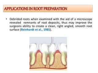
PRESENTED BY
DR.J.VELKUMAR, IIIYR PG
ANNAMALAI UNIVERSITY
GUIDED BY
DR.R.MYTHILI MDS
PROF & HOD
ANNAMALAI UNIVERSITY,
CHIDAMBARAM
• Periodontal microsurgery is defined as refinement in
existing basic surgical techniques that are made
possible by use of the surgical microscope and
subsequent improved visual acuity.
INTRODUCTION
HISTORY
 Carl Nylen, considered the father of microsurgery.
 Apotheker & Jako first introduced the microscope to dentistry
in 1978.
 In 1993, Shanelec & Tibbetts presented a continuing education
course on periodontal microsurgery at the annual meeting of AAP
• Improved tissue handling
• Optimize defect debridement and root instrumentation;
• Ensure optimal delivery of the regenerative technology
• Precise suturing.
• Reduced incision size and surgical retraction are directly
related to reduced postoperative pain and rapid healing
(Tonetti et al.,)
• The ergonomics of hand position and body posture is closely
related to the improved motor skills made possible
OBJECTIVES
• Microsurgery is a surgical philosophy that embraces three
distinct values.
1. The first value is the improvement of motor skills to enhance
surgical ability -Internal Precision Grip.
2. The second basic value of microsurgery is the application of
microsurgical instruments to reduce tissue trauma.
3. The third basic value of microsurgery is an emphasis on
passive wound closure with exact primary apposition of the
wound edge
CLINICALPHILOSOPHY OF MICROSURGERY
(Belcher 2001)
Illumination
Magnification Increased precision
THE MICROSURGICAL
TRIAD
• The magnified image that is formed has stereoscopic
properties that are created by the use of convergent lens
systems. also called a Keplerian optical system (Caplan).
Loupes
• SIMPLE LOUPES
• COMPOUND LOUPES
• PRISM LOUPES
Kanc J, Jordan PG et al.,
TYPES OF KePLerIANLOUPES
• Designed on Galilean principles
( Apotheker et al., 1981).
• Stereoscopic viewing of the operative
field without eye convergence.
• Rotating variable magnification element
• Foot controlled focus and magnification
2.5x to 8x- Low magnification
for orientation to the surgical field
10x to 16x Medium magnification
for operating.
20x to 30x- High magnification
for observing fine details.
Magnification
Barraquer Jl, hoerenz et al.,
OPERATING MICROSCOPE
• A basic set comprises of a needle holder,microscopic
scissors, micro scalpel holder, anatomical and
surgical forceps and a set of various elevators.
Instrumentation
Knives are chemically etched rather than
ground, their sharper blades produce a more
precise wound edge. (Somerville)
microsurgical knives: 1, blade
breaker;2, crescent; 3,
minicrescent; 4, 260° spoon; 5,
lamella,and 6, sclera
SUTURE MATERIALS
•Gauge 9-0 (0.03 mm) to 12-0 (0.001mm)
•Finer needles (3/8),1/2 curvature and suture
materials are used in microsurgery. (Halstead
1993)
MICROSURGICAL INSTRUMENTATION
• To augment the dimensions of attached
gingival tissue
• To gain root coverage
• To eliminate the aberrant frenum
• Ridge augmentation
• Crown lengthening
• Creation of interdental papillae
• Extremely predictable root coverage in millers class I and II
and enhanced results in class III and class IV compared to
conventional surgery
APPLICATIONS IN MUCOGINGIVAL
SURGERIES
Microsurgical technique for augmentationof the interdental papilla
W. Peter Nordland et al.,
Gingival recession management
1 2 3
4
Dr.Scott Kissel et al.,
• Debrided roots when examined with the aid of a microscope
revealed remnants of root deposits, thus may improve the
surgeons ability to create a clean, right angled, smooth root
surface (Reinhardt et al., 1985).
APPLICATIONS IN ROOT PREPARATION
• One of the novel applications of microsurgery is in
the sinus lift procedure.
• The surgical microscope can aid in visualization of the
sinus membrane.
• Magnification achieved by the surgical microscope is
used in implant site development and placement.
IN IMPLANT THERAPY
Microsurgical Approach to Periodontal Regeneration- Initial Evaluationin a
Case Cohort
Dr. Pierpaolo Cortellini
.
Cohort study involved 26 patients with one deep interdental intrabony
defect each. They were treated with periodontal regeneration using
guided tissue regeneration membranes and papilla preservation flaps
performed with the aid of an operating microscope and microsurgical
instruments. The procedure resulted in clinically important amounts of
CAL gains and minimal recessions. J Periodontol 2001;72:559-569.
• Less tissue trauma
• Less mobility
• Less patient anxiety
• Atraumatic tissue management
• Accurate primary wound closure
• Improved cosmetic result
• Increased diagnostic skill
ADVANTAGES
•Technique sensitive
•Loss of depth of field as magnification increases and loss
of visual reference points.
•Higher patient cost
•Time consuming
LIMITATIONS
• Periodontal microsurgery provides a natural
evolution in the progression of periodontics.
• However the choice of micro and macro
surgical approaches must be seen in different
lights,cost and treatment outcomes, and
patient centered parameters.
CONCLUSION
• Atlas of cosmetic and reconstructive surgery-cohen 3rd edition
• clinical periodontology_10th edition Carranza, Newmann
• Periodontics: medicine, surgery and implants. Rose, mealey, genco,
cohen.
• A perspective on the future of periodontal microsurgery-perio 2000,vol
11 1996.
• MICROSURGERY: A CLINICAL PHILOSOPHY FOR SURGICAL
CRAFTSMANSHIP-www.ejournalofdentistry.com
• A Perspective on Periodontal Microsurgery-(Int J Periodontics
Restorative Dent 2001;21:191–196.)
• Perio 2000--vol 11, Issue 1 58–64, June 1996
• Practical Periodontal Plastic Surgery-Serge Dibart • Mamdouh Karima
• PERIODONTAL MICROSURGERYAnnals and Essences of Dentistry-Vol. - III
Issue 1 Jan – Mar 2011

micro seminar.ppt

  • 1.
    PRESENTED BY DR.J.VELKUMAR, IIIYRPG ANNAMALAI UNIVERSITY GUIDED BY DR.R.MYTHILI MDS PROF & HOD ANNAMALAI UNIVERSITY, CHIDAMBARAM
  • 2.
    • Periodontal microsurgeryis defined as refinement in existing basic surgical techniques that are made possible by use of the surgical microscope and subsequent improved visual acuity. INTRODUCTION
  • 3.
    HISTORY  Carl Nylen,considered the father of microsurgery.  Apotheker & Jako first introduced the microscope to dentistry in 1978.  In 1993, Shanelec & Tibbetts presented a continuing education course on periodontal microsurgery at the annual meeting of AAP
  • 4.
    • Improved tissuehandling • Optimize defect debridement and root instrumentation; • Ensure optimal delivery of the regenerative technology • Precise suturing. • Reduced incision size and surgical retraction are directly related to reduced postoperative pain and rapid healing (Tonetti et al.,) • The ergonomics of hand position and body posture is closely related to the improved motor skills made possible OBJECTIVES
  • 5.
    • Microsurgery isa surgical philosophy that embraces three distinct values. 1. The first value is the improvement of motor skills to enhance surgical ability -Internal Precision Grip. 2. The second basic value of microsurgery is the application of microsurgical instruments to reduce tissue trauma. 3. The third basic value of microsurgery is an emphasis on passive wound closure with exact primary apposition of the wound edge CLINICALPHILOSOPHY OF MICROSURGERY
  • 6.
    (Belcher 2001) Illumination Magnification Increasedprecision THE MICROSURGICAL TRIAD
  • 7.
    • The magnifiedimage that is formed has stereoscopic properties that are created by the use of convergent lens systems. also called a Keplerian optical system (Caplan). Loupes
  • 8.
    • SIMPLE LOUPES •COMPOUND LOUPES • PRISM LOUPES Kanc J, Jordan PG et al., TYPES OF KePLerIANLOUPES
  • 9.
    • Designed onGalilean principles ( Apotheker et al., 1981). • Stereoscopic viewing of the operative field without eye convergence. • Rotating variable magnification element • Foot controlled focus and magnification 2.5x to 8x- Low magnification for orientation to the surgical field 10x to 16x Medium magnification for operating. 20x to 30x- High magnification for observing fine details. Magnification Barraquer Jl, hoerenz et al., OPERATING MICROSCOPE
  • 10.
    • A basicset comprises of a needle holder,microscopic scissors, micro scalpel holder, anatomical and surgical forceps and a set of various elevators. Instrumentation
  • 11.
    Knives are chemicallyetched rather than ground, their sharper blades produce a more precise wound edge. (Somerville) microsurgical knives: 1, blade breaker;2, crescent; 3, minicrescent; 4, 260° spoon; 5, lamella,and 6, sclera SUTURE MATERIALS •Gauge 9-0 (0.03 mm) to 12-0 (0.001mm) •Finer needles (3/8),1/2 curvature and suture materials are used in microsurgery. (Halstead 1993) MICROSURGICAL INSTRUMENTATION
  • 12.
    • To augmentthe dimensions of attached gingival tissue • To gain root coverage • To eliminate the aberrant frenum • Ridge augmentation • Crown lengthening • Creation of interdental papillae • Extremely predictable root coverage in millers class I and II and enhanced results in class III and class IV compared to conventional surgery APPLICATIONS IN MUCOGINGIVAL SURGERIES
  • 13.
    Microsurgical technique foraugmentationof the interdental papilla W. Peter Nordland et al.,
  • 14.
    Gingival recession management 12 3 4 Dr.Scott Kissel et al.,
  • 15.
    • Debrided rootswhen examined with the aid of a microscope revealed remnants of root deposits, thus may improve the surgeons ability to create a clean, right angled, smooth root surface (Reinhardt et al., 1985). APPLICATIONS IN ROOT PREPARATION
  • 16.
    • One ofthe novel applications of microsurgery is in the sinus lift procedure. • The surgical microscope can aid in visualization of the sinus membrane. • Magnification achieved by the surgical microscope is used in implant site development and placement. IN IMPLANT THERAPY
  • 17.
    Microsurgical Approach toPeriodontal Regeneration- Initial Evaluationin a Case Cohort Dr. Pierpaolo Cortellini . Cohort study involved 26 patients with one deep interdental intrabony defect each. They were treated with periodontal regeneration using guided tissue regeneration membranes and papilla preservation flaps performed with the aid of an operating microscope and microsurgical instruments. The procedure resulted in clinically important amounts of CAL gains and minimal recessions. J Periodontol 2001;72:559-569.
  • 18.
    • Less tissuetrauma • Less mobility • Less patient anxiety • Atraumatic tissue management • Accurate primary wound closure • Improved cosmetic result • Increased diagnostic skill ADVANTAGES
  • 19.
    •Technique sensitive •Loss ofdepth of field as magnification increases and loss of visual reference points. •Higher patient cost •Time consuming LIMITATIONS
  • 20.
    • Periodontal microsurgeryprovides a natural evolution in the progression of periodontics. • However the choice of micro and macro surgical approaches must be seen in different lights,cost and treatment outcomes, and patient centered parameters. CONCLUSION
  • 22.
    • Atlas ofcosmetic and reconstructive surgery-cohen 3rd edition • clinical periodontology_10th edition Carranza, Newmann • Periodontics: medicine, surgery and implants. Rose, mealey, genco, cohen. • A perspective on the future of periodontal microsurgery-perio 2000,vol 11 1996. • MICROSURGERY: A CLINICAL PHILOSOPHY FOR SURGICAL CRAFTSMANSHIP-www.ejournalofdentistry.com • A Perspective on Periodontal Microsurgery-(Int J Periodontics Restorative Dent 2001;21:191–196.) • Perio 2000--vol 11, Issue 1 58–64, June 1996 • Practical Periodontal Plastic Surgery-Serge Dibart • Mamdouh Karima • PERIODONTAL MICROSURGERYAnnals and Essences of Dentistry-Vol. - III Issue 1 Jan – Mar 2011

Editor's Notes