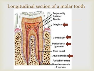
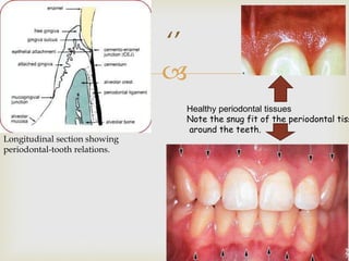
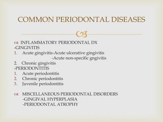
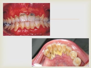
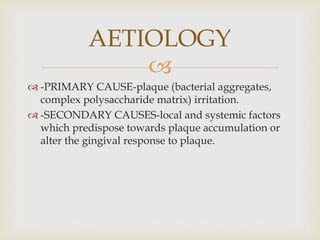
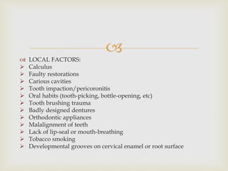
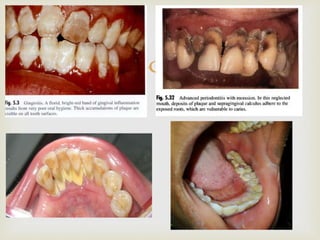
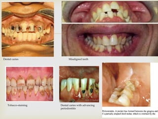
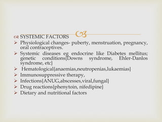
BY
THE DENTAL TEAM
CENTRAL HOSPITAL AGBOR
23/06/15

 INTRODUCTION
 DISEASES OF THE PERIODONTIUM
 COMMON PERIODONTAL DISEASE
 AETIOLOGY
 PATHOGENESIS
 CLINICAL PRESENTATION/EFFECTS OF
PERIODONTAL DISEASES
 MANAGEMENT
 CONCLUSION
OUTLINE

 Periodontal tissues otherwise referred to as the
periodontium are the surrounding soft and hard
tissues that support the tooth. They are specialized
tissues that both surround and support the teeth,
maintaining them in their anatomical positions in the
mandible (lower jaw)and maxilla (upper jaw).
 It comes from the Greek words peri- meaning
“around” and –odons meaning tooth.
INTRODUCTION

 The periodontal tissues are four namely:
-gingiva(gum)
-cementum
-alveolar bone
-periodontal ligament
 The disease conditions that affect these tissues are
therefore referred to as periodontal diseases.
INTRODUCTION cont’d

Longitudinal section of a molar tooth

‘’
Healthy periodontal tissues
Note the snug fit of the periodontal tiss
around the teeth.
Longitudinal section showing
periodontal-tooth relations.
International Workshop for a Classification of Periodontal
Diseases and Conditions in 1999 classified these diseases as
follows:
 Gingival diseases (non-plaque induced and plaque
induced)
 Chronic periodontitis.
 Aggressive periodontitis.
 Periodontitis as a manifestation of a systemic disease,
physiological changes, infection, drug reactions, DM,
AIDS, dietary and nutritional factors
DISEASES OF THE PERIODONTIUM

 Necrotizing periodontal diseases.
 Abscesses of the periodontium.
 Periodontitis associated with endodontic lesions.
 Developmental or aquired deformities and
conditions e.g. genetic conditions like Downs
syndrome, Ehlers-Danlos syndrome, hereditary
gingival fibromatosis etc; hematological conditions
such as anaemia, leukaemia etc

 INFLAMMATORY PERIODONTAL DX
-GINGIVITIS
1. Acute gingivitis-Acute ulcerative gingivitis
-Acute non-specific gngivitis
2. Chronic gingivitis
-PERIODONTITIS
1. Acute periodontitis
2. Chronic periodontitis
3. Juvenile periodontitis
 MISCELLANEOUS PERIODONTAL DISORDERS
-GINGIVAL HYPERPLASIA
-PERIODONTAL ATROPHY
COMMON PERIODONTAL DISEASES


 -PRIMARY CAUSE-plaque (bacterial aggregates,
complex polysaccharide matrix) irritation.
 -SECONDARY CAUSES-local and systemic factors
which predispose towards plaque accumulation or
alter the gingival response to plaque.
AETIOLOGY

 LOCAL FACTORS:
 Calculus
 Faulty restorations
 Carious cavities
 Tooth impaction/pericoronitis
 Oral habits (tooth-picking, bottle-opening, etc)
 Tooth brushing trauma
 Badly designed dentures
 Orthodontic appliances
 Malalignment of teeth
 Lack of lip-seal or mouth-breathing
 Tobacco smoking
 Developmental grooves on cervical enamel or root surface


Dental caries Misaligned teeth
Tobacco-staining Dental caries with advancing
periodontitis

Jaw fractures
Orthodontic brackets Dentures
 SYSTEMIC FACTORS
 Physiological changes- puberty, menstruation, pregnancy,
oral contraceptives.
 Systemic diseases eg endocrine like Diabetes mellitus;
genetic conditions{Downs syndrome, Ehler-Danlos
syndrome, etc}
 Hematological{anaemias,neutropenias,lukaemias}
 Immunosuppressive therapy,
 Infections{ANUG,abscesses,viral,fungal}
 Drug reactions{phenytoin, nifedipine}
 Dietary and nutritional factors

Cervical abrasionApical periodontitis
 Healthy gingivae are firm , pink, knife-edged and do not
bleed on probing.
 Periodontal disease primarily begins usually from plaque
accumulation leading to gingivitis which when sustained
can progress to chronic periodontitis
 This disease process is mostly painless and can go
unnoticed for years.
 Note that periodontal destruction when established is not
continuous but progresses in an episodic manner with
bursts of destructive activity alternating with periods of
quiescence and possibly repair.
PATHOGENESIS
Foot note: plaque= bacterial aggregation + complex polysaccharide matrix
calculus= calcified plaque

Plaque-induced periodontal lesions can be divided into four stages:
1. Initial lesion
2. Early lesion
3. Established lesion
4. Advanced lesion
1. Initial lesion: here host-response mechanisms are raised in
response to causative micro-organisms {major offenders being
porphyromonas gingivalis & Aggregatibacter
actinomycetemcomitans} in accumulating dental plaque within 2-
4 days.
False pockets are formed as marginal gingiva and interdental
papilla become bulbous and bright red as connective tissue and
collagen surrounding blood
vessels in the area dissolve,
leaking out fluid into the tissues.

2. Early lesion: occurs about 6-12 days later; here features
of the initial lesion are accelerated.
Up to 60-70 percent of collagen is lost.
Formation of micro-abcesses at the junctional epithelium.
3. Established lesion: begins about 2-3 weeks post-plaque
accumulation.
Plasma cell accumulation in gingival sulci (no bone loss
yet); presence of complement and antigen-antibody
complexes is marked.
Apical migration of junction epithelium (gum recession
makes teeth appear longer)
This stage can remain indefinitely or progress to an
advanced lesion.

4. Advanced lesion: here the features are better described
clinically than histologically. Typically there is
 Periodontal pocket formation.
 Gingival ulceration and suppuration.
 Destruction of alveolar bone and periodontal ligament
 Tooth mobility, drifting and eventual tooth loss
Because the bone loss appears here, it is equated as
periodontitis.
The earlier lesions can be classified as gingivitis of
increasing severity.
The advanced lesion spreads apically, laterally around
the tooth and deep into the gum tissue papilla.
Bone resorption produces scarring and fibrous change.


 Swelling and hyperemia of interdental papilla and gingival
margins
 Plaque and calculus deposits
 Halitosis
 Bleeding
 Pocketing
 Tooth mobility
 Sensitivity
 Pain
 Pulpal disease(perio-endodontic lesions, pulpitis, apical
abscesses)
 Tooth fractures
 Tooth loss
CLINICAL
PRESENTATION/EFFECTS OF
PERIODONTAL DISEASE

 Establish a diagnosis.
 Aim to create a healthy mouth which the patient is
capable of, and willing to maintain.
These principles can be divided as follows:
Initial (cause-related) phase
Corrective phase
Maintenance (supportive phase)
MANAGEMENT

1) Initial (cause-related) phase : here we aim to control or
eliminate gingivitis and stop any further progression of
periodontal disease by removing plaque and other
contributory factors. This is key and can cause a failure of
more complex treatments.
MANAGEMENT

2) The corrective phase designed to restore function and
sometimes, aesthetics. This includes procedures like
- scaling and polishing
- root debridement
- periodontal access surgery
- regenerative surgery
- muco-gingival surgery
-selected use of local and systemic antibiotics
-furcation lesion treatments
-restorative procedures(overhanging fillings, fitting
of crowns, bridges etc)
- endodontic treatment
- occlusal adjustment

 All the aforementioned procedures are aimed at:
- Elimination of pathological pockets and the creation
of tight epithelial attachments.
- To arrest bone loss and in some cases improve
alveolar bone support(bone augmentation
procedures).
- Create an oral environment that the patient can
easily keep plaque-free.

3) The maintenance (supportive) phase that aims at
reinforcing patient motivation so their oral hygiene is
kept at a level that prevents a reoccurrence of disease.
Patient should be instructed on proper dental hygiene
practices: toothbrush and tooth brushing techniques,
flossing.
Proper balanced diet for overall health.
Regular dental checkups ( every 3 or 6 months).
 Oral hygiene practices (brushing, flossing habits), dental and
medical history
 Examine the teeth for:
-plaque control,
-calculus,
- staining;
-gingival colour change,
-swelling,
-recession,
-pocketing;
-furcation involvement,
-tooth mobility,
-bleeding on probing.
Dental check ups
 Radiographs
- Full mouth periapicals, horizontal bitewings: to show degree
bone loss(vertical, horizontal), root surface deposits, furcation
involvement, perio-endo lesions.
- Sequential radiographs can be used to monitor the disease.
Dental check ups
 Periodontal diseases are largely preventable hence the
dental team prescribes the following:
 Effective tooth brushing technique with medium
bristled toothbrush and fluoridated toothpaste twice
daily.
 The proper use of floss, plastic toothpicks and other
interdental aids.
 The use of mouthwashes as prescribed by dentist.
 Proper nutrition.
Visit your dentist for consultation at least every 6
months for routine oral examination. There, scaling and
polishing amongst other lines of management can be
instituted.
CONCLUSION

Dental flossing(with handl
Ultrasonic scaling and polishing
Manual scaling and polishing
Toothbrush and toothpaste
Dental flossing

Good nutrition

 Laura mitchell & David A. Mitchell Oxford
Handbook of Clinical Dentistry 5th Edition. Pg 173-
214
 Cawsons Essentials of Oral Pathology and Oral
Medicine 7th edition. Pg 68-89
 Outline of Periodontics by J.D Manson and B.M Eley
 www.dentallecnotes.blogspot.com
 www.en.m.wikipedia.org/wiki/Periodontium
 www.shutterstock.com
REFERENCES

Thanks for listening

PERIODONTAL DXS PPT final

  • 1.
    BY THE DENTAL TEAM CENTRALHOSPITAL AGBOR 23/06/15
  • 2.
      INTRODUCTION  DISEASESOF THE PERIODONTIUM  COMMON PERIODONTAL DISEASE  AETIOLOGY  PATHOGENESIS  CLINICAL PRESENTATION/EFFECTS OF PERIODONTAL DISEASES  MANAGEMENT  CONCLUSION OUTLINE
  • 3.
      Periodontal tissuesotherwise referred to as the periodontium are the surrounding soft and hard tissues that support the tooth. They are specialized tissues that both surround and support the teeth, maintaining them in their anatomical positions in the mandible (lower jaw)and maxilla (upper jaw).  It comes from the Greek words peri- meaning “around” and –odons meaning tooth. INTRODUCTION
  • 4.
      The periodontaltissues are four namely: -gingiva(gum) -cementum -alveolar bone -periodontal ligament  The disease conditions that affect these tissues are therefore referred to as periodontal diseases. INTRODUCTION cont’d
  • 5.
  • 6.
     ‘’ Healthy periodontal tissues Notethe snug fit of the periodontal tiss around the teeth. Longitudinal section showing periodontal-tooth relations.
  • 7.
    International Workshop fora Classification of Periodontal Diseases and Conditions in 1999 classified these diseases as follows:  Gingival diseases (non-plaque induced and plaque induced)  Chronic periodontitis.  Aggressive periodontitis.  Periodontitis as a manifestation of a systemic disease, physiological changes, infection, drug reactions, DM, AIDS, dietary and nutritional factors DISEASES OF THE PERIODONTIUM
  • 8.
      Necrotizing periodontaldiseases.  Abscesses of the periodontium.  Periodontitis associated with endodontic lesions.  Developmental or aquired deformities and conditions e.g. genetic conditions like Downs syndrome, Ehlers-Danlos syndrome, hereditary gingival fibromatosis etc; hematological conditions such as anaemia, leukaemia etc
  • 9.
      INFLAMMATORY PERIODONTALDX -GINGIVITIS 1. Acute gingivitis-Acute ulcerative gingivitis -Acute non-specific gngivitis 2. Chronic gingivitis -PERIODONTITIS 1. Acute periodontitis 2. Chronic periodontitis 3. Juvenile periodontitis  MISCELLANEOUS PERIODONTAL DISORDERS -GINGIVAL HYPERPLASIA -PERIODONTAL ATROPHY COMMON PERIODONTAL DISEASES
  • 10.
  • 11.
      -PRIMARY CAUSE-plaque(bacterial aggregates, complex polysaccharide matrix) irritation.  -SECONDARY CAUSES-local and systemic factors which predispose towards plaque accumulation or alter the gingival response to plaque. AETIOLOGY
  • 12.
      LOCAL FACTORS: Calculus  Faulty restorations  Carious cavities  Tooth impaction/pericoronitis  Oral habits (tooth-picking, bottle-opening, etc)  Tooth brushing trauma  Badly designed dentures  Orthodontic appliances  Malalignment of teeth  Lack of lip-seal or mouth-breathing  Tobacco smoking  Developmental grooves on cervical enamel or root surface
  • 13.
  • 14.
     Dental caries Misalignedteeth Tobacco-staining Dental caries with advancing periodontitis
  • 15.
  • 16.
     SYSTEMIC FACTORS Physiological changes- puberty, menstruation, pregnancy, oral contraceptives.  Systemic diseases eg endocrine like Diabetes mellitus; genetic conditions{Downs syndrome, Ehler-Danlos syndrome, etc}  Hematological{anaemias,neutropenias,lukaemias}  Immunosuppressive therapy,  Infections{ANUG,abscesses,viral,fungal}  Drug reactions{phenytoin, nifedipine}  Dietary and nutritional factors
  • 17.
  • 18.
     Healthy gingivaeare firm , pink, knife-edged and do not bleed on probing.  Periodontal disease primarily begins usually from plaque accumulation leading to gingivitis which when sustained can progress to chronic periodontitis  This disease process is mostly painless and can go unnoticed for years.  Note that periodontal destruction when established is not continuous but progresses in an episodic manner with bursts of destructive activity alternating with periods of quiescence and possibly repair. PATHOGENESIS Foot note: plaque= bacterial aggregation + complex polysaccharide matrix calculus= calcified plaque
  • 19.
     Plaque-induced periodontal lesionscan be divided into four stages: 1. Initial lesion 2. Early lesion 3. Established lesion 4. Advanced lesion 1. Initial lesion: here host-response mechanisms are raised in response to causative micro-organisms {major offenders being porphyromonas gingivalis & Aggregatibacter actinomycetemcomitans} in accumulating dental plaque within 2- 4 days. False pockets are formed as marginal gingiva and interdental papilla become bulbous and bright red as connective tissue and collagen surrounding blood vessels in the area dissolve, leaking out fluid into the tissues.
  • 20.
     2. Early lesion:occurs about 6-12 days later; here features of the initial lesion are accelerated. Up to 60-70 percent of collagen is lost. Formation of micro-abcesses at the junctional epithelium. 3. Established lesion: begins about 2-3 weeks post-plaque accumulation. Plasma cell accumulation in gingival sulci (no bone loss yet); presence of complement and antigen-antibody complexes is marked. Apical migration of junction epithelium (gum recession makes teeth appear longer) This stage can remain indefinitely or progress to an advanced lesion.
  • 21.
     4. Advanced lesion:here the features are better described clinically than histologically. Typically there is  Periodontal pocket formation.  Gingival ulceration and suppuration.  Destruction of alveolar bone and periodontal ligament  Tooth mobility, drifting and eventual tooth loss Because the bone loss appears here, it is equated as periodontitis. The earlier lesions can be classified as gingivitis of increasing severity. The advanced lesion spreads apically, laterally around the tooth and deep into the gum tissue papilla. Bone resorption produces scarring and fibrous change.
  • 22.
  • 23.
      Swelling andhyperemia of interdental papilla and gingival margins  Plaque and calculus deposits  Halitosis  Bleeding  Pocketing  Tooth mobility  Sensitivity  Pain  Pulpal disease(perio-endodontic lesions, pulpitis, apical abscesses)  Tooth fractures  Tooth loss CLINICAL PRESENTATION/EFFECTS OF PERIODONTAL DISEASE
  • 24.
      Establish adiagnosis.  Aim to create a healthy mouth which the patient is capable of, and willing to maintain. These principles can be divided as follows: Initial (cause-related) phase Corrective phase Maintenance (supportive phase) MANAGEMENT
  • 25.
     1) Initial (cause-related)phase : here we aim to control or eliminate gingivitis and stop any further progression of periodontal disease by removing plaque and other contributory factors. This is key and can cause a failure of more complex treatments. MANAGEMENT
  • 26.
     2) The correctivephase designed to restore function and sometimes, aesthetics. This includes procedures like - scaling and polishing - root debridement - periodontal access surgery - regenerative surgery - muco-gingival surgery -selected use of local and systemic antibiotics -furcation lesion treatments -restorative procedures(overhanging fillings, fitting of crowns, bridges etc) - endodontic treatment - occlusal adjustment
  • 27.
      All theaforementioned procedures are aimed at: - Elimination of pathological pockets and the creation of tight epithelial attachments. - To arrest bone loss and in some cases improve alveolar bone support(bone augmentation procedures). - Create an oral environment that the patient can easily keep plaque-free.
  • 28.
     3) The maintenance(supportive) phase that aims at reinforcing patient motivation so their oral hygiene is kept at a level that prevents a reoccurrence of disease. Patient should be instructed on proper dental hygiene practices: toothbrush and tooth brushing techniques, flossing. Proper balanced diet for overall health. Regular dental checkups ( every 3 or 6 months).
  • 29.
     Oral hygienepractices (brushing, flossing habits), dental and medical history  Examine the teeth for: -plaque control, -calculus, - staining; -gingival colour change, -swelling, -recession, -pocketing; -furcation involvement, -tooth mobility, -bleeding on probing. Dental check ups
  • 30.
     Radiographs - Fullmouth periapicals, horizontal bitewings: to show degree bone loss(vertical, horizontal), root surface deposits, furcation involvement, perio-endo lesions. - Sequential radiographs can be used to monitor the disease. Dental check ups
  • 31.
     Periodontal diseasesare largely preventable hence the dental team prescribes the following:  Effective tooth brushing technique with medium bristled toothbrush and fluoridated toothpaste twice daily.  The proper use of floss, plastic toothpicks and other interdental aids.  The use of mouthwashes as prescribed by dentist.  Proper nutrition. Visit your dentist for consultation at least every 6 months for routine oral examination. There, scaling and polishing amongst other lines of management can be instituted. CONCLUSION
  • 32.
     Dental flossing(with handl Ultrasonicscaling and polishing Manual scaling and polishing Toothbrush and toothpaste Dental flossing
  • 33.
  • 34.
      Laura mitchell& David A. Mitchell Oxford Handbook of Clinical Dentistry 5th Edition. Pg 173- 214  Cawsons Essentials of Oral Pathology and Oral Medicine 7th edition. Pg 68-89  Outline of Periodontics by J.D Manson and B.M Eley  www.dentallecnotes.blogspot.com  www.en.m.wikipedia.org/wiki/Periodontium  www.shutterstock.com REFERENCES
  • 35.