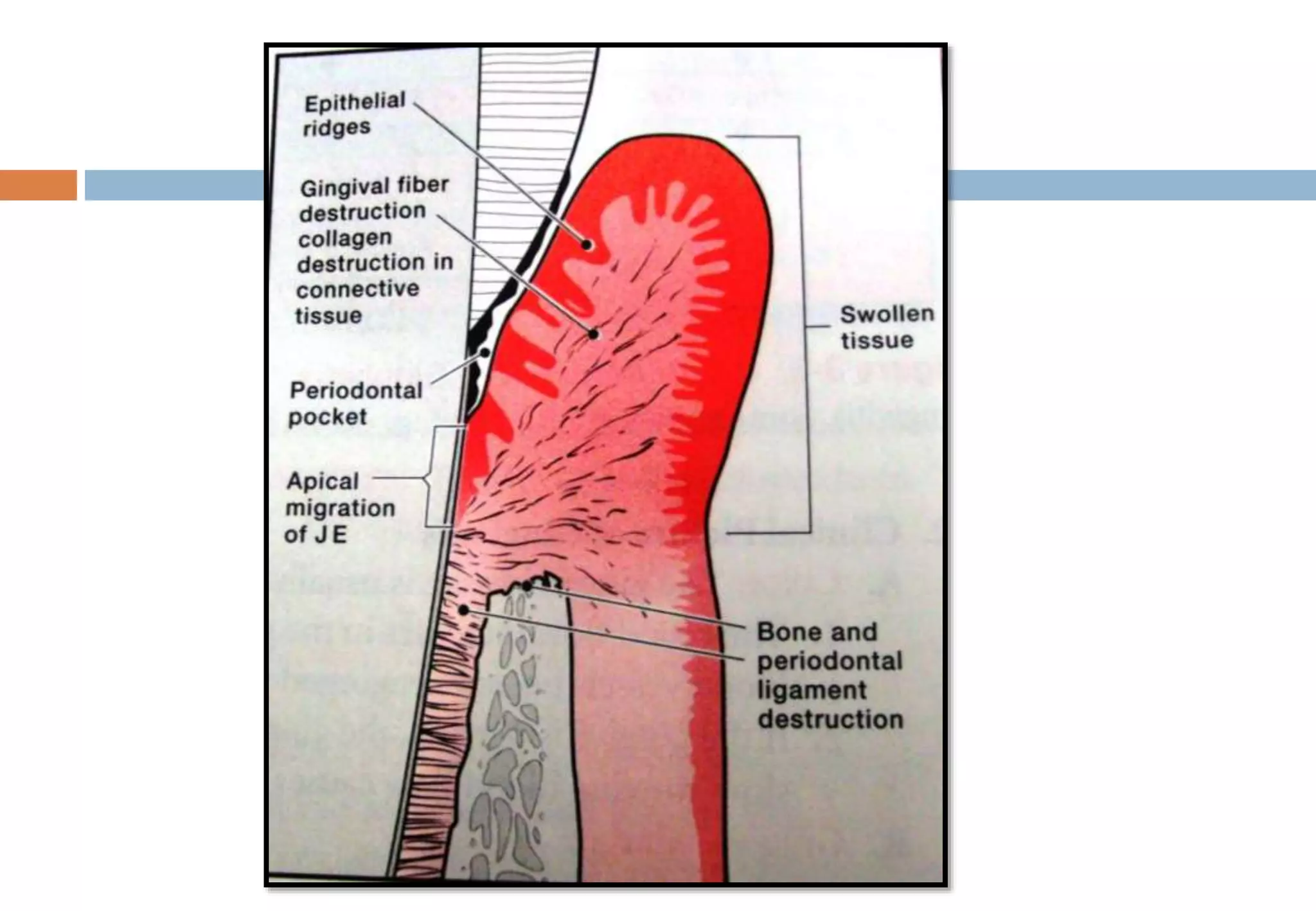
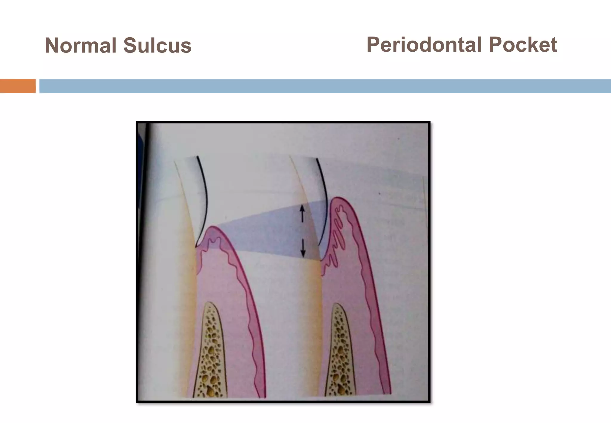
THE PERIODONTAL POCKET
T.SIVASANKARI.
1ST YEAR POSTGRADUATE.
INTRODUCTION
 The periodontal pocket, defined as a pathologically deepened
gingival sulcus.
 The most characteristic feature distinguishing periodontitis from
gingivitis is loss of connective tissue attachments & bone in
conjunction with formation of pocket due to apical migration of the
junctional epithelium.
 As a consequence of this apical migration, junctional epithelium
becomes attached to the root Cementum & the lateral wall of the
pocket is covered by epithelium-pocket epithelium.
Normal Sulcus Periodontal Pocket
Characteristics of periodontal pockets
 Large intercellular space.
 Its is not attached to the tooth.
 Micro ulcerations.
 Numerous retepegs.
Classification
Pockets can be classified as:
Gingival pocket
Periodontal pocket
Suprabony
Intrabony.
 Classification of pocket according to the involved tooth surface:
Simple pocket
Compound pocket
Complex pocket.
Classification
Pockets can be classified as:
Gingival pocket (pseudopocket). - formed by gingival
enlargement without destruction of the underlying periodontal
tissues. The sulcus is deepened because of the increased
bulk of the gingiva.
Periodontal Pocket: (absolute), occurs with destruction of the
supporting periodontal tissues. progressive pocket deepening
leads to destruction of the supportive periodontal tissues &
loosening & exfoliation of the teeth.
Suprabony.
Intrabony.
Suprabony (supracrestal or
supraalveolar),the bottom of
the pocket is coronal to the
underlining alveolar bone.
Intrabony
(infrabony,subcrestal,
intraalveolar),the bottom of
the pocket is apical to the
level of the adjacent alveolar
bone.
The lateral pocket walls lies
between the tooth surface &
the alveolar bone.
Classification Of Pockets According To The Involved
Tooth Surfaces
PATHOGENESIS
The initial lesion in the development of periodontitis is the
inflammation of the ginigiva in response to a bacterial
challenge.
Pocket formation starts as an inflammatory change in the
connective tissue wall of the gingival sulcus.
Just apical to JE, collagen fibers are destroyed,& the area
becomes occupied by inflammatory cells & edema.
 Collagenous & other enzymes secreted by various cells in healthy &
inflamed tissues, such as fibroblasts, PMNs, macrophages becomes
extracellular & destroy collagen-matrix metalloproteinase.
 Fibroblasts phagocytize collagen fibers by extending cytoplasmic
processes to the ligament Cementum interface & degrade the inserted
collagen fibrils & the fibrils of the Cementum matrix.(Deporter DA et al
1980).
 The apical cells of the JE proliferate along the root, extending finger
like projections two or three cells in thickness.
 The coronal portion detaches from the root as the apical
portion migrates.
 As a result of inflammation the PMNs invade the coronal end
of the JE increasing nos.
PMN infiltration & loss of cell cohesiveness
 60% volume of junctional epithelium cells, losses
cohesiveness & detach from tooth surface & migrate apically
Extension of JE along the root requires the presence of healthy
epithelial cells.
Degenerative changes seen in the JE at the base of
periodontal pocket are usually less severe than those in the
epithelium of the lateral pocket wall.
Because migration of JE requires healthy viable cells, it is
reasonable to assume that the degenerative changes seen in
this area occurs after the JE reaches its position on the
Cementum.
HISTOPATHOLOGY
Soft tissue wall:
The connective tissue is edematous & densely infiltrated with
plasma cells, lymphocytes, PMNs.
The blood vessels are increased in nos. particularly in the
subepithelial connective tissue layer.
In addition to exudation & degenerative changes, the CT
shows proliferation of the endothelial cells, with newly formed
capillaries, fibroblasts, collagen fibers.
Lateral Wall Of Periodontal Pocket Showing Epithelial
Proliferation And Atrophic Changes
 The most severe degenerative changes in the periodontal
pocket occur along the lateral wall.
 Epithelial buds or interlacing cords of epithelial cells
projects from the lateral wall into the adjacent inflamed
connective tissue & may extend further apically than the JE.
 These epithelial projections, reminder of the lateral
epithelium, are densely infiltrated by leukocytes & edema
from the inflamed connective tissue.
 The cells undergo vacuolar degeneration& rupture to form
vesicles
 Ulceration of the lateral wall, exposure of the underlying
inflamed connective tissue,& suppuration.
 A comparative study of gingival histopathological changes in
aggressive & chronic periodontitis revealed more pronounced
degenerative changes in the epithelium of aggressive cases with
more open intercellular space, with micro clefts & necrotic
areas.(Hillmann G et al 1998).
 Ulceration of the lateral wall may occur in shallow pockets,& deep
pockets are occasionally observed in which the lateral epithelium
is relatively intact or shows only slight degeneration.
 The epithelium at the gingival crest of a periodontal pocket is
generally intact & thickened with prominent retepegs.
BACTERIAL INVASION:
 Filaments, rods, cocciod organisms with predominent gram
negative cell wall has been found in intercellular spaces of
the epithelium.(Frank RM et al 1980).
 Hillmann et al reported the presence of
P.gingivalis,P.intermedia in the ginigiva of aggressive
periodontitis cases.
 AA has also been found in the tissue(Chrisersson LA et al
1987)
 Bacteria may invade the intercellular space under exfoliating
epithelial cells, but they are also found to be deeper epithelial
cells & accumulating on the basement lamina.
 Some bacteria transverse the basement lamina & invade the
subepithelial connective tissue (Saglie FR et al 1982)
Electron Micrograph Of A Section Of Pocket Wall Showing
Bacterial Penetration Into The Epitheliem And Connective
Tissue
MICROTOPOGRAPHY OF GINGIVAL WALL OF THE
PERIODONTAL POCKET
 These areas are irregularly oval or elongated & adjacent to one
another & measure about 50-200mm.
 Areas of relative quiescence: showing a relatively flat surface with
minor depression & mounds & occasional shedding of cells.
 Areas of bacterial accumulation: which appears as depression on
the epithelial surface, with abundant debris & bacterial clumps
penetrating into the enlarged intercellular spaces. These are
mainly cocci, rods,filaments with few spirochetes.
 Areas of emergence of leukocytes, where leukocytes appear
in the pocket wall through holes located in the intercellular
spaces
Areas of leukocyte bacterial interaction ,where numerous
leukocytes are present & are covered with bacteria in an
apparent process of phagocytosis. bacterial plaque associated
with the epithelium is seen either as an organized matrix
covered by a fibrin like material in contact with the surface of
cells or as bacteria penetrating into the intercellular space.
Areas of intense epithelial desquamation:which consist of semi
attached & folded epithelial squames, sometimes partially
covered with bacteria
 Areas of ulceration: with exposed connective tissue.
 Areas of hemorrhage, with numerous erythrocytes.
 The transition from one area to another could result from bacteria
accumulating in previously quiescent areas & triggering the
emergence of leukocytes & the leukocyte-bacterial interaction.
 This will lead to intense epithelial desquamation & finally to
ulceration & hemorrhage.
PERIODONTAL POCKET AS HEALING LESION
 Periodontal pocket are chronic inflammatory lesions, constantly undergoing
repair.
 Complete healing does not occur because of persistence of bacterial attack-
causing degeneration of the new tissue elements.
 The condition of the soft tissue wall of the periodontal pocket results from the
interplay of the destructive & constructive tissue changes.
 If the inflammatory fluid & cellular exudate predominate, the pocket wall is
bluish red, soft, spongy,& friable, with a smooth, shiny surface, at the clinical
level- edematous pocket wall.
 If there is relative predominance of newly formed connective
tissue cells & fibers, the pocket wall is more firm & pink-fibrotic
pocket wall.
 The more severe degenerative changes in periodontal tissues
occur adjacent to the tooth surface & subgingival plaque.
 In some cases, inflammation & ulceration on the inside of the
pocket wall are walled off by fibrous tissue on the outer aspect.
 Externally the pocket appears pink & fibrotic, despite the
inflammatory changes occurring internally.
POCKET CONTENTS
 Periodontal pocket contain debris consisting principally of
microorganisms & their products, gingival fluids, food
remnants, salivary mucins, desqumated epithelial cells &
leukocytes
 Plaque-covered calculus usually projects from the tooth
surface.
 Purulent exudate if present consisting of living, degenerated
& necrotic leukocytes, living & dead bacteria, serum,a scant
amount of fibrin.(McMillan et al 1958)
SIGNIFICANCE OF PUS FORMATION
 Pus is a common feature of periodontal disease ,but it is only
a secondary sign.
 The presence of pus or the ease with which it can be
expressed from the pocket merely express the nature of
inflammatory change in the pocket wall.
 Not an indication of the depth of the pocket or the severity of
the destruction.
 Extensive pus formation may occur in shallow pockets, where
as deep pockets may exhibit little or no pus.
ROOT SURFACE WALL
 As the pocket deepens, collagen fibers embedded in the
cementum are destroyed & cementum becomes exposed to
oral environment.
 Collagenous remnants of sharpey’s fibersin the cementum
undergo degeneration, creating an environment favorable to
the penetration of bacteria.
 Bacteria penetration into the cementum can be found as deep
as the cementodentinal junction,& may also enter the dentinal
tubules.
Penetration & growth of bacteria leads to fragmentation &
breakdown of the cementum surface & results in areas of
necrotic cementum, separated from the tooth by masses of
bacteria.
 Endotoxins are also detected in the Cementum wall of
periodontal pocket.
 Diseased root fragments also prevent the in vitro attachment of
human gingival fibroblasts, where as normal roots allow the cells
to attach freely.(Aleo JJ et al 1980).
 Clinically as softening of the Cementum surface, painful when
probing.
 These necrotic areas are removed by root planning until a hard,
smooth surface is reached.
 Cementum is very thin in the cervical areas, scaling & RP
remove it entirely, exposing the underlying dentin
DECALCIFICATION & REMINERALIZATION OF
CEMENTUM
 Areas of increased mineralization :result of an exchange, on
exposure to the oral cavity, of minerals & organic components at
the cementum saliva interface.
 Minerals that are increased in diseased root surface include
calcium, magnesium, phosphorous,fluoride.
 The hyperminerlized zones-increased proliferation of the crystal
structures & organic changes suggestive of a subsurface
cuticle.(Selvig KA et al 1969).
 These zones have also been seen in micro radiographic studies
as a layer of 10-20 mm thick, with areas as thick as 50 mm.
 No decrease in mineralization was found in deeper areas,
indicating mineralization does not come from the adjacent
areas.
 Areas of demineralization -root caries. unlike enamel caries, root
surface caries tend to progress around rather than in to the tooth.
 Active root caries lesion appear as well-defined yellowish or
light-brown areas, are frequently covered by plaque,& have
softened or leathery consistency on probing.
 Inactive lesion are well defined darker lesions with a smooth
surface & harder consistency on probing.(Fejerskov O et al
1986).
 The dominant microorganisms in root surface caries is
Actinomyces viscosus,others,S.mutans,S.salivarius,S.sanguis &
B.cereus have been found to produce root caries in animal
model.
 Quirynen et al reported that when pocket level & probing depth
decreases after periodontal therapy, a shift in the oral bacteria
occurs.
 Root caries may be the cause of toothache in patients with
periodontal disease & no evidence of coronal decay.
 Caries of Cementum requires special attention when the pocket
is treated.
 The necrotic cementum must be removed by scaling & root
planning until firm tooth surface is reached.
 Areas of cellular resorption of cementum & dentin: common in
roots exposed to periodontal diseases.
 If the root is exposed by progressive pocket formation before
repair occurs, these areas appear like isolated cavitations that
penetrate into the dentin.
 These areas can be differentiated from the caries of cementum
by their clear cut outline & hard surface.
 They may be the sources of considerable pain, requiring
placement of a restoration.
Surface morphology of tooth wall
The following zones can be found in the bottom of a
periodontal pocket
Cementum covered by calculus:
Attached plaque :covers calculus & extend apically from it to a
variable degree, probably 100-500 mm.
The zone of unattached plaque:surrounds the attached plaque &
extend apically.
The zone of attachment of JE to the tooth :the extension of this
zone, which in normal sulci is more than 500 mm, is usually
reduced in periodontal pocket to less than 100mm.
A zone of semi destroyed CT fibers: apical to the junctional
epithelium.
 Zone 3,4,5-plaque free zone.
 The total width of the plaque free zone varies according to the
type of tooth & depth of pocket.
 Unattached plaque-gram positive cocci & various gram negative
morphotypes, including cocci, rods, filaments, fusiform
& spirochetes.
 The most apical zone contains predominantly gram negative
rods & cocci.
Periodontal Disease Activity
 More recently, as a result of studies on the specificity of plaque
bacteria, the concept of periodontal disease activity was
evolved.
 According to this concept, periodontal pockets go through
periods of exacerbation & quiescence, resulting from episodic
bursts of activity followed by periods of remission.
 Periods of quiescence -reduced inflammatory response & little
or no loss of bone & CT attachments.
 A buildup of unattached plaque, with its gram negative,
motile,& anaerobic bacteria starts a period of exacerbation in
which bone & CT attachments are lost & the pocket deepens.
 Based on study of radioiodine l absorptiometry, McHenry et al
confirmed that bone loss in untreated periodontitis disease
occurs in an episodic manner.
 Periods of inactivity
 Periods of activity
Histologically,the pocket epithelium appears thin & ulcerated &
an infiltrate composed predominantly composed of plasma
cells,(Davenport RH et al1982),PMNs, or both are seen.
Bacterial samples from the pocket lumen analyzed with dark
field microscopy shows high proportion of motile organisms &
spirochetes(Listgarten MA et al1978).
Site Specificity
 Periodontal destruction does not occur in all parts of the
mouth at the sametime but rather on a few teeth at a time or
even only some aspects of some teeth at any given time-site
specificity of periodontal disease.
Pulp Changes Associated With Periodontal
Pocket
 The spread of infections from the periodontal pocket may
cause pathological changes in the pulp.
 Involvement of the pulp in the periodontal disease occurs
through either apical foramen or the lateral canals in the root
after infection spreads from the pocket through the
periodontal ligament
Relationship Of Attachment Loss & Bone Loss To
Pocket Depth
 Pocket formation causes loss of attachment of gingiva &
denudation of the root surface.
 The severity of the attachment loss is generally, but not always,
correlated with the depth of the pocket.
 This is because the degree of attachment loss depends on the
location of the base of the pocket on the root surface.
 Pocket depth is the distance between the base of the pocket &
the crest of the gingival margin.
Clinical diagnosis of pocket
Clinical Features
 Bluish red thickened marginal gingiva.
 Bluish red vertical zone from the gingival margin to the
alveolar mucosa.
 Gingival bleeding & suppuration
 Tooth mobility
 Diastema formation
 Localized pain or pain deep in the bone
Correlation Of Clinical & Histopathological
Features Of The Periodontal Pocket.
Gingival wall of the pocket
presents various degrees of
bluish red discoloration
;flaccidity;a smooth,shiny
surface;& pitting on
pressure.
 The discolaration is caused by
circulatory stangulation;the
flaccidity;by destruction of
gingival fibers & surrounding
tissues;the smooth,shiny
surface,by atrophy of
epithelium & edema;& the
pitting on pressure,by edema &
degeneration.
Clinical features Histopathologic features
Less frequently, gingival wall
may be pink & firm.
Fibrotic changes
predominate over
exudation& degeneration,
particularly in relation to
outer surface wall of the
pocket wall
Bleeding is elicited by gently
probing soft tissue wall of
pocket.
Ease of bleeding results
from increased vascularity,
thinning & degeneration of
epithelium,& proximity of
engorged vessels to inner
surface.
When explored with a probe,
inner aspect of pocket is
generally painful.
Pus may be expressed by
applying digital pressure.
Pain on tactile stimulation is
caused by ulceration of inner
aspect of pocket wall.
Pus occurs with suppurative
inflammation of inner wall.
DISTINGUISHING FEATURES
Base of pocket is coronal to
level alveolar bone.
Destruction of the underlying
bone is horizontal.
Base of pocket is apical to
crest of alveolar bone so
that the bone is adjacent to
the soft tissue wall.
Vertical(angular)
Suprabony pocket Intrabony pocket
Transseptal fibers that are
restored during progressive
periodontal disease are
arranged horizontally in
space between base of
pocket & alveolar bone
Oblique rather than
horizontal.
On the facial & lingual
surfaces, periodontal
ligament fibers beneath
pocket follow their normal
horizontal-oblique course
between the tooth & bone.
Follow angular pattern of
adjacent bone. They extend
from Cementum beneath
base of pocket along
alveolar bone & over crest to
join with outer periosteum.
Detection Of Pockets.
 The only accurate method of detecting & measuring
periodontal pocket is careful exploration with the periodontal
probe.
 Pockets are not detected by radiographic examinations.
Pocket probing
The biological depth is the
distance between the
gingival margin & the base
of the pocket.
The probing depth is the
distance to which an probe
penetrates to the pocket.
 Armitage et al –beagle dogs-standardized force of 25 grams.
 In healthy specimens, probe penetrated the epithelium to about
two-third of its length.
 In gingivitis, it stopped 0.1 mm short of its apical end.
 In periodontitis, the probe tip consistently went past the most
apical cells of the junctional epithelium
 In humans the probe tip penetrates to the most coronal intact
fibers of the connective tissue attachment(Listgarten et al
1976).
 The depth of penetration of probe in the connective tissue
apical to the junctional epithelium in a periodontal pocket is
about 0.3mm.
 Forces of 0.75 N have been found to be well tolerated &
accurate(Tibbetts 1969)
Determining The Level Of Attachment
When the gingival margin is located on the anatomic crown, the
level of attachment is determined by subtracting from the depth of
the pocket the distance from the gingival margin to the CEJ. if both
are the same, the loss of attachment is zero.
When the gingival margin coincides with the CEJ, the loss of
attachment equals the pocket depth.
When the gingival margin is located apical to CEJ, the loss of
attachment is greater than pocket depth-distance between the CEJ
& the gingival margin should be added to the pocket depth.
Bleeding On Probing
 The insertion of probe to the bottom of the pocket elicits bleeding
if the gingiva is inflamed & pocket epithelium is atrophic or
ulcerated.
 Depending on the severity of inflammation, bleeding can vary
from tenacious red line along the gingival surface to profuse
bleeding. after treatment BOP ceases.
 Armitage concluded that the presence of BOP in a “treated &
maintained patient population” is an important risk predictor for
increased loss of attachment.
Area Between Base Of Pocket & Alveolar Bone
 Distance between the apical end of the JE & the alveolar bone
is relatively constant.
 The distance between the apical extent of the calculus & the
alveolar crest is constant, having a mean length of 1.97mm
(Stanley HR 1955).
 The distance from the attached plaque to bone is never less
than 0.5mm & never more than 2.7mm(Wade 1960).
 These finding suggest that bone resorbing activity induced by
the bacteria is exerted within these distances.
Relation in response to treatment
planning
Critical Zones In Pocket Surgery
 Criteria for the selection of one of the different surgical
techniques for pocket therapy are based on clinical findings in
the soft tissue pocket wall, tooth surface, underlying bone,
attached gingiva.
 Zone 1: soft tissue pocket wall: morphologic features,
thickness,& topography of the soft tissue pocket wall&
persistence of inflammatory changes in the wall.
 Zone 2:tooth surface.
presence of deposits & alterations on the cementum surface &
determine the accessibility of the root surface to
instrumentation.
evaluation of the results of phase 1 therapy should determine
the need for the further therapy & the method to be used.
 Zone 3:underlying bone
shape & the height of the alveolar bone next to the pocket wall
through careful probing.
bony craters, horizontal or angular bone losses,& other bone
deformities are important criteria in selection of treatment
technique
 Zone 4:attached gingiva
 presence or absence of adequate band of attached gingiva
when selecting the pocket treatment method.
 An inadequate attached gingiva is caused by high frenum
attachment, marked gingival recession or a deep pocket that
reaches the level of mucogingival junction
 All these conditions should be explored & their influence on
pocket therapy determined.
Methods Of Pocket Therapy
The methods of pocket therapy can be classified as:
 New attachment technique: offers the ideal result
because they eliminate the pocket depth by reuniting the
gingiva to the tooth at a position coronal to the bottom of
the pre-existing pocket.
 New attachment is usually associated with filling in of
bone& regeneration of periodontal ligament & cementum.
 Removal of the pocket wall: the wall of the pocket consist of sift
tissue & may include bone in the case of intrabony pockets.
 It can be removed by the following
 Retraction or shrinkage, in which scaling & root planning
procedures resolve the inflammatory process, and the gingiva
therefore shrinks, reducing the pocket depth.
 Surgical removal performed by the gingivectomy technique or
by means of an undisplaced flap.
 Apical displacement with an apically displaced flap.
 Removal of the tooth side of the pocket: which is
accomplished by tooth extraction or by partial tooth
extraction(hemisection or root resection)
Peri Implant Probing
 Periimplantitis can create pockets around implants, so
probing around the implants becomes part of the
examination & diagnosis.
 To prevent scratching of the implant surface, plastic
periodontal probe should be used.
Peri-implant Mucosa
 The wound healing that occurs following the closure of
mucoperiosteal flaps during implant surgery results in the
establishment of a mucosal attachment(transmucosal
attachment) to the implant.
 The transmucosal attachment serves as a seal that prevents the
products from the oral cavity reaching the bone that anchors the
implant.
 In an early study by Berglundh et al 1991,a comparison of some
features of gingiva & mucosa around implant in dog model.
 Berglundh et al 1991 described the peri –implant mucosa
features. the outer surface is covered by a well keratinized
oral epithelium, which in the marginal border connects with
the barrier epithelium that is facing the abutment part of the
implant.
 This barrier epithelium has many features in common with the
junctional epithelium found on the tooth site.
 The barrier epithelium is only a few cell layer thick and
terminates about 2mm apical to soft tissue margin.
 In A Zone About 1-1.5 Mm High Between The Apical Level Of
Barrier Epithelium & The Alveolar Crest, The Connective
Tissue Appears To Be In Direct Contact With Titanium Dioxide
Layer Of The Implant.
 The Collagen Fibers Seems To Originate From The
Periosteum Of The Bone Crest & Extend Towards The Margin
Of The Soft Tissue In Directions Parallel To The Surface Of
The Abutment.
Composition Of The Connective Tissue
 The composition of the connective tissue in the supra alveolar
compartments at teeth & implants were examined by Bergludh
et al 1991.
 He stated that the main difference between the mesenchymal
tissue around the tooth & at an implant site is the occurrence of
a cementum on the root surface.
 At the implant site, the collagen fiber bundles are oriented in a
direction parallel to the implant surface. These horizontal fibers
appear to bend in a vertical direction (Buser et al 1992).
 The connective tissue in the attachment zone at implant
contains more collagen but fewer fibroblast & vascular
structures than the tissue in the corresponding location at teeth.
 Moon et al 1999 -dog model-reported that this border tissue
could be divided into two zones.
 Zone A:-40mm wide & is present next to implant surface. in this
zone there is no blood vessels but a large no. Fibroblasts that
are oriented with their long axis parallel with the implant surface.
 Collagen content was found to be 67%,vascular structure
0.3%,fibroblasts 32%
ZONE B:in lateral direction this zone is continuous with zone A &
is 160mm wide there are fewer fibroblasts & more collagen
fibers & more vascular structures.
Collagen content was found to be 85%,vasular structure
3%,fibroblasts 11%.
Plaque Formation On Implants
 In order to elict an inflammatory response which may(or) may
not lead to periimplant infection, bacteria must colonize implant
surfaces.
 The transmucosal penetration of the oral implants results in a
complex interface that involves the implant surface, the peri-
implant soft tissue % the ecology of the oral cavity including
bacterial plaque accumulation.
 A clinically stable per-implant seal may result from an equilibrium
between the host epithelium & bacterial plaque accumulation.
 Mombelli et al 1988 studied early plaque formation & using an
anaerobic culture technique.
 Over 85% of the micro-organisms were identified as coccoid
cells & over 80% of the cultivated bacteria were gram-positive
facultative cocci.
 Clinical signs of infection, including pocket development & pus
formation emerged 120 days after implantation.& probing depth
of 6mm was recorded.
 Hence the permanent presence of the putative periodontal
pathogens even at low levels, may be of concern in patients with
a documented increased susceptibility for periodontitis.
 The competent & successful reduction of pathogens by
periodontal therapy, represents a must for higher predictability to
avoid peri-implant infections.
Pocket Around Implants
 In natural dentition the junctional epithelium is believed to
provide a seal at the base of a periodontal sulcus against
penetration of substances(bacterial & chemical).
 Disruption of this seal by the lysis of connective tissue fibers
inserted into the rot cementum apical to the junctional
epithelium leads to rapid migration of the crevicular epithelium
forming a pathological pocket.
 As no cementum (or) fiber insertion is reported on the
surface of titanium transmucosal abutments,an epithelial
permucosal seal may provide the only barrier against
pathological insults to deeper tissues.
 Destruction of the integrity of the perimucosal titanium
surface seal could lead to extension of the pathologic
process to the underlying osseous structure.
Histopathology
 The response of the gingiva & the peri-implant mucosa to
early & more long standing periods of plaque formation was
analysed both in studies in man & in experiments in animals.
 Zitzmann et al (2001)-response to plaque formation in the soft
tissues at implant & tooth sites in humans.
 It was demonstrated that plaque build up was associated with
clinical signs of soft tissue inflammation.
 In addition,the proportion of B-cells & neutrophils increased
more in the lesion in the gingiva than the peri-implant mucosa.
 Berglundh et al (1992)-in beagle dogs,compared the reaction of
the gingiva & the peri-implant mucosa to 3 weeks of plaque
formation.
 During the course of the study,it was observed that similar
amounts of plaque formed on the tooth & implant segment of the
dentition.
 Leonhardt et al (1992) concluded that early microbial colonization
on titanium implants followed the same pattern as that on teeth.
 Ericsson et al (1992) with the increasing duration of plaque build
up(3mths) in dog,the lesions in the peri-implant mucosa seemed
to have expanded & to have progressed further apically,while the
gingival lesions remained unchanged.
 Sanz et al (1991) analyzed soft tissue biopsies from 6 patients with
peri-implantitis-65% of the connective tissue portion was occupied
by an inflammatory lesion.
 It was reported that at sites where implants were removed due to
periimplantitis, “an inflammatory infiltrate, composed of
macrophages, lymphocytes & plasma cells,were found in the
connective tissue around implants.
 Berglundh et al (2004) –mucosa contained very large
lesioninwhichnumerousplasmscells,lymphocytes,macrophages
were present.
 It was observed that the inflammatory cell infiltrate
consistently extended to an apical area to the pocket
epithelium & that the apical part of the soft tissue lesion
frequently reached the bone tissue.
 In the apical part of the lesion the inflamed connective tissue
appeared to be in direct contact with the biofilm on the implant
surface.
 Lindhe et al (1992)-dog-premolars were extracted in one side of
the mandible, fixtures were inserted & abutment connection
performed 3mths later.
 Periodontitis & peri-implantitis lesions were induced.
 A “pocket” between the tooth/gingiva & implant/mucosa was
created,a submarginal microbiota rapidly formed.
 Leonhardt et al (1992)-observed that plaque that had formed in the
deep pockets were similar at tooth & implant sites & was
dominated by gram-negative anaerobic species.
 These finding indicate that,in humans,the microbiota at teeth &
implants has many features in common.
 Implants,& teeth that are surrounded by healthy soft tissue are
associated with biofilm including small amounts of gram-positive
coccoid cells & rods.
 Sites with extensive periodontal & peri-implant inflammation harbour
biofilms with large numbers of gram-negative anaerobic bacteria.
 Lindhe et al 1992 -concluded that the pattern of spread of inflammation
was different in periodontal & per-implant tissues.
 The lesion in plaque associated periodontitis were limited to the
connective tissue,while in peri-implant tissues the lesions extend to the
alveolar bone.
Probing The Mucosa At The Implant Site
 Ericsson & Lindhe 1993:a probe with a tip diameter of 0.5mm was
inserted into the buccal pockets using a standard force of 0.5N.
 At the implant site probing caused both compression & lateral discoloration
of the peri-implant mucosa & the average histologic probing depth was
markedly deeper than at the tooth site namely 0.2mm.
 The distance between the probe tip & bone crest implant site was about
0.2mm which was less than in teeth sites 1.2mm.
 Concluded that the attachment between implant surface & the mucosa was
weeker than the corresponding attachment between tooth & the gingiva.
Probing Around Implants
 Lang et al (1994)-beagle dog-reported a mean histologic
probing depth of 1.75mm at healthy sites. They also
determined the probing depth at sites with mucositis& peri-
implantitis was 1.62 & 3.8mm.
 They further stated that at healthy & mucositis sites the probe
tip identifies the connective tissue adhesion level(base of the
barrier epithelium).but in peri-implantitis the probe exceeded
the base of the ulcerated pocket epithelium by 0.5mm
Schou et al (2002) - compared the probing measurements at
implant & teeth in eight monkeys.
An electronic probe with a tip diameter of 0.5mm& a standardized
probing force of 0.3-0.4N used. it was demonstrated that the
probe tip was located at a similar distance from the bone in
healthy tooth sites & implant sites.
In peri-implantitis,the probe tip was constantly identified at a more
apical position that at corresponding(gingivitis& periodontitis) sites
at teeth.
 It was concluded that the application of force more than 0.5N
can result in mechanical displacement into tissue adjacent to
implant.
 In the presence of inflammation at implant sites, the probe
penetrates to a more apical position than at inflammed sites at
teeth.
REFERENCES
carranza’s clinical periodontology tenth edition.
Clinical periodontology & implant dentistry-jan lindhe.
Foundation of periodontics-nield-gehrig
Periodontics-garant
Fundamentals of periodontics-kornman.
International journal of periodontics & restorative
dentistry 2000.

The periodontal pocket.pptx

  • 1.
  • 2.
    INTRODUCTION  The periodontalpocket, defined as a pathologically deepened gingival sulcus.  The most characteristic feature distinguishing periodontitis from gingivitis is loss of connective tissue attachments & bone in conjunction with formation of pocket due to apical migration of the junctional epithelium.  As a consequence of this apical migration, junctional epithelium becomes attached to the root Cementum & the lateral wall of the pocket is covered by epithelium-pocket epithelium.
  • 4.
  • 5.
    Characteristics of periodontalpockets  Large intercellular space.  Its is not attached to the tooth.  Micro ulcerations.  Numerous retepegs.
  • 6.
    Classification Pockets can beclassified as: Gingival pocket Periodontal pocket Suprabony Intrabony.  Classification of pocket according to the involved tooth surface: Simple pocket Compound pocket Complex pocket.
  • 7.
    Classification Pockets can beclassified as: Gingival pocket (pseudopocket). - formed by gingival enlargement without destruction of the underlying periodontal tissues. The sulcus is deepened because of the increased bulk of the gingiva.
  • 9.
    Periodontal Pocket: (absolute),occurs with destruction of the supporting periodontal tissues. progressive pocket deepening leads to destruction of the supportive periodontal tissues & loosening & exfoliation of the teeth. Suprabony. Intrabony.
  • 10.
    Suprabony (supracrestal or supraalveolar),thebottom of the pocket is coronal to the underlining alveolar bone.
  • 11.
    Intrabony (infrabony,subcrestal, intraalveolar),the bottom of thepocket is apical to the level of the adjacent alveolar bone. The lateral pocket walls lies between the tooth surface & the alveolar bone.
  • 12.
    Classification Of PocketsAccording To The Involved Tooth Surfaces
  • 15.
    PATHOGENESIS The initial lesionin the development of periodontitis is the inflammation of the ginigiva in response to a bacterial challenge. Pocket formation starts as an inflammatory change in the connective tissue wall of the gingival sulcus. Just apical to JE, collagen fibers are destroyed,& the area becomes occupied by inflammatory cells & edema.
  • 17.
     Collagenous &other enzymes secreted by various cells in healthy & inflamed tissues, such as fibroblasts, PMNs, macrophages becomes extracellular & destroy collagen-matrix metalloproteinase.  Fibroblasts phagocytize collagen fibers by extending cytoplasmic processes to the ligament Cementum interface & degrade the inserted collagen fibrils & the fibrils of the Cementum matrix.(Deporter DA et al 1980).  The apical cells of the JE proliferate along the root, extending finger like projections two or three cells in thickness.
  • 18.
     The coronalportion detaches from the root as the apical portion migrates.  As a result of inflammation the PMNs invade the coronal end of the JE increasing nos.
  • 19.
    PMN infiltration &loss of cell cohesiveness  60% volume of junctional epithelium cells, losses cohesiveness & detach from tooth surface & migrate apically
  • 21.
    Extension of JEalong the root requires the presence of healthy epithelial cells. Degenerative changes seen in the JE at the base of periodontal pocket are usually less severe than those in the epithelium of the lateral pocket wall. Because migration of JE requires healthy viable cells, it is reasonable to assume that the degenerative changes seen in this area occurs after the JE reaches its position on the Cementum.
  • 22.
    HISTOPATHOLOGY Soft tissue wall: Theconnective tissue is edematous & densely infiltrated with plasma cells, lymphocytes, PMNs. The blood vessels are increased in nos. particularly in the subepithelial connective tissue layer. In addition to exudation & degenerative changes, the CT shows proliferation of the endothelial cells, with newly formed capillaries, fibroblasts, collagen fibers.
  • 23.
    Lateral Wall OfPeriodontal Pocket Showing Epithelial Proliferation And Atrophic Changes
  • 24.
     The mostsevere degenerative changes in the periodontal pocket occur along the lateral wall.  Epithelial buds or interlacing cords of epithelial cells projects from the lateral wall into the adjacent inflamed connective tissue & may extend further apically than the JE.  These epithelial projections, reminder of the lateral epithelium, are densely infiltrated by leukocytes & edema from the inflamed connective tissue.  The cells undergo vacuolar degeneration& rupture to form vesicles  Ulceration of the lateral wall, exposure of the underlying inflamed connective tissue,& suppuration.
  • 25.
     A comparativestudy of gingival histopathological changes in aggressive & chronic periodontitis revealed more pronounced degenerative changes in the epithelium of aggressive cases with more open intercellular space, with micro clefts & necrotic areas.(Hillmann G et al 1998).  Ulceration of the lateral wall may occur in shallow pockets,& deep pockets are occasionally observed in which the lateral epithelium is relatively intact or shows only slight degeneration.  The epithelium at the gingival crest of a periodontal pocket is generally intact & thickened with prominent retepegs.
  • 26.
    BACTERIAL INVASION:  Filaments,rods, cocciod organisms with predominent gram negative cell wall has been found in intercellular spaces of the epithelium.(Frank RM et al 1980).  Hillmann et al reported the presence of P.gingivalis,P.intermedia in the ginigiva of aggressive periodontitis cases.  AA has also been found in the tissue(Chrisersson LA et al 1987)
  • 27.
     Bacteria mayinvade the intercellular space under exfoliating epithelial cells, but they are also found to be deeper epithelial cells & accumulating on the basement lamina.  Some bacteria transverse the basement lamina & invade the subepithelial connective tissue (Saglie FR et al 1982)
  • 28.
    Electron Micrograph OfA Section Of Pocket Wall Showing Bacterial Penetration Into The Epitheliem And Connective Tissue
  • 29.
    MICROTOPOGRAPHY OF GINGIVALWALL OF THE PERIODONTAL POCKET  These areas are irregularly oval or elongated & adjacent to one another & measure about 50-200mm.  Areas of relative quiescence: showing a relatively flat surface with minor depression & mounds & occasional shedding of cells.  Areas of bacterial accumulation: which appears as depression on the epithelial surface, with abundant debris & bacterial clumps penetrating into the enlarged intercellular spaces. These are mainly cocci, rods,filaments with few spirochetes.
  • 30.
     Areas ofemergence of leukocytes, where leukocytes appear in the pocket wall through holes located in the intercellular spaces
  • 32.
    Areas of leukocytebacterial interaction ,where numerous leukocytes are present & are covered with bacteria in an apparent process of phagocytosis. bacterial plaque associated with the epithelium is seen either as an organized matrix covered by a fibrin like material in contact with the surface of cells or as bacteria penetrating into the intercellular space. Areas of intense epithelial desquamation:which consist of semi attached & folded epithelial squames, sometimes partially covered with bacteria
  • 33.
     Areas ofulceration: with exposed connective tissue.  Areas of hemorrhage, with numerous erythrocytes.  The transition from one area to another could result from bacteria accumulating in previously quiescent areas & triggering the emergence of leukocytes & the leukocyte-bacterial interaction.  This will lead to intense epithelial desquamation & finally to ulceration & hemorrhage.
  • 35.
    PERIODONTAL POCKET ASHEALING LESION  Periodontal pocket are chronic inflammatory lesions, constantly undergoing repair.  Complete healing does not occur because of persistence of bacterial attack- causing degeneration of the new tissue elements.  The condition of the soft tissue wall of the periodontal pocket results from the interplay of the destructive & constructive tissue changes.  If the inflammatory fluid & cellular exudate predominate, the pocket wall is bluish red, soft, spongy,& friable, with a smooth, shiny surface, at the clinical level- edematous pocket wall.
  • 36.
     If thereis relative predominance of newly formed connective tissue cells & fibers, the pocket wall is more firm & pink-fibrotic pocket wall.  The more severe degenerative changes in periodontal tissues occur adjacent to the tooth surface & subgingival plaque.  In some cases, inflammation & ulceration on the inside of the pocket wall are walled off by fibrous tissue on the outer aspect.  Externally the pocket appears pink & fibrotic, despite the inflammatory changes occurring internally.
  • 37.
    POCKET CONTENTS  Periodontalpocket contain debris consisting principally of microorganisms & their products, gingival fluids, food remnants, salivary mucins, desqumated epithelial cells & leukocytes  Plaque-covered calculus usually projects from the tooth surface.  Purulent exudate if present consisting of living, degenerated & necrotic leukocytes, living & dead bacteria, serum,a scant amount of fibrin.(McMillan et al 1958)
  • 38.
    SIGNIFICANCE OF PUSFORMATION  Pus is a common feature of periodontal disease ,but it is only a secondary sign.  The presence of pus or the ease with which it can be expressed from the pocket merely express the nature of inflammatory change in the pocket wall.  Not an indication of the depth of the pocket or the severity of the destruction.  Extensive pus formation may occur in shallow pockets, where as deep pockets may exhibit little or no pus.
  • 39.
    ROOT SURFACE WALL As the pocket deepens, collagen fibers embedded in the cementum are destroyed & cementum becomes exposed to oral environment.  Collagenous remnants of sharpey’s fibersin the cementum undergo degeneration, creating an environment favorable to the penetration of bacteria.  Bacteria penetration into the cementum can be found as deep as the cementodentinal junction,& may also enter the dentinal tubules.
  • 41.
    Penetration & growthof bacteria leads to fragmentation & breakdown of the cementum surface & results in areas of necrotic cementum, separated from the tooth by masses of bacteria.
  • 43.
     Endotoxins arealso detected in the Cementum wall of periodontal pocket.  Diseased root fragments also prevent the in vitro attachment of human gingival fibroblasts, where as normal roots allow the cells to attach freely.(Aleo JJ et al 1980).  Clinically as softening of the Cementum surface, painful when probing.  These necrotic areas are removed by root planning until a hard, smooth surface is reached.  Cementum is very thin in the cervical areas, scaling & RP remove it entirely, exposing the underlying dentin
  • 44.
    DECALCIFICATION & REMINERALIZATIONOF CEMENTUM  Areas of increased mineralization :result of an exchange, on exposure to the oral cavity, of minerals & organic components at the cementum saliva interface.  Minerals that are increased in diseased root surface include calcium, magnesium, phosphorous,fluoride.  The hyperminerlized zones-increased proliferation of the crystal structures & organic changes suggestive of a subsurface cuticle.(Selvig KA et al 1969).
  • 45.
     These zoneshave also been seen in micro radiographic studies as a layer of 10-20 mm thick, with areas as thick as 50 mm.  No decrease in mineralization was found in deeper areas, indicating mineralization does not come from the adjacent areas.
  • 46.
     Areas ofdemineralization -root caries. unlike enamel caries, root surface caries tend to progress around rather than in to the tooth.  Active root caries lesion appear as well-defined yellowish or light-brown areas, are frequently covered by plaque,& have softened or leathery consistency on probing.  Inactive lesion are well defined darker lesions with a smooth surface & harder consistency on probing.(Fejerskov O et al 1986).  The dominant microorganisms in root surface caries is Actinomyces viscosus,others,S.mutans,S.salivarius,S.sanguis & B.cereus have been found to produce root caries in animal model.
  • 47.
     Quirynen etal reported that when pocket level & probing depth decreases after periodontal therapy, a shift in the oral bacteria occurs.  Root caries may be the cause of toothache in patients with periodontal disease & no evidence of coronal decay.  Caries of Cementum requires special attention when the pocket is treated.  The necrotic cementum must be removed by scaling & root planning until firm tooth surface is reached.
  • 48.
     Areas ofcellular resorption of cementum & dentin: common in roots exposed to periodontal diseases.  If the root is exposed by progressive pocket formation before repair occurs, these areas appear like isolated cavitations that penetrate into the dentin.  These areas can be differentiated from the caries of cementum by their clear cut outline & hard surface.  They may be the sources of considerable pain, requiring placement of a restoration.
  • 49.
    Surface morphology oftooth wall The following zones can be found in the bottom of a periodontal pocket
  • 50.
    Cementum covered bycalculus: Attached plaque :covers calculus & extend apically from it to a variable degree, probably 100-500 mm. The zone of unattached plaque:surrounds the attached plaque & extend apically. The zone of attachment of JE to the tooth :the extension of this zone, which in normal sulci is more than 500 mm, is usually reduced in periodontal pocket to less than 100mm. A zone of semi destroyed CT fibers: apical to the junctional epithelium.
  • 51.
     Zone 3,4,5-plaquefree zone.  The total width of the plaque free zone varies according to the type of tooth & depth of pocket.  Unattached plaque-gram positive cocci & various gram negative morphotypes, including cocci, rods, filaments, fusiform & spirochetes.  The most apical zone contains predominantly gram negative rods & cocci.
  • 52.
    Periodontal Disease Activity More recently, as a result of studies on the specificity of plaque bacteria, the concept of periodontal disease activity was evolved.  According to this concept, periodontal pockets go through periods of exacerbation & quiescence, resulting from episodic bursts of activity followed by periods of remission.  Periods of quiescence -reduced inflammatory response & little or no loss of bone & CT attachments.
  • 53.
     A buildupof unattached plaque, with its gram negative, motile,& anaerobic bacteria starts a period of exacerbation in which bone & CT attachments are lost & the pocket deepens.  Based on study of radioiodine l absorptiometry, McHenry et al confirmed that bone loss in untreated periodontitis disease occurs in an episodic manner.  Periods of inactivity  Periods of activity
  • 54.
    Histologically,the pocket epitheliumappears thin & ulcerated & an infiltrate composed predominantly composed of plasma cells,(Davenport RH et al1982),PMNs, or both are seen. Bacterial samples from the pocket lumen analyzed with dark field microscopy shows high proportion of motile organisms & spirochetes(Listgarten MA et al1978).
  • 55.
    Site Specificity  Periodontaldestruction does not occur in all parts of the mouth at the sametime but rather on a few teeth at a time or even only some aspects of some teeth at any given time-site specificity of periodontal disease.
  • 56.
    Pulp Changes AssociatedWith Periodontal Pocket  The spread of infections from the periodontal pocket may cause pathological changes in the pulp.  Involvement of the pulp in the periodontal disease occurs through either apical foramen or the lateral canals in the root after infection spreads from the pocket through the periodontal ligament
  • 57.
    Relationship Of AttachmentLoss & Bone Loss To Pocket Depth  Pocket formation causes loss of attachment of gingiva & denudation of the root surface.  The severity of the attachment loss is generally, but not always, correlated with the depth of the pocket.  This is because the degree of attachment loss depends on the location of the base of the pocket on the root surface.  Pocket depth is the distance between the base of the pocket & the crest of the gingival margin.
  • 58.
  • 59.
    Clinical Features  Bluishred thickened marginal gingiva.  Bluish red vertical zone from the gingival margin to the alveolar mucosa.  Gingival bleeding & suppuration  Tooth mobility  Diastema formation  Localized pain or pain deep in the bone
  • 62.
    Correlation Of Clinical& Histopathological Features Of The Periodontal Pocket. Gingival wall of the pocket presents various degrees of bluish red discoloration ;flaccidity;a smooth,shiny surface;& pitting on pressure.  The discolaration is caused by circulatory stangulation;the flaccidity;by destruction of gingival fibers & surrounding tissues;the smooth,shiny surface,by atrophy of epithelium & edema;& the pitting on pressure,by edema & degeneration. Clinical features Histopathologic features
  • 63.
    Less frequently, gingivalwall may be pink & firm. Fibrotic changes predominate over exudation& degeneration, particularly in relation to outer surface wall of the pocket wall
  • 64.
    Bleeding is elicitedby gently probing soft tissue wall of pocket. Ease of bleeding results from increased vascularity, thinning & degeneration of epithelium,& proximity of engorged vessels to inner surface.
  • 65.
    When explored witha probe, inner aspect of pocket is generally painful. Pus may be expressed by applying digital pressure. Pain on tactile stimulation is caused by ulceration of inner aspect of pocket wall. Pus occurs with suppurative inflammation of inner wall.
  • 66.
    DISTINGUISHING FEATURES Base ofpocket is coronal to level alveolar bone. Destruction of the underlying bone is horizontal. Base of pocket is apical to crest of alveolar bone so that the bone is adjacent to the soft tissue wall. Vertical(angular) Suprabony pocket Intrabony pocket
  • 67.
    Transseptal fibers thatare restored during progressive periodontal disease are arranged horizontally in space between base of pocket & alveolar bone Oblique rather than horizontal.
  • 68.
    On the facial& lingual surfaces, periodontal ligament fibers beneath pocket follow their normal horizontal-oblique course between the tooth & bone. Follow angular pattern of adjacent bone. They extend from Cementum beneath base of pocket along alveolar bone & over crest to join with outer periosteum.
  • 69.
    Detection Of Pockets. The only accurate method of detecting & measuring periodontal pocket is careful exploration with the periodontal probe.  Pockets are not detected by radiographic examinations.
  • 72.
    Pocket probing The biologicaldepth is the distance between the gingival margin & the base of the pocket.
  • 73.
    The probing depthis the distance to which an probe penetrates to the pocket.
  • 74.
     Armitage etal –beagle dogs-standardized force of 25 grams.  In healthy specimens, probe penetrated the epithelium to about two-third of its length.  In gingivitis, it stopped 0.1 mm short of its apical end.  In periodontitis, the probe tip consistently went past the most apical cells of the junctional epithelium
  • 76.
     In humansthe probe tip penetrates to the most coronal intact fibers of the connective tissue attachment(Listgarten et al 1976).  The depth of penetration of probe in the connective tissue apical to the junctional epithelium in a periodontal pocket is about 0.3mm.  Forces of 0.75 N have been found to be well tolerated & accurate(Tibbetts 1969)
  • 77.
    Determining The LevelOf Attachment When the gingival margin is located on the anatomic crown, the level of attachment is determined by subtracting from the depth of the pocket the distance from the gingival margin to the CEJ. if both are the same, the loss of attachment is zero. When the gingival margin coincides with the CEJ, the loss of attachment equals the pocket depth. When the gingival margin is located apical to CEJ, the loss of attachment is greater than pocket depth-distance between the CEJ & the gingival margin should be added to the pocket depth.
  • 78.
    Bleeding On Probing The insertion of probe to the bottom of the pocket elicits bleeding if the gingiva is inflamed & pocket epithelium is atrophic or ulcerated.  Depending on the severity of inflammation, bleeding can vary from tenacious red line along the gingival surface to profuse bleeding. after treatment BOP ceases.  Armitage concluded that the presence of BOP in a “treated & maintained patient population” is an important risk predictor for increased loss of attachment.
  • 79.
    Area Between BaseOf Pocket & Alveolar Bone  Distance between the apical end of the JE & the alveolar bone is relatively constant.  The distance between the apical extent of the calculus & the alveolar crest is constant, having a mean length of 1.97mm (Stanley HR 1955).  The distance from the attached plaque to bone is never less than 0.5mm & never more than 2.7mm(Wade 1960).  These finding suggest that bone resorbing activity induced by the bacteria is exerted within these distances.
  • 80.
    Relation in responseto treatment planning
  • 81.
    Critical Zones InPocket Surgery  Criteria for the selection of one of the different surgical techniques for pocket therapy are based on clinical findings in the soft tissue pocket wall, tooth surface, underlying bone, attached gingiva.  Zone 1: soft tissue pocket wall: morphologic features, thickness,& topography of the soft tissue pocket wall& persistence of inflammatory changes in the wall.
  • 82.
     Zone 2:toothsurface. presence of deposits & alterations on the cementum surface & determine the accessibility of the root surface to instrumentation. evaluation of the results of phase 1 therapy should determine the need for the further therapy & the method to be used.  Zone 3:underlying bone shape & the height of the alveolar bone next to the pocket wall through careful probing. bony craters, horizontal or angular bone losses,& other bone deformities are important criteria in selection of treatment technique
  • 83.
     Zone 4:attachedgingiva  presence or absence of adequate band of attached gingiva when selecting the pocket treatment method.  An inadequate attached gingiva is caused by high frenum attachment, marked gingival recession or a deep pocket that reaches the level of mucogingival junction  All these conditions should be explored & their influence on pocket therapy determined.
  • 84.
    Methods Of PocketTherapy The methods of pocket therapy can be classified as:  New attachment technique: offers the ideal result because they eliminate the pocket depth by reuniting the gingiva to the tooth at a position coronal to the bottom of the pre-existing pocket.  New attachment is usually associated with filling in of bone& regeneration of periodontal ligament & cementum.
  • 85.
     Removal ofthe pocket wall: the wall of the pocket consist of sift tissue & may include bone in the case of intrabony pockets.  It can be removed by the following  Retraction or shrinkage, in which scaling & root planning procedures resolve the inflammatory process, and the gingiva therefore shrinks, reducing the pocket depth.  Surgical removal performed by the gingivectomy technique or by means of an undisplaced flap.  Apical displacement with an apically displaced flap.
  • 86.
     Removal ofthe tooth side of the pocket: which is accomplished by tooth extraction or by partial tooth extraction(hemisection or root resection)
  • 88.
    Peri Implant Probing Periimplantitis can create pockets around implants, so probing around the implants becomes part of the examination & diagnosis.  To prevent scratching of the implant surface, plastic periodontal probe should be used.
  • 89.
    Peri-implant Mucosa  Thewound healing that occurs following the closure of mucoperiosteal flaps during implant surgery results in the establishment of a mucosal attachment(transmucosal attachment) to the implant.  The transmucosal attachment serves as a seal that prevents the products from the oral cavity reaching the bone that anchors the implant.  In an early study by Berglundh et al 1991,a comparison of some features of gingiva & mucosa around implant in dog model.
  • 90.
     Berglundh etal 1991 described the peri –implant mucosa features. the outer surface is covered by a well keratinized oral epithelium, which in the marginal border connects with the barrier epithelium that is facing the abutment part of the implant.  This barrier epithelium has many features in common with the junctional epithelium found on the tooth site.  The barrier epithelium is only a few cell layer thick and terminates about 2mm apical to soft tissue margin.
  • 92.
     In AZone About 1-1.5 Mm High Between The Apical Level Of Barrier Epithelium & The Alveolar Crest, The Connective Tissue Appears To Be In Direct Contact With Titanium Dioxide Layer Of The Implant.  The Collagen Fibers Seems To Originate From The Periosteum Of The Bone Crest & Extend Towards The Margin Of The Soft Tissue In Directions Parallel To The Surface Of The Abutment.
  • 93.
    Composition Of TheConnective Tissue  The composition of the connective tissue in the supra alveolar compartments at teeth & implants were examined by Bergludh et al 1991.  He stated that the main difference between the mesenchymal tissue around the tooth & at an implant site is the occurrence of a cementum on the root surface.  At the implant site, the collagen fiber bundles are oriented in a direction parallel to the implant surface. These horizontal fibers appear to bend in a vertical direction (Buser et al 1992).
  • 94.
     The connectivetissue in the attachment zone at implant contains more collagen but fewer fibroblast & vascular structures than the tissue in the corresponding location at teeth.  Moon et al 1999 -dog model-reported that this border tissue could be divided into two zones.  Zone A:-40mm wide & is present next to implant surface. in this zone there is no blood vessels but a large no. Fibroblasts that are oriented with their long axis parallel with the implant surface.  Collagen content was found to be 67%,vascular structure 0.3%,fibroblasts 32%
  • 95.
    ZONE B:in lateraldirection this zone is continuous with zone A & is 160mm wide there are fewer fibroblasts & more collagen fibers & more vascular structures. Collagen content was found to be 85%,vasular structure 3%,fibroblasts 11%.
  • 96.
    Plaque Formation OnImplants  In order to elict an inflammatory response which may(or) may not lead to periimplant infection, bacteria must colonize implant surfaces.  The transmucosal penetration of the oral implants results in a complex interface that involves the implant surface, the peri- implant soft tissue % the ecology of the oral cavity including bacterial plaque accumulation.  A clinically stable per-implant seal may result from an equilibrium between the host epithelium & bacterial plaque accumulation.
  • 97.
     Mombelli etal 1988 studied early plaque formation & using an anaerobic culture technique.  Over 85% of the micro-organisms were identified as coccoid cells & over 80% of the cultivated bacteria were gram-positive facultative cocci.  Clinical signs of infection, including pocket development & pus formation emerged 120 days after implantation.& probing depth of 6mm was recorded.
  • 98.
     Hence thepermanent presence of the putative periodontal pathogens even at low levels, may be of concern in patients with a documented increased susceptibility for periodontitis.  The competent & successful reduction of pathogens by periodontal therapy, represents a must for higher predictability to avoid peri-implant infections.
  • 99.
    Pocket Around Implants In natural dentition the junctional epithelium is believed to provide a seal at the base of a periodontal sulcus against penetration of substances(bacterial & chemical).  Disruption of this seal by the lysis of connective tissue fibers inserted into the rot cementum apical to the junctional epithelium leads to rapid migration of the crevicular epithelium forming a pathological pocket.
  • 100.
     As nocementum (or) fiber insertion is reported on the surface of titanium transmucosal abutments,an epithelial permucosal seal may provide the only barrier against pathological insults to deeper tissues.  Destruction of the integrity of the perimucosal titanium surface seal could lead to extension of the pathologic process to the underlying osseous structure.
  • 101.
    Histopathology  The responseof the gingiva & the peri-implant mucosa to early & more long standing periods of plaque formation was analysed both in studies in man & in experiments in animals.  Zitzmann et al (2001)-response to plaque formation in the soft tissues at implant & tooth sites in humans.  It was demonstrated that plaque build up was associated with clinical signs of soft tissue inflammation.  In addition,the proportion of B-cells & neutrophils increased more in the lesion in the gingiva than the peri-implant mucosa.
  • 103.
     Berglundh etal (1992)-in beagle dogs,compared the reaction of the gingiva & the peri-implant mucosa to 3 weeks of plaque formation.  During the course of the study,it was observed that similar amounts of plaque formed on the tooth & implant segment of the dentition.  Leonhardt et al (1992) concluded that early microbial colonization on titanium implants followed the same pattern as that on teeth.  Ericsson et al (1992) with the increasing duration of plaque build up(3mths) in dog,the lesions in the peri-implant mucosa seemed to have expanded & to have progressed further apically,while the gingival lesions remained unchanged.
  • 105.
     Sanz etal (1991) analyzed soft tissue biopsies from 6 patients with peri-implantitis-65% of the connective tissue portion was occupied by an inflammatory lesion.  It was reported that at sites where implants were removed due to periimplantitis, “an inflammatory infiltrate, composed of macrophages, lymphocytes & plasma cells,were found in the connective tissue around implants.  Berglundh et al (2004) –mucosa contained very large lesioninwhichnumerousplasmscells,lymphocytes,macrophages were present.
  • 106.
     It wasobserved that the inflammatory cell infiltrate consistently extended to an apical area to the pocket epithelium & that the apical part of the soft tissue lesion frequently reached the bone tissue.
  • 107.
     In theapical part of the lesion the inflamed connective tissue appeared to be in direct contact with the biofilm on the implant surface.  Lindhe et al (1992)-dog-premolars were extracted in one side of the mandible, fixtures were inserted & abutment connection performed 3mths later.  Periodontitis & peri-implantitis lesions were induced.  A “pocket” between the tooth/gingiva & implant/mucosa was created,a submarginal microbiota rapidly formed.  Leonhardt et al (1992)-observed that plaque that had formed in the deep pockets were similar at tooth & implant sites & was dominated by gram-negative anaerobic species.
  • 109.
     These findingindicate that,in humans,the microbiota at teeth & implants has many features in common.  Implants,& teeth that are surrounded by healthy soft tissue are associated with biofilm including small amounts of gram-positive coccoid cells & rods.  Sites with extensive periodontal & peri-implant inflammation harbour biofilms with large numbers of gram-negative anaerobic bacteria.  Lindhe et al 1992 -concluded that the pattern of spread of inflammation was different in periodontal & per-implant tissues.  The lesion in plaque associated periodontitis were limited to the connective tissue,while in peri-implant tissues the lesions extend to the alveolar bone.
  • 110.
    Probing The MucosaAt The Implant Site  Ericsson & Lindhe 1993:a probe with a tip diameter of 0.5mm was inserted into the buccal pockets using a standard force of 0.5N.  At the implant site probing caused both compression & lateral discoloration of the peri-implant mucosa & the average histologic probing depth was markedly deeper than at the tooth site namely 0.2mm.  The distance between the probe tip & bone crest implant site was about 0.2mm which was less than in teeth sites 1.2mm.  Concluded that the attachment between implant surface & the mucosa was weeker than the corresponding attachment between tooth & the gingiva.
  • 113.
    Probing Around Implants Lang et al (1994)-beagle dog-reported a mean histologic probing depth of 1.75mm at healthy sites. They also determined the probing depth at sites with mucositis& peri- implantitis was 1.62 & 3.8mm.  They further stated that at healthy & mucositis sites the probe tip identifies the connective tissue adhesion level(base of the barrier epithelium).but in peri-implantitis the probe exceeded the base of the ulcerated pocket epithelium by 0.5mm
  • 114.
    Schou et al(2002) - compared the probing measurements at implant & teeth in eight monkeys. An electronic probe with a tip diameter of 0.5mm& a standardized probing force of 0.3-0.4N used. it was demonstrated that the probe tip was located at a similar distance from the bone in healthy tooth sites & implant sites. In peri-implantitis,the probe tip was constantly identified at a more apical position that at corresponding(gingivitis& periodontitis) sites at teeth.
  • 115.
     It wasconcluded that the application of force more than 0.5N can result in mechanical displacement into tissue adjacent to implant.  In the presence of inflammation at implant sites, the probe penetrates to a more apical position than at inflammed sites at teeth.
  • 116.
    REFERENCES carranza’s clinical periodontologytenth edition. Clinical periodontology & implant dentistry-jan lindhe. Foundation of periodontics-nield-gehrig Periodontics-garant Fundamentals of periodontics-kornman. International journal of periodontics & restorative dentistry 2000.