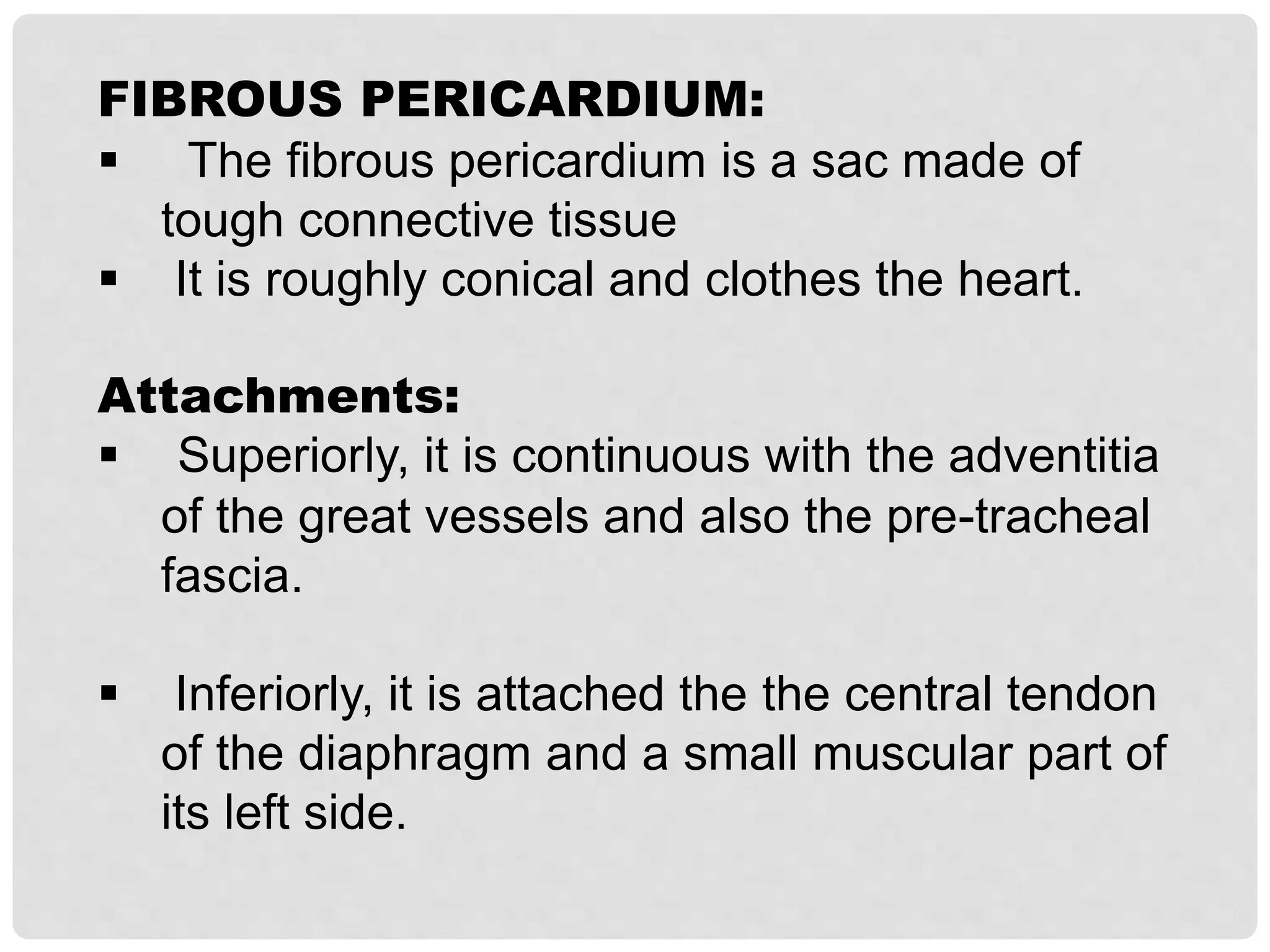
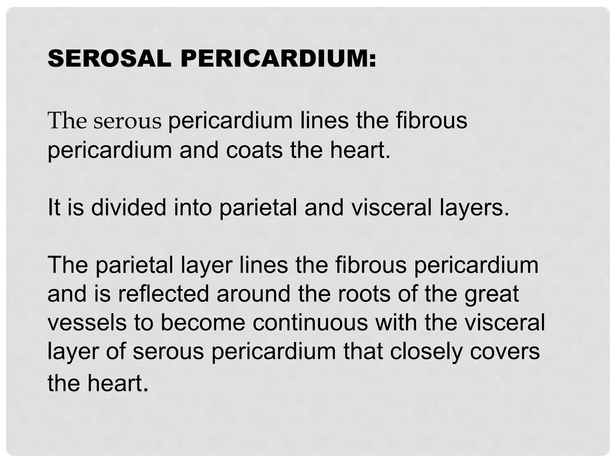
The pericardium
Pericardium: (Peri-around, Cardium-heart)
The pericardium is a fibro-serous sac that encloses the
heart and the roots of the great vessels. Its function is:
1) to restrict excessive movements of the heart.
2) serve as a lubricated container to the heart.
3) Acts as shock absorber with the help of the fluid filled
sac.
The pericardium lies within the middle mediastinum,
posterior to the body of the sternum (2 – 6th costal
cartilages) and anterior to the (5th – 8th thoracic vertebrae).
Pericardium
Inner Serous
layer
Inner visceral
Outer
Parietal
Outer
Fibrous layer
Single Layer
FIBROUS PERICARDIUM:
 The fibrous pericardium is a sac made of
tough connective tissue
 It is roughly conical and clothes the heart.
Attachments:
 Superiorly, it is continuous with the adventitia
of the great vessels and also the pre-tracheal
fascia.
 Inferiorly, it is attached the the central tendon
of the diaphragm and a small muscular part of
its left side.
 Anteriorly, it is attached to the posterior
surface of the sternum by superior and inferior
sternopericardial ligaments.
 The pericardium is securely anchored by
these attachments and maintains the general
thoracic position of the heart, serving as the
cardiac seat belt’.
Relations
Anteriorly:
 Seperated from the thoracic wall by the lungs
and the pleural coverings.
 But, in a small area behind the lower left half
of the body of the sternum and the sternal ends
of the left 4th and 5th costal cartilages , the
pericardium is in direct contact with the thoracic
wall.
 the lower end of the thymus.
Inferiorly:
 the pericardium is seperated from the liver and
the fundus of stomach by the diaphragm.
Posteriorly:
 The principal bronchi, the esophagus, the
esophageal plexus, the descending thoracic
aorta, and the posterior parts of the mediastinal
surface of both lungs.
Laterally:
 Mediastinal Pleura.
 The phrenic nerve with its accompaning
vessels, descends between the mediastinal
pleura and the fibrous pericardium on either
side.
SEROSAL PERICARDIUM:
The serous pericardium lines the fibrous
pericardium and coats the heart.
It is divided into parietal and visceral layers.
The parietal layer lines the fibrous pericardium
and is reflected around the roots of the great
vessels to become continuous with the visceral
layer of serous pericardium that closely covers
the heart.
The visceral layer is closely applied to the
heart and is often called the epicardium.
The slitlike space between the parietal and
visceral layers is referred to as the pericardial
cavity.
Normally, the cavity contains a small amount
of tissue fluid (about 50 mL), the pericardial
fluid, which acts as a lubricant to facilitate
movements of the heart.
Pericardial Sinuses
On the posterior surface of the heart, the reflection of
the serous pericardium around the large veins forms
a recess called 1. oblique sinus.
2. transverse sinus, which is a short passage that lies
between the reflection of serous pericardium around
the aorta and pulmonary trunk (post.surface of
heart).
The pericardial sinuses formed as a consequence of
the way the heart bends during development. They
have no clinical significance.
Vascular supply:
• 1. internal thoracic.
• 2. musculophrenic arteries.
• 3. descending thoracic aorta.
• The veins are tributaries of the azygous
system.
Innervation:
1) phrenic nerves:
(fibrous pericardium and the parietal layer of the
serous pericardium).
2) branches of the sympathetic trunks and the
vagus nerves (visceral layer of the serous
pericardium) .
Clinical Notes
Pericarditis: (inflammation of the serous pericardium).
pericardial fluid may accumulate excessively, which can
compress the atria and interfere with the filling of the
heart during diastole. This compression of the heart is
called cardiac tamponade.
Cardiac tamponade can also occur secondary to stab or
gunshot wounds when the chambers of the heart have
been penetrated. The blood escapes into the pericardial
cavity and can restrict the filling of the heart.
in acute pericarditis : Roughening of the visceral and parietal
layers of serous pericardium by inflammatory exudate
produces pericardial friction rub, which can be felt on
palpation and heard through a stethoscope.
Pericardial fluid can be aspirated from the pericardial cavity by
process called paracentesis.
The needle can be introduced to the left of the xiphoid
process in an upward and backward direction at an angle of
45° to the skin.
When paracentesis is performed at this site, the pleura and
lung are not damaged because of the presence of the cardiac
notch in this area.
 Autoimmune disorders that can cause
pericarditis include rheumatoid arthritis, lupus,
and scleroderma.
 Pericarditis occurs in up to 15% of patients who
have acute myocardial infarctions (heart
attacks).
• Some of the drugs that can produce pericarditis
include procainamide, hydralazine, phenytoin,
and isoniazid.

pericaduim anatomy pre - clinical (MBBS)

  • 1.
  • 3.
    Pericardium: (Peri-around, Cardium-heart) Thepericardium is a fibro-serous sac that encloses the heart and the roots of the great vessels. Its function is: 1) to restrict excessive movements of the heart. 2) serve as a lubricated container to the heart. 3) Acts as shock absorber with the help of the fluid filled sac. The pericardium lies within the middle mediastinum, posterior to the body of the sternum (2 – 6th costal cartilages) and anterior to the (5th – 8th thoracic vertebrae).
  • 4.
  • 6.
    FIBROUS PERICARDIUM:  Thefibrous pericardium is a sac made of tough connective tissue  It is roughly conical and clothes the heart. Attachments:  Superiorly, it is continuous with the adventitia of the great vessels and also the pre-tracheal fascia.  Inferiorly, it is attached the the central tendon of the diaphragm and a small muscular part of its left side.
  • 7.
     Anteriorly, itis attached to the posterior surface of the sternum by superior and inferior sternopericardial ligaments.  The pericardium is securely anchored by these attachments and maintains the general thoracic position of the heart, serving as the cardiac seat belt’.
  • 8.
    Relations Anteriorly:  Seperated fromthe thoracic wall by the lungs and the pleural coverings.  But, in a small area behind the lower left half of the body of the sternum and the sternal ends of the left 4th and 5th costal cartilages , the pericardium is in direct contact with the thoracic wall.  the lower end of the thymus.
  • 9.
    Inferiorly:  the pericardiumis seperated from the liver and the fundus of stomach by the diaphragm.
  • 10.
    Posteriorly:  The principalbronchi, the esophagus, the esophageal plexus, the descending thoracic aorta, and the posterior parts of the mediastinal surface of both lungs. Laterally:  Mediastinal Pleura.  The phrenic nerve with its accompaning vessels, descends between the mediastinal pleura and the fibrous pericardium on either side.
  • 11.
    SEROSAL PERICARDIUM: The serouspericardium lines the fibrous pericardium and coats the heart. It is divided into parietal and visceral layers. The parietal layer lines the fibrous pericardium and is reflected around the roots of the great vessels to become continuous with the visceral layer of serous pericardium that closely covers the heart.
  • 12.
    The visceral layeris closely applied to the heart and is often called the epicardium. The slitlike space between the parietal and visceral layers is referred to as the pericardial cavity. Normally, the cavity contains a small amount of tissue fluid (about 50 mL), the pericardial fluid, which acts as a lubricant to facilitate movements of the heart.
  • 13.
    Pericardial Sinuses On theposterior surface of the heart, the reflection of the serous pericardium around the large veins forms a recess called 1. oblique sinus. 2. transverse sinus, which is a short passage that lies between the reflection of serous pericardium around the aorta and pulmonary trunk (post.surface of heart). The pericardial sinuses formed as a consequence of the way the heart bends during development. They have no clinical significance.
  • 16.
    Vascular supply: • 1.internal thoracic. • 2. musculophrenic arteries. • 3. descending thoracic aorta. • The veins are tributaries of the azygous system.
  • 17.
    Innervation: 1) phrenic nerves: (fibrouspericardium and the parietal layer of the serous pericardium). 2) branches of the sympathetic trunks and the vagus nerves (visceral layer of the serous pericardium) .
  • 18.
    Clinical Notes Pericarditis: (inflammationof the serous pericardium). pericardial fluid may accumulate excessively, which can compress the atria and interfere with the filling of the heart during diastole. This compression of the heart is called cardiac tamponade. Cardiac tamponade can also occur secondary to stab or gunshot wounds when the chambers of the heart have been penetrated. The blood escapes into the pericardial cavity and can restrict the filling of the heart.
  • 19.
    in acute pericarditis: Roughening of the visceral and parietal layers of serous pericardium by inflammatory exudate produces pericardial friction rub, which can be felt on palpation and heard through a stethoscope. Pericardial fluid can be aspirated from the pericardial cavity by process called paracentesis. The needle can be introduced to the left of the xiphoid process in an upward and backward direction at an angle of 45° to the skin. When paracentesis is performed at this site, the pleura and lung are not damaged because of the presence of the cardiac notch in this area.
  • 21.
     Autoimmune disordersthat can cause pericarditis include rheumatoid arthritis, lupus, and scleroderma.  Pericarditis occurs in up to 15% of patients who have acute myocardial infarctions (heart attacks). • Some of the drugs that can produce pericarditis include procainamide, hydralazine, phenytoin, and isoniazid.