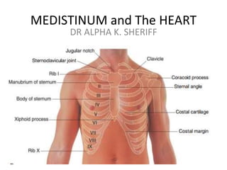
SlideShare a Scribd company logo
1 of 72
MEDISTINUM and The HEART
DR ALPHA K. SHERIFF
THE MEDIASTINUM
• The mediastinum-is a broad central partition that
separate the laterally placed pleural cavities . It extends:
• From the sternum to the bodies of the vertebrae, and
• From the superior thoracic aperture to the diaphragm.
• It contains the following:
The thymus gland The pericardia sac
The trachea The heart
The major arteries of the vein.
• It serve as a passageway for :
The esophagus
The thoracic duct
• Various component of the nervous system as they
traverse the thorax on their way to the abdomen
• The mediastinum contains all the thoracic Viscera
and structures expect the lungs.
• The mediastinum can be subdivided into smaller
regions by a transverse plane extending from the
sternal angle to the intervertebral disc between TIV
and TV separating the mediastinum into
• Superior mediastinum
• Inferior mediastinum -further divides into
• Anterior mediastinum
• Middle mediastinum
• Posterior mediastinum
• The Anterior Mediastinum:- is the area
anterior to the pericardia! sac and posterior to
the body of the sternum.
• The Posterior Mediastinum:-is the region
posterior to the pericardial sac and the
diaphragm and anterior to the bodies of the
vertebrae.
• The Middle Mediastinum:- is centrally located
in the thoracic cavity. It contains the
pericardium, heart, origins of the great
vessels, various nerves, and smaller vessels.
Superior Mediastinum
The superior mediastinum is posterior to the manubrium of the sternum and
anterior to the bodies of the first four thoracic vertebrae.
• Its superior boundary is an oblique plane passing from the jugular notch upward
and posteriorly to the superior border of vertebra TI.
• Inferiorly, a transverse plane passing from the sternal angle to the intervertebral
disc between vertebra TIV/V separates it from the inferior mediastinum.
• Laterally, it is bordered by the mediastinal part of the parietal pleura on either
side.
The superior mediastinum is continuous with the neck above and with the inferior
mediastinum below.
The major structures found in the superior mediastinum include the:
1. thymus, .7. esophagus
2. right and left brachiocephalic veins, .8. phrenic nerves,
3. left superior intercostal vein, .9. vagus nerves,
4. superior vena cava, .10. left recurrent laryngeal branch
5. arch of the aorta with its three of the left vagus nerve
large branches . 11. thoracic duct
6.trachea, other small nerves, blood vessels .12. lymphatics.
Thymus Gland
• The Thymus is an endocrine and lymphatic gland situated in the anterior
component of the superior mediastinum, lying immediately posterior to the
manubrium of the sternum. It is an asymmetrical, bilobed structure.
• The upper extent of the thymus can reach into the neck as high as the
thyroid gland;
• a lower portion typically extends into the anterior mediastinum over the
pericardial sac.
• It involved in the early development of the immune system, the thymus is a
large structure in the child, begins to atrophy after puberty, and shows
considerable size variation in the adult.
• In the elderly adult, it is barely identifiable as an organ, consisting mostly of
fatty tissue that is sometimes arranged as two lobulated fatty structures.
• Arteries to the thymus consist of small branches originating from the
internal thoracic arteries.
• Venous drainage is usually into the left brachiocephalic vein and possibly
into the internal thoracic veins.
• Lymphatic drainage returns to multiple groups of nodes at one or more of
the following locations:
• along the internal thoracic arteries (parasternal) ;
• at the tracheal bifurcation (tracheobronchial); and
• in the root of the neck.
Pericardium
The Pericardium is a fibroserous sac surrounding the
heart and the roots of the great vessels. It consists of two
components:
1. The Fibrous Pericardium 2.the Serous Pericardium.
The Fibrous Pericardium is a tough connective tissue
outer layer that defines the boundaries of the middle
mediastinum.
The Serous Pericardium is thin and consists of
two parts:
 The parietal layer of serous pericardium lines the
inner surface of the fibrous pericardium.
 The Visceral layer (epicardium) of serous pericardium adheres
to the heart and forms its outer covering.
Pericardial cavity-is the space between the two
layers of serous pericardium, containing a small
amount of fluid. This potential space allows the
relatively movement of the heart.
Fibrous pericardium
The fibrous pericardium is a cone-shaped bag. It base rest on the
central tendon of the diaphragm and
It apex continuous with the adventitia of the great vessels.
Anteriorly, it is attached to the posterior surface of the sternum by
sternopericardial ligaments.
These attachments help to retain the heart in its position in the
thoracic cavity. The sac also limits cardiac distention.
The phrenic nerves, which innervate the diaphragm, originating
from spinal cord levels C3 to C5, innervate the fibrous pericardium.
.
Their location, within the fibrous pericardium, directly related to
the embryological origin of the diaphragm and the changes that
occur during the formation of the pericardia! cavity.
The Pericardiacophrenic vessels are also located within and supply
the fibrous pericardium.
Serous Pericardium
The parietal layer of the serous pericardium is continuous with the visceral layer of
serous pericardium around the roots of the great vessels.
These reflections of serous pericardium occur in two locations:
• one superiorly, surrounding the arteries, the aorta, and
the pulmonary trunk;
• the second , posteriorly, surrounding the veins, the superior and inferior vena
cava, and the pulmonary veins.
Vessels and nerves
The pericardium is supplied by branches from the internal thoracic,
pericardiacophrenic, musculophrenic, and inferior phrenic arteries, and the
thoracic aorta.
Veins from the pericardium enter the azygos system of veins and the internal
thoracic and superior phrenic veins.
Nerves supply arise from the vagus nerve [X], the sympathetic trunks, and the
phrenic
nerves.
Note: The source of somatic sensation (pain) from the parietal pericardium is
carried by somatic afferent fibers in the phrenic nerves.
For this reason, "pain" related to a pericardia! problem may be referred to the
supraclavicular region of the shoulder or lateral neck area dermatomes for spinal
cord segments C3, C4, and C5.
• Pericarditis
Pericarditis is an inflammatory condition of the
pericardium.
Common causes are viral and bacterial infections,
systemic illnesses (e.g., chronic renal failure),and after
myocardial infarction.
Note: Pericarditis is different from myocardial
infarction and the treatment and prognosis are
different.
Pericarditis pt complain of continuous central chest
pain that may radiate to one or both arms. This pain
may be relieved by sitting and leaning forward. An
electrocardiogram (ECG) is used to help differentiate
between the two conditions.
In myocardial infarction, however, the pain central but
does not radiate to any place. Pt may not need to lean
forward.
Pericardial effusion
Normally, only a tiny amount of fluid is present between
the visceral and parietal layers of the serous
pericardium.
In certain situations, this space can be filled with excess
fluid (PericardiaI Effusion).
The fibrous pericardium is a "relatively fixed"
structure that cannot expand easily.
Rapid accumulation of excess fluid within the pericardial
sac compresses the heart (cardiac tamponade), resulting in
biventricular failure.
Treatment: by removing the fluid with a needle inserted
into the pericardial sac can relieve the symptoms.
Constrictive Pericarditis
Constrictive Peicarditis:- is the abnormal thickening
of the pericardiaI sac which compresses the heart,
impairing heart function and resulting in heart
failure.
Diagnosis: is made by inspecting for the jugular
venous pulse in the neck.
In normal individuals, the jugular venous pulse
drops on inspiration.
In patients with Constrictive Pericarditis, the reverse
happens and this is called Kussmaul's sign.
Treatment: Involves surgical opening of the
pericardial sac
The HEART
The HEART is a fist-sized muscular organ that pumps blood
throughout the body.
It is the primary organ in the circulatory system.
Cardiac orientation
The general shape and orientation of the heart are that of a
pyramid that has fallen over and is resting on one of its sides.
In the thoracic cavity, the apex of this pyramid projects forward,
downward, and to the left,
whereas the base is opposite the apex and faces in a posterior
direction.
The sides of the pyramid consist of:
• a diaphragmatic (inferior) surface on which the pyramid rests,
• an anterior (sternocostal) surface oriented anteriorly,
• a right pulmonary surface, and
• a left pulmonary surface.
Cont’
BASE (Posterior surface) and APEX
The base of the heart is quadrilateral and
directed posteriorly. It consists of:
• the left atrium,
• a small portion of the right atrium, and
• the proximal parts of the great veins (superior
and inferior venae cavae and the pulmonary
veins).
Note: The great veins enter the base of the heart,
with the pulmonary veins entering the right and
left sides of the left atrium
Cont’
The superior and inferior venae cavae at the upper
and lower ends of the right atrium, the base
of the heart is fixed posteriorly to the pericardial
wall, opposite the bodies of vertebrae TV to TVIII
{TVI to TIX when standing) .
• The esophagus lies immediately posterior to the
base.
• The apex of the heart is formed by the inferolateral
part of the left ventricle and is positioned deep to
the left fifth intercostal
space, 8-9 cm from the midsternal line.
Other Surfaces o the Heart
• Anterior Surface- consists mostly of the right ventricle, with
some of the right atrium on the right and some of the left
ventricle on the left.
• Diaphragmatic Surface- is that part of the heart in it
anatomical position resting on the diaphragm. It consist of
the left ventricle and a small portion of the right ventricle
separated by the posterior interventricular groove.
• Left Pulmonary Surface faces the left lung, is broad and
convex, and consists of the left ventricle and a portion of the
left atrium.
• Right Pulmonary Surface faces the right lung, is broad and
convex, and consists of the right atrium.
Margins and Borders
The Right and Left margins- are the same as the right
and left pulmonary surfaces of the heart.
The inferior margin- is the sharp edge
between the anterior and diaphragmatic surfaces of
the heart which is formed mostly by the right ventricle
and a small portion of the left ventricle near the apex.
The obtuse margin. It separates the anterior and left
pulmonary surfaces. It is round and extends from the
left auricle to the cardiac apex and is formed mostly by
the left ventricle and superiorly by a small portion of
the left auricle.
Partitions of the HEART
There are two partitioning of the heart. EXTERNAL and INTERNAL
PARTITIONS
Internal partitions divide the heart into four chambers (2 Atria and 2
Ventricles) and produce surface or external grooves referred to as sulci.
External Partitions
The coronary sulcus circles the heart, separating the atria from the
ventricles. As it circles the heart, it contains the right coronary artery,
the small cardiac vein, the coronary sinus, and the circumflex branch of
the left coronary artery.
The anterior and posterior interventricular sulci separate the two
ventricles –
The anterior interventricular sulcus which is on the anterior surface
of the heart and contains the anterior interventricular artery and the
great cardiac vein, and
The posterior interventricular sulcus is on the diaphragmatic surface
of the heart and contains the posterior interventricular artery and the
middle cardiac vein.
These sulci are continuous inferiorly, just to the right of the apex of the
heart
Cardiac Chambers
The heart functionally consists of two pumps separated by a partition.
The RIGHT PUMP receives deoxygenated blood from the body and sends
it to the lungs.
The LEFT PUMP receives oxygenated blood from the lungs and sends it
to the body.
Each pump consists of an atrium and a ventricle separated by a valve.
The THIN-WALLED ATRIA receive blood coming into the heart, whereas the
relatively THICK-WALED VENTRICLES pump blood out of the heart.
More force is required to pump blood through the body than through the
lungs, so the muscular wall of the left ventricle is thicker than the right.
Interatrial, interventricular, and atrioventricular septa separate the four
(4) chambers of the heart.
Right Atrium
The right atrium:- This chamber serves as the right border of the heart i.e
the heart's anterior surface.
Blood returning to the right atrium enters through one of three vessels.
These are:
• the superior and inferior venae cavae, which together
deliver blood to the heart from the body; and
• the coronary sinus, which returns blood from the walls of the heart
itself.
Blood coming from the right atrium passes into the right ventricle through
the right atrioventricular orifice.
The atrioventricular opening faces forward and medially and is closed
during ventricular contraction by the tricuspid valve
The interior of the right atrium is divided into two continuous spaces.
Externally, this separation is indicated by a shallow, vertical groove the
sulcus terminalis cordis, extends from the right side of the opening of the
superior vena cava to the right side of the opening of the inferior vena
cava.
Cont’
• Internally, this division is indicated by the crista terminalis ,
which is a smooth, muscular ridge that begins just in front
of the opening of the superior vena cava and extends to the
anterior inferior vena cava.
• The space posterior to the crista is the sinus of venae cavae
derived embryologically from the right horn of the sinus
venosus.
• This component of the right atrium
has smooth, thin walls, and both venae cavae empty into
this space.
• The cardiac veins sends blood into the Rt atrium through the coronary
sinus (opens into the right atrium).
• The embryonic sinus venosus are associated with the valve of the coronary
sinus and the valve of inferior vena cava, respectively.
• During development, the valve of the inferior vena cava helps direct
incoming oxygenated blood through the foramen ovale and into the left
atrium.
• The interatrial septum separates the right atrium from the left atrium.
• The fossa ovalis (oval fossa), is a depression clearly visible in the septum
just above the orifice of the inferior vena cava with its prominent margin,
the limbus fossa ovalis (border of the oval fossa).
• The fossa ovalis marks the location of the embryonic foramen ovale, which
is an important part of fetal circulation.
• The foramen ovale allows oxygenated blood entering the right atrium
through the inferior vena cava to pass directly to the left atrium and so
bypass the lungs, which are nonfunctional before birth.
• Openings of the smallest cardiac veins (the
foramina of the venae cordis minimae)-on the
walls of the right atrium,drains the
myocardium directly into the right atrium
Right Ventricle
• The right ventricle forms most of the anterior surface of the
heart and a portion of the diaphragmatic surface.
• The right atrium is to the right of the right ventricle and the
right ventricle is located in front of and to the left of the
right atrioventricular orifice.
• Blood entering the right ventricle from the right atrium
therefore
moves in a horizontal and forward direction.
• The outflow tract of the right ventricle, which leads to
the pulmonary trunk, is the conus arteriosus
(infundibulum). This area has smooth walls and derives
from the embryonic bulbus cordis.
• The walls of the inflow portion of the right ventricle have
numerous muscular, irregular structures called trabeculae
carneae
• The Trabeculae carneae (papillary muscles): are attached to
the ventricular surface, and it other end serves as the point of
attachment for tendon-like fibrous cords (the chordae
tendineae), connecting to the free edges of the cusps of the
tricuspid valve.
• There are three papillary muscles in the right ventricle.
They are named relative to their point of origin on the
ventricular surface:
(i)Anterior, (ii) Posterior, and (iii) Septal papillary muscles
• Anterior papillary muscle: is the largest and most constant
papillary muscle, and arises from the anterior wall of the
ventricle.
• Posterior papillary muscle: may consist of one, two, or
three structures, with some chordae tendineae arising
directly from the ventricular wall.
• Septal papillary muscle: is the most inconsistent papillary
muscle, being either small or absent, with chordae tendineae
emerging directly from the septal wall.
Tricuspid Valve
The tricuspid valve (right atrioventricular valve) is responsible for the
closure of the right atrioventricular orifice during ventricular contraction .
The tricuspid valve is three cusps or leaflets organ.
These are: the anterior, septal, and posterior cusps, based/positioned in
the right ventricle.
The free margins of the cusps are attached to the chordae tendineae, which
arise from the tips of the papillary muscles.
During filling of the right ventricle, the tricuspid valve opens, and the three
cusps project into the right ventricle allowing blood to flow into the right
ventricle. Howevere, when this fails to happened, blood move back into the
right atrium.
The papillary muscles prevents thees cusps from being everted into the
right atrium.
Note: The papillary muscles and associated chordae tendineae keep the
valves properly closed during ventriclar contraction causing blood to exit
the right ventricle and move into the pulmonary trunk.
Necrosis of this papillary muscle following a myocardial infarction (heart
attack) may result in prolapse of the related valve.
Pulmonary Valve
The Pulmonary valve serves as the gateway to blood flowing to the
lungs through the pulmonary trunk from the right.
The pulmonary valve consists of three semilunar cusps with free
edges projecting upward into the lumen of the pulmonary trunk.
The free superior edge of each cusp has a middle cusp, the nodule
of the semilunar cusp, and a thin lateral lunula of the semilunar
cusp.
The cusps are the left, right, and anterior semilunar cusps, relative
to their fetal position before rotation of the outflow tracts from
the ventricles is complete.
Each cusp forms a sinus in the pulmonary trunk. After ventricular
contraction, the blood fills these pulmonary sinuses and forces the
cusps closed.
This prevents blood in the pulmonary trunk from refilling the right
ventricle
Left Atrium
The left atrium forms most of the base and posterior surface of the heart.
The left atrium is derived embryologically from two structures.
• The posterior half, or inflow portion, receives the four pulmonary veins . It
has smooth walls and derives from the proximal parts of the pulmonary
veins that are incorporated into the left atrium during development.
• The anterior half is continuous with the left auricle. It contains musculi
pectinati and derives from the embryonic primitive atrium.
The interatrial septum is part of the anterior wall of the left atrium.
The thin area or depression in the septum is the valve of the foramen ovale
and is opposite the floor of the fossa ovalis in the right atrium.
During development, the valve of the foramen ovale prevents blood from
passing from the left atrium to the right atrium.
This valve may not be completely fused in some adults, leaving a "probe
patent" passage between the right atrium and the left atrium.
Left Ventricle
The left ventricle anatomically, lies anterior to the left atrium.
Blood enters the ventricle through the left atrioventricular
orifice and flows in a forward direction to the apex. The
chamber itself is conical, is longer than the right ventricle,
and has the thickest layer of myocardium.
It has two Papillary muscles (the anterior and posterior
papillary muscles), together with chordae tendineae, are
similar to that in the right ventricle.
The interventricular septum separating the two
ventricles,forms the anterior wall and some of the wall on the
right side of the left ventricle.
This septum is having two parts:
• a muscular part, and
• a membranous part.
The muscular part is thick and forms the major part of the
septum, whereas the membranous part is the thin.
Mitral Valve
The Mitral Valve (left atrioventricular valve) also
referred to as the bicuspid valve because it has two
cusps, the anterior and posterior cusps, closes the left
atrioventricular orifice during ventricular contraction.
The bases of the cusps are secured to a fibrous ring
surrounding the opening, and the cusps are continuous
with each other at the commissures.
The coordinated action of the papillary muscles and
chordae tendineae is as described for the right
ventricle.
Aortic Valve
The Aortic valve close the opening from the left ventricle into the
aorta.
The aortic vestibule, or outflow tract of the left ventricle, is
continuous superiorly with the ascending aorta.
The valve is similar in structure to the pulmonary valve.
It consists of three semilunar cusps each projecting upward into
the lumen of the ascending aorta .
Between the semilunar cusps and the wall of the ascending aorta
are pocket-like sinuses-the right, left, and posterior aortic sinuses.
The right and left coronary arteries originate from the right and
left aortic sinuses.
The functioning of the aortic valve is similar to that of the
pulmonary valve with one important additional process: as blood
recoils after ventricular contraction and fills the aortic sinuses, it
automatically force the blood into the coronary arteries because
these vessels originate from the right and left aortic sinuses.
Valve Disease
Valve problems consist of two basic types:
• Incompetence (insufficiency), which results from poorly functioning
valves; and
• Stenosis, a narrowing of the orifice, caused by the valve's inability to open
fully.
Mitral valve disease is usually a mixed pattern of stenosis and
incompetence, one of which usually predominates. Both stenosis and
incompetence lead to a poorly functioning valve and subsequent heart
changes, which include:
• left ventricular hypertrophy (this is appreciably less marked in patients
with mitral stenosis);
• increased pulmonary venous pressure;
• pulmonary edema; and
• enlargement (dilation) and hypertrophy of the left atrium.
Aortic valve disease-both aortic stenosis and aortic regurgitation (backflow)
can produce marked heart failure.
Valve disease in the right side of the heart (affecting the tricuspid or
pulmonary valve) is most likely caused by infection. The resulting valve
dysfunction produces abnormal pressure changes in the right atrium and
right ventricle, and these can induce cardiac failure.
Cardiac Skeleton
The cardiac skeleton is a collection of dense, fibrous connective tissue in the form of four
rings with interconnecting areas in a plane between the atria and the ventricles.
The four rings of the cardiac skeleton surrounds:
the two atrioventricular orifices,
the aortic orifice and
opening of the pulmonary trunks.
The interconnecting areas include:
• the right fibrous trigone, which is a thickened area of connective tissue between the
aortic ring and right atrioventricular ring; and
• the left fibrous trigone, which is a thickened area of connective tissue between the aortic
ring and the left atrioventricular ring.
The cardiac skeleton helps maintain the integrity of the openings it surrounds and provides
points of attachment for the cusps.
It also separates the atrial musculature from the ventricular musculature.
The atrial myocardium originates from the upper border of the rings, whereas the
ventricular myocardium originates from the lower border of the rings.
The cardiac skeleton also serves as a dense connective tissue partition that electrically
isolates the atria from the ventricles.
The atrioventricular bundle, which passes through the anulus, is the single connection
between these two groups of myocardium.
Coronary Vasculature
There are two coronary arteries arising from the aortic sinuses
of the ascending aorta and supply the heart muscles and other
tissues of the heart.
These arteries circled the heart in the coronary sulcus, with
marginal and interventricular branches, in the interventricular
sulci, converging toward the apex of the heart.
The returning venous blood passes through cardiac veins, most
of which empty into the coronary sinus.
This large venous structure is located in the coronary sulcus on
the posterior surface of the heart between the left atrium and
left ventricle.
The coronary sinus empties into the right atrium between the
opening of the inferior vena cava and the right atrioventricular
orifice.
Coronary Arteries
The right coronary artery :-originates from the right aortic sinus of the ascending
aorta. It passes anteriorly and then descends vertically in the coronary sulcus,
between the right atrium and right ventricle.
Upon reaching the inferior margin of the heart, it turns posteriorly and continues in
the sulcus onto the diaphragmatic surface and base of the heart.
During it course, it gives arise to several branches:
1. An atrial branch ,
2. A right marginal branch is given off as it approaches the inferior (acute) margin
of the heart, and
3. the posterior interventricular branch(Its final major branch), which lies in the
posterior interventricular sulcu.
The right coronary artery supplies:
the right atrium and right ventricle,
the sinu-atrial and atrioventricular nodes,
the interatrial septum, a portion of the left atrium,
the posteroinferior one third of the interventricular septum, and
a portion of the posterior part of the left ventricle.
Cont’
The left coronary artery originates from the left aortic sinus of the
ascending aorta. It passes between the pulmonary trunk and the left auricle
before entering the coronary sulcus.
The artery divides into its two terminal branches:
the anterior interventricular branch and the circumflex branch.
• The anterior interventricular branch (left anterior descending artery-LAD)
continues around the left side of the pulmonary trunk and descends
obliquely toward the apex of the heart in the anterior interventricular
sulcus.
During its course, one or two large diagonal branches may arise and
descend diagonally across the anterior surface of the left ventricle.
• The circumflex branch courses toward the left, in the coronary sulcus and
onto the base/ diaphragmatic surface of the heart, and usually ends before
reaching the posterior interventricular sulcus.
The left marginal artery ,arises from it and continues across the rounded
margin of the heart.
The left coronary artery supply :
1. most of the left atrium and left ventricle, and
2. most of the interventricular septum, including the atrioventricular
bundle and its branches.
Clinical Correlation
• Heart attack:- occurs when the perfusion to the myocardium is insufficient
to meet the metabolic needs of the tissue, leading to irreversible tissue
damage.
• The most common cause is a total occlusion of a major coronary artery.
Coronary Artery Disease
Occlusion of a major coronary artery, usually due to atherosclerosis, leads
to inadequate oxygenation of an area of myocardium and cell death.
• The severity of the problem will be related to:
1. the size and location of the artery involved,
2. whether or not the blockage is complete, and
3. whether there are collateral vessels to provide perfusion to the territory
from other vessels.
Depending on the severity, patients can develop Chest pain
(angina) or a myocardial infarction (MI)
Cardiac Veins
The coronary sinus receives four major tributaries:
The great cardiac vein
The middle cardiac vein,
The small cardiac vein and
The posterior cardiac veins.
Great Cardiac vein: It starts at the apex of the heart. It is
related to the anterior interventricular artery and is often
termed the anterior interventricular vein.
Continuing along its path in the coronary sulcus, the great
cardiac vein enlarges to form the coronary sinus, which
enters the right atrium.
• The middle cardiac vein (posterior interventricular vein)
begins near the apex of the heart and ascends in the
posterior interventricular sulcus toward the coronary sinus.
• It is associated with the posterior interventricular branch of
the right or left coronary artery throughout its course.
• The Small cardiac vein begins in the lower anterior section of
the coronary sulcus between the right atrium and right
ventricle.
• It continues in this groove onto the base/diaphragmatic
surface and enters the coronary sinus in the right atrium
directly.
• It is a companion of the right coronary artery throughout its
course and may receive the right marginal vein.
• The posterior cardiac vein lies on the posterior surface of the
left ventricle just to the left of the middle cardiac vein. It
either enters the coronary sinus directly or joins the great
cardiac vein.
Other cardiac veins.
Two additional groups of cardiac veins are also involved in the venous
drainage of the heart.
• The anterior veins of the right ventricle (anterior cardiac veins) are small
veins that arise on the anterior surface of the right ventricle. They drain the
anterior portion of the right ventricle. The right marginal vein may be part
of this group if it does not enter the small cardiac vein
• A group of smallest cardiac veins (venae cordis minimae or veins of
Thebesius) have also been described.
They drain directly into the right atrium and right ventricle, they are
occasionally associated with the left atrium, and are rarely associated with
the left ventricle.
Coronary Lymphatics.
The lymphatic vessels of the heart follow the coronary arteries and drain
mainly into:
• brachiocephalic nodes, anterior to the brachiocephalic veins; and
• tracheobronchial nodes, at the inferior end of the trachea.
Conducting System of the Heart
The musculature of the atria and ventricles is capable of contracting
spontaneously.
The cardiac conduction system initiates and coordinates contraction.
The conduction system consists of nodes and networks of specialized
cardiac muscle cells organized into four basic components:
• the sinu-atrial node,
• the atrioventricular node,
• the atrioventricular bundle with its right and left bundle branches,
and
• the subendocardial plexus of conduction cells (the Purkinje fibers) .
This system is an important unidirectional pathway of
excitation/contraction that are insulated from the surrounding
myocardium by connective tissue.
Thus, a unidirectional wave of excitation and contraction is
established, which moves from the papillary muscles and apex of the
ventricles to the arterial outflow tracts.
Sinu-atrial node(SA Node):- Impulses begin at the sinu-
atrial node, the cardiac pacemaker.
• The SA Node are collection of cells located at the
superior end of the crista terminalis at the junction of
the superior vena cava and the right atrium.
The excitation signals generated by the SA node spread
across the atria, causing the muscle to contract.
Atrioventricular node (AV Node):-Concurrently, the
excited wave from the atria stimulates trough the AV
node, which is located near the opening of the
coronary sinus, close to the tricuspid valve, and within
the atrioventricular septum.
• The AV node is a collection of specialized cells that
forms the beginning of an complex system of
conducting tissue, the atrioventricular bundle, which
extends the excitatory impulse to all ventricular
musculature.
• The Atrioventricular bundle:-is a direct continuation of the
atrioventricular node .
• It follows along the lower border of the membranous part of the
interventricular septum before splitting into right and left bundles.
• The right bundle branch continues on the right side of the
interventricular septum toward the apex of the right ventricle.
• From the septum it reach the base of the anterior papillary
muscle.
At this point, it divides and continuous with the final component of
the cardiac conduction system, the subendocardial plexus of
ventricular conduction cells or Purkinje fibers.
• This network of specialized cells spreads throughout the ventricle
to supply the ventricular musculature, including the papillary
muscles.
• The left bundle branch passes to the left side of the muscular
interventricular septum and descends to the apex of the left
ventricle. Along its course it gives off branches that eventually
become continuous
with the Purkinje fibers.
• As described on the right side, this network of specialized cells
spreads the excitation impulses throughout the left ventricle.
Cardiac Innervation
Cardiac innervation is under the influence of the Autonomic Nervous
System controlled by the division of the Peripheral Nervous System (PNS)
which directly regulates:
• Heart Rate (Chronotropic effect),
• force of each contraction (Inotropic effect) and
• Cardiac Output.
Branches from both the parasympathetic and sympathetic systems
contribute to the formation of the cardiac
plexus.
This plexus consists of :
 a superficial part, inferior to the aortic arch and between it and the
pulmonary trunk and
 a deep part, between the aortic arch and the tracheal bifurcation.
From the cardiac plexus, small branches that are mixed nerves containing
both sympathetic and parasympathetic fibers supply the heart.
These branches affect nodal tissue and other components of the conduction
system, coronary blood vessels, and atrial and ventricular musculature.
Parasympathetic Innervation
Stimulation of the parasympathetic system leads to:
• decreases heart rate,
• reduces force of contraction, and
• constricts the coronary arteries.
The preganglionic parasympathetic fibers reach the heart as cardiac
branches from the right and left vagus nerves.
They enter the cardiac plexus and synapse in ganglia located either within
the plexus or in the walls of the atria.
Sympathetic innervation
Stimulation of the sympathetic system leads to:
• increases heart rate, and
• increases the force of contraction.
Sympathetic fibers reach the cardiac plexus through the cardiac nerves from
the sympathetic trunk.
Preganglionic sympathetic fibers from the upper four or five segments of
the thoracic spinal cord enter and move through the sympathetic trunk.
They synapse in the cervical and upper thoracic sympathetic ganglia, and
the postganglionic fibers proceed as bilateral branches from the
sympathetic trunk to the cardiac plexus.
Visceral Afferents
Visceral afferents from the heart are component of the cardiac plexus.
These fibers pass through the cardiac plexus and return to the central
nervous system in the cardiac nerves from the sympathetic trunk and in the
vagal cardiac branches.
• The afferents associated with the vagal cardiac nerves return to the vagus
nerve [X] They sense alterations in blood pressure and blood chemistry and
are therefore primarily concerned with cardiac reflexes.
• The afferents associated with the cardiac nerves from the sympathetic
trunks return to either the cervical or the thoracic portions of the
sympathetic trunk.
• If they are in the cervical portion of the trunk, they normally descend to
the thoracic region, where they reenter the upper four or five thoracic
spinal cord segments, along with the afferents from the thoracic region of
the sympathetic trunk.
• Visceral afferents associated with the sympathetic system conduct pain
sensation from the heart, which is detected at the cellular level as tissue-
damaging events (i.e. , cardiac ischemia). This pain is often “a Referred
Pain" to cutaneous regions supplied by the same spinal cord levels.
MEDISTINUM and The HEART.pptx1111111111111111111

More Related Content

Similar to MEDISTINUM and The HEART.pptx1111111111111111111

Pericardium and heart
Pericardium and heartPericardium and heart
Pericardium and heartEgoist el
 
Pericardium and heart
Pericardium and heartPericardium and heart
Pericardium and heartDr.B.B. Gosai
 
Mediastinum 2020.pdf
Mediastinum 2020.pdfMediastinum 2020.pdf
Mediastinum 2020.pdfFatimakabara1
 
cardiac anatomy for students
cardiac anatomy for studentscardiac anatomy for students
cardiac anatomy for studentsAnees Hasnain
 
10. ANATOMY OF THE MEDIASTINUM MATERIAL.pptx
10. ANATOMY OF THE MEDIASTINUM MATERIAL.pptx10. ANATOMY OF THE MEDIASTINUM MATERIAL.pptx
10. ANATOMY OF THE MEDIASTINUM MATERIAL.pptxLivingBeing
 
Anatomy of heart presentation
Anatomy of heart presentationAnatomy of heart presentation
Anatomy of heart presentationRajeshkumarm16
 
Cardiovascular Anatomy.pptx
Cardiovascular Anatomy.pptxCardiovascular Anatomy.pptx
Cardiovascular Anatomy.pptxxamdathisbad
 
Cardiovascular Anatomy.pptx
Cardiovascular Anatomy.pptxCardiovascular Anatomy.pptx
Cardiovascular Anatomy.pptxxamdathisbad
 
The pericardium and the pericardial sinuses
The pericardium and the pericardial sinusesThe pericardium and the pericardial sinuses
The pericardium and the pericardial sinusesMohana Sekar
 
3-pericardium & Heart.pptx
3-pericardium & Heart.pptx3-pericardium & Heart.pptx
3-pericardium & Heart.pptxGulAndama2
 
THORAX OS powerpoint
THORAX OS powerpointTHORAX OS powerpoint
THORAX OS powerpointMarime S
 
anatomy and physiology of heart..ppt....
anatomy and physiology of heart..ppt....anatomy and physiology of heart..ppt....
anatomy and physiology of heart..ppt....DeepikaLingam2
 
(Tannu Tanya )anatomy of heart.pdf.besics
(Tannu Tanya )anatomy of heart.pdf.besics(Tannu Tanya )anatomy of heart.pdf.besics
(Tannu Tanya )anatomy of heart.pdf.besicssamaryadavkz435
 
anatomyofheart-160826060632 2.pdf
anatomyofheart-160826060632 2.pdfanatomyofheart-160826060632 2.pdf
anatomyofheart-160826060632 2.pdfraksha318928
 
anatomyofheart-160826060632.pdf
anatomyofheart-160826060632.pdfanatomyofheart-160826060632.pdf
anatomyofheart-160826060632.pdfraksha318928
 
anatomyofheart-160826060632.pdf
anatomyofheart-160826060632.pdfanatomyofheart-160826060632.pdf
anatomyofheart-160826060632.pdfDiamondKumar2
 

Similar to MEDISTINUM and The HEART.pptx1111111111111111111 (20)

Pericardium and heart
Pericardium and heartPericardium and heart
Pericardium and heart
 
Heart Anatomy- Dr. Sonalika.pdf
Heart Anatomy- Dr. Sonalika.pdfHeart Anatomy- Dr. Sonalika.pdf
Heart Anatomy- Dr. Sonalika.pdf
 
Pericardium and heart
Pericardium and heartPericardium and heart
Pericardium and heart
 
Lecture 4 Heart Anatomy
Lecture 4 Heart AnatomyLecture 4 Heart Anatomy
Lecture 4 Heart Anatomy
 
Mediastinum 2020.pdf
Mediastinum 2020.pdfMediastinum 2020.pdf
Mediastinum 2020.pdf
 
cardiac anatomy for students
cardiac anatomy for studentscardiac anatomy for students
cardiac anatomy for students
 
10. ANATOMY OF THE MEDIASTINUM MATERIAL.pptx
10. ANATOMY OF THE MEDIASTINUM MATERIAL.pptx10. ANATOMY OF THE MEDIASTINUM MATERIAL.pptx
10. ANATOMY OF THE MEDIASTINUM MATERIAL.pptx
 
Anatomy of heart presentation
Anatomy of heart presentationAnatomy of heart presentation
Anatomy of heart presentation
 
Cardiovascular Anatomy.pptx
Cardiovascular Anatomy.pptxCardiovascular Anatomy.pptx
Cardiovascular Anatomy.pptx
 
Cardiovascular Anatomy.pptx
Cardiovascular Anatomy.pptxCardiovascular Anatomy.pptx
Cardiovascular Anatomy.pptx
 
The pericardium and the pericardial sinuses
The pericardium and the pericardial sinusesThe pericardium and the pericardial sinuses
The pericardium and the pericardial sinuses
 
3-pericardium & Heart.pptx
3-pericardium & Heart.pptx3-pericardium & Heart.pptx
3-pericardium & Heart.pptx
 
THORAX OS powerpoint
THORAX OS powerpointTHORAX OS powerpoint
THORAX OS powerpoint
 
anatomy and physiology of heart..ppt....
anatomy and physiology of heart..ppt....anatomy and physiology of heart..ppt....
anatomy and physiology of heart..ppt....
 
(Tannu Tanya )anatomy of heart.pdf.besics
(Tannu Tanya )anatomy of heart.pdf.besics(Tannu Tanya )anatomy of heart.pdf.besics
(Tannu Tanya )anatomy of heart.pdf.besics
 
Heart.pptx
Heart.pptxHeart.pptx
Heart.pptx
 
anatomyofheart-160826060632 2.pdf
anatomyofheart-160826060632 2.pdfanatomyofheart-160826060632 2.pdf
anatomyofheart-160826060632 2.pdf
 
anatomyofheart-160826060632.pdf
anatomyofheart-160826060632.pdfanatomyofheart-160826060632.pdf
anatomyofheart-160826060632.pdf
 
Anatomy of heart
Anatomy of heartAnatomy of heart
Anatomy of heart
 
anatomyofheart-160826060632.pdf
anatomyofheart-160826060632.pdfanatomyofheart-160826060632.pdf
anatomyofheart-160826060632.pdf
 

More from marrahmohamed33

5. KELOIDS for presentation.pptwwwwwwwwww
5. KELOIDS for presentation.pptwwwwwwwwww5. KELOIDS for presentation.pptwwwwwwwwww
5. KELOIDS for presentation.pptwwwwwwwwwwmarrahmohamed33
 
5. Immunology.pptxcccccccccccccccccccccccccc
5. Immunology.pptxcccccccccccccccccccccccccc5. Immunology.pptxcccccccccccccccccccccccccc
5. Immunology.pptxccccccccccccccccccccccccccmarrahmohamed33
 
4. Gastric Cancer.pptxaaaaaaaaaaaaaaaaaaaaaaa
4. Gastric Cancer.pptxaaaaaaaaaaaaaaaaaaaaaaa4. Gastric Cancer.pptxaaaaaaaaaaaaaaaaaaaaaaa
4. Gastric Cancer.pptxaaaaaaaaaaaaaaaaaaaaaaamarrahmohamed33
 
5.Iron deficiency anemia ok.pptdddddddddddddd
5.Iron deficiency anemia ok.pptdddddddddddddd5.Iron deficiency anemia ok.pptdddddddddddddd
5.Iron deficiency anemia ok.pptddddddddddddddmarrahmohamed33
 
2. ERYTHROPOIESIS.ppt11111111111111111111
2. ERYTHROPOIESIS.ppt111111111111111111112. ERYTHROPOIESIS.ppt11111111111111111111
2. ERYTHROPOIESIS.ppt11111111111111111111marrahmohamed33
 
1. HAEMATOPOIESIS.ppt1111111111111111111
1. HAEMATOPOIESIS.ppt11111111111111111111. HAEMATOPOIESIS.ppt1111111111111111111
1. HAEMATOPOIESIS.ppt1111111111111111111marrahmohamed33
 
4. Lecture 3 - Classification of anemias.ppt
4. Lecture 3 - Classification of anemias.ppt4. Lecture 3 - Classification of anemias.ppt
4. Lecture 3 - Classification of anemias.pptmarrahmohamed33
 
ACUTE LEUKEMIA.pptxsalah.pptx111111111111111
ACUTE LEUKEMIA.pptxsalah.pptx111111111111111ACUTE LEUKEMIA.pptxsalah.pptx111111111111111
ACUTE LEUKEMIA.pptxsalah.pptx111111111111111marrahmohamed33
 
10 OVERVIEW OF WHITE BLOOD CE111111111LLS.ppt
10 OVERVIEW OF WHITE BLOOD CE111111111LLS.ppt10 OVERVIEW OF WHITE BLOOD CE111111111LLS.ppt
10 OVERVIEW OF WHITE BLOOD CE111111111LLS.pptmarrahmohamed33
 
Medical_Mycology.ppt11111111111111111111
Medical_Mycology.ppt11111111111111111111Medical_Mycology.ppt11111111111111111111
Medical_Mycology.ppt11111111111111111111marrahmohamed33
 
2 Updated COVID 19 introduction V21.pptx
2 Updated COVID 19 introduction V21.pptx2 Updated COVID 19 introduction V21.pptx
2 Updated COVID 19 introduction V21.pptxmarrahmohamed33
 
Diseases of fungi.pptx111111111111111111
Diseases of fungi.pptx111111111111111111Diseases of fungi.pptx111111111111111111
Diseases of fungi.pptx111111111111111111marrahmohamed33
 
Lipid Soluble Vitamins.ppt1111111111111111111
Lipid Soluble Vitamins.ppt1111111111111111111Lipid Soluble Vitamins.ppt1111111111111111111
Lipid Soluble Vitamins.ppt1111111111111111111marrahmohamed33
 
Water soluble vitamins.ppt1111111111111111
Water soluble vitamins.ppt1111111111111111Water soluble vitamins.ppt1111111111111111
Water soluble vitamins.ppt1111111111111111marrahmohamed33
 
Team Holy Spirit - C.Pathology.pptx11111
Team Holy Spirit - C.Pathology.pptx11111Team Holy Spirit - C.Pathology.pptx11111
Team Holy Spirit - C.Pathology.pptx11111marrahmohamed33
 
PROTEINURIA.pptxqqqqqqqqqqqqqqqqqqqqqqqqqqqqq
PROTEINURIA.pptxqqqqqqqqqqqqqqqqqqqqqqqqqqqqqPROTEINURIA.pptxqqqqqqqqqqqqqqqqqqqqqqqqqqqqq
PROTEINURIA.pptxqqqqqqqqqqqqqqqqqqqqqqqqqqqqqmarrahmohamed33
 
Basic surgical instruments 2.ppt112²2222222
Basic surgical instruments 2.ppt112²2222222Basic surgical instruments 2.ppt112²2222222
Basic surgical instruments 2.ppt112²2222222marrahmohamed33
 
Kidneys CLINICAL biochemistry.ppt111111111
Kidneys CLINICAL biochemistry.ppt111111111Kidneys CLINICAL biochemistry.ppt111111111
Kidneys CLINICAL biochemistry.ppt111111111marrahmohamed33
 
Enzymes1.ppt1111111111111111111111111111111
Enzymes1.ppt1111111111111111111111111111111Enzymes1.ppt1111111111111111111111111111111
Enzymes1.ppt1111111111111111111111111111111marrahmohamed33
 
PHYSIOLOGY blood rog.4.pptx111111111111111111111
PHYSIOLOGY blood rog.4.pptx111111111111111111111PHYSIOLOGY blood rog.4.pptx111111111111111111111
PHYSIOLOGY blood rog.4.pptx111111111111111111111marrahmohamed33
 

More from marrahmohamed33 (20)

5. KELOIDS for presentation.pptwwwwwwwwww
5. KELOIDS for presentation.pptwwwwwwwwww5. KELOIDS for presentation.pptwwwwwwwwww
5. KELOIDS for presentation.pptwwwwwwwwww
 
5. Immunology.pptxcccccccccccccccccccccccccc
5. Immunology.pptxcccccccccccccccccccccccccc5. Immunology.pptxcccccccccccccccccccccccccc
5. Immunology.pptxcccccccccccccccccccccccccc
 
4. Gastric Cancer.pptxaaaaaaaaaaaaaaaaaaaaaaa
4. Gastric Cancer.pptxaaaaaaaaaaaaaaaaaaaaaaa4. Gastric Cancer.pptxaaaaaaaaaaaaaaaaaaaaaaa
4. Gastric Cancer.pptxaaaaaaaaaaaaaaaaaaaaaaa
 
5.Iron deficiency anemia ok.pptdddddddddddddd
5.Iron deficiency anemia ok.pptdddddddddddddd5.Iron deficiency anemia ok.pptdddddddddddddd
5.Iron deficiency anemia ok.pptdddddddddddddd
 
2. ERYTHROPOIESIS.ppt11111111111111111111
2. ERYTHROPOIESIS.ppt111111111111111111112. ERYTHROPOIESIS.ppt11111111111111111111
2. ERYTHROPOIESIS.ppt11111111111111111111
 
1. HAEMATOPOIESIS.ppt1111111111111111111
1. HAEMATOPOIESIS.ppt11111111111111111111. HAEMATOPOIESIS.ppt1111111111111111111
1. HAEMATOPOIESIS.ppt1111111111111111111
 
4. Lecture 3 - Classification of anemias.ppt
4. Lecture 3 - Classification of anemias.ppt4. Lecture 3 - Classification of anemias.ppt
4. Lecture 3 - Classification of anemias.ppt
 
ACUTE LEUKEMIA.pptxsalah.pptx111111111111111
ACUTE LEUKEMIA.pptxsalah.pptx111111111111111ACUTE LEUKEMIA.pptxsalah.pptx111111111111111
ACUTE LEUKEMIA.pptxsalah.pptx111111111111111
 
10 OVERVIEW OF WHITE BLOOD CE111111111LLS.ppt
10 OVERVIEW OF WHITE BLOOD CE111111111LLS.ppt10 OVERVIEW OF WHITE BLOOD CE111111111LLS.ppt
10 OVERVIEW OF WHITE BLOOD CE111111111LLS.ppt
 
Medical_Mycology.ppt11111111111111111111
Medical_Mycology.ppt11111111111111111111Medical_Mycology.ppt11111111111111111111
Medical_Mycology.ppt11111111111111111111
 
2 Updated COVID 19 introduction V21.pptx
2 Updated COVID 19 introduction V21.pptx2 Updated COVID 19 introduction V21.pptx
2 Updated COVID 19 introduction V21.pptx
 
Diseases of fungi.pptx111111111111111111
Diseases of fungi.pptx111111111111111111Diseases of fungi.pptx111111111111111111
Diseases of fungi.pptx111111111111111111
 
Lipid Soluble Vitamins.ppt1111111111111111111
Lipid Soluble Vitamins.ppt1111111111111111111Lipid Soluble Vitamins.ppt1111111111111111111
Lipid Soluble Vitamins.ppt1111111111111111111
 
Water soluble vitamins.ppt1111111111111111
Water soluble vitamins.ppt1111111111111111Water soluble vitamins.ppt1111111111111111
Water soluble vitamins.ppt1111111111111111
 
Team Holy Spirit - C.Pathology.pptx11111
Team Holy Spirit - C.Pathology.pptx11111Team Holy Spirit - C.Pathology.pptx11111
Team Holy Spirit - C.Pathology.pptx11111
 
PROTEINURIA.pptxqqqqqqqqqqqqqqqqqqqqqqqqqqqqq
PROTEINURIA.pptxqqqqqqqqqqqqqqqqqqqqqqqqqqqqqPROTEINURIA.pptxqqqqqqqqqqqqqqqqqqqqqqqqqqqqq
PROTEINURIA.pptxqqqqqqqqqqqqqqqqqqqqqqqqqqqqq
 
Basic surgical instruments 2.ppt112²2222222
Basic surgical instruments 2.ppt112²2222222Basic surgical instruments 2.ppt112²2222222
Basic surgical instruments 2.ppt112²2222222
 
Kidneys CLINICAL biochemistry.ppt111111111
Kidneys CLINICAL biochemistry.ppt111111111Kidneys CLINICAL biochemistry.ppt111111111
Kidneys CLINICAL biochemistry.ppt111111111
 
Enzymes1.ppt1111111111111111111111111111111
Enzymes1.ppt1111111111111111111111111111111Enzymes1.ppt1111111111111111111111111111111
Enzymes1.ppt1111111111111111111111111111111
 
PHYSIOLOGY blood rog.4.pptx111111111111111111111
PHYSIOLOGY blood rog.4.pptx111111111111111111111PHYSIOLOGY blood rog.4.pptx111111111111111111111
PHYSIOLOGY blood rog.4.pptx111111111111111111111
 

Recently uploaded

Premium Call Girls Cottonpet Whatsapp 7001035870 Independent Escort Service
Premium Call Girls Cottonpet Whatsapp 7001035870 Independent Escort ServicePremium Call Girls Cottonpet Whatsapp 7001035870 Independent Escort Service
Premium Call Girls Cottonpet Whatsapp 7001035870 Independent Escort Servicevidya singh
 
Low Rate Call Girls Patna Anika 8250192130 Independent Escort Service Patna
Low Rate Call Girls Patna Anika 8250192130 Independent Escort Service PatnaLow Rate Call Girls Patna Anika 8250192130 Independent Escort Service Patna
Low Rate Call Girls Patna Anika 8250192130 Independent Escort Service Patnamakika9823
 
College Call Girls Pune Mira 9907093804 Short 1500 Night 6000 Best call girls...
College Call Girls Pune Mira 9907093804 Short 1500 Night 6000 Best call girls...College Call Girls Pune Mira 9907093804 Short 1500 Night 6000 Best call girls...
College Call Girls Pune Mira 9907093804 Short 1500 Night 6000 Best call girls...Miss joya
 
Bangalore Call Girls Hebbal Kempapura Number 7001035870 Meetin With Bangalor...
Bangalore Call Girls Hebbal Kempapura Number 7001035870  Meetin With Bangalor...Bangalore Call Girls Hebbal Kempapura Number 7001035870  Meetin With Bangalor...
Bangalore Call Girls Hebbal Kempapura Number 7001035870 Meetin With Bangalor...narwatsonia7
 
Bangalore Call Girls Nelamangala Number 7001035870 Meetin With Bangalore Esc...
Bangalore Call Girls Nelamangala Number 7001035870  Meetin With Bangalore Esc...Bangalore Call Girls Nelamangala Number 7001035870  Meetin With Bangalore Esc...
Bangalore Call Girls Nelamangala Number 7001035870 Meetin With Bangalore Esc...narwatsonia7
 
(Rocky) Jaipur Call Girl - 9521753030 Escorts Service 50% Off with Cash ON De...
(Rocky) Jaipur Call Girl - 9521753030 Escorts Service 50% Off with Cash ON De...(Rocky) Jaipur Call Girl - 9521753030 Escorts Service 50% Off with Cash ON De...
(Rocky) Jaipur Call Girl - 9521753030 Escorts Service 50% Off with Cash ON De...indiancallgirl4rent
 
Call Girls Service Pune Vaishnavi 9907093804 Short 1500 Night 6000 Best call ...
Call Girls Service Pune Vaishnavi 9907093804 Short 1500 Night 6000 Best call ...Call Girls Service Pune Vaishnavi 9907093804 Short 1500 Night 6000 Best call ...
Call Girls Service Pune Vaishnavi 9907093804 Short 1500 Night 6000 Best call ...Miss joya
 
Call Girls Service Jaipur Grishma WhatsApp ❤8445551418 VIP Call Girls Jaipur
Call Girls Service Jaipur Grishma WhatsApp ❤8445551418 VIP Call Girls JaipurCall Girls Service Jaipur Grishma WhatsApp ❤8445551418 VIP Call Girls Jaipur
Call Girls Service Jaipur Grishma WhatsApp ❤8445551418 VIP Call Girls Jaipurparulsinha
 
VIP Call Girls Pune Vani 9907093804 Short 1500 Night 6000 Best call girls Ser...
VIP Call Girls Pune Vani 9907093804 Short 1500 Night 6000 Best call girls Ser...VIP Call Girls Pune Vani 9907093804 Short 1500 Night 6000 Best call girls Ser...
VIP Call Girls Pune Vani 9907093804 Short 1500 Night 6000 Best call girls Ser...Miss joya
 
Best Rate (Hyderabad) Call Girls Jahanuma ⟟ 8250192130 ⟟ High Class Call Girl...
Best Rate (Hyderabad) Call Girls Jahanuma ⟟ 8250192130 ⟟ High Class Call Girl...Best Rate (Hyderabad) Call Girls Jahanuma ⟟ 8250192130 ⟟ High Class Call Girl...
Best Rate (Hyderabad) Call Girls Jahanuma ⟟ 8250192130 ⟟ High Class Call Girl...astropune
 
VIP Service Call Girls Sindhi Colony 📳 7877925207 For 18+ VIP Call Girl At Th...
VIP Service Call Girls Sindhi Colony 📳 7877925207 For 18+ VIP Call Girl At Th...VIP Service Call Girls Sindhi Colony 📳 7877925207 For 18+ VIP Call Girl At Th...
VIP Service Call Girls Sindhi Colony 📳 7877925207 For 18+ VIP Call Girl At Th...jageshsingh5554
 
Bangalore Call Girl Whatsapp Number 100% Complete Your Sexual Needs
Bangalore Call Girl Whatsapp Number 100% Complete Your Sexual NeedsBangalore Call Girl Whatsapp Number 100% Complete Your Sexual Needs
Bangalore Call Girl Whatsapp Number 100% Complete Your Sexual NeedsGfnyt
 
Aspirin presentation slides by Dr. Rewas Ali
Aspirin presentation slides by Dr. Rewas AliAspirin presentation slides by Dr. Rewas Ali
Aspirin presentation slides by Dr. Rewas AliRewAs ALI
 
Call Girls Ludhiana Just Call 9907093804 Top Class Call Girl Service Available
Call Girls Ludhiana Just Call 9907093804 Top Class Call Girl Service AvailableCall Girls Ludhiana Just Call 9907093804 Top Class Call Girl Service Available
Call Girls Ludhiana Just Call 9907093804 Top Class Call Girl Service AvailableDipal Arora
 
💎VVIP Kolkata Call Girls Parganas🩱7001035870🩱Independent Girl ( Ac Rooms Avai...
💎VVIP Kolkata Call Girls Parganas🩱7001035870🩱Independent Girl ( Ac Rooms Avai...💎VVIP Kolkata Call Girls Parganas🩱7001035870🩱Independent Girl ( Ac Rooms Avai...
💎VVIP Kolkata Call Girls Parganas🩱7001035870🩱Independent Girl ( Ac Rooms Avai...Taniya Sharma
 
Bangalore Call Girls Marathahalli 📞 9907093804 High Profile Service 100% Safe
Bangalore Call Girls Marathahalli 📞 9907093804 High Profile Service 100% SafeBangalore Call Girls Marathahalli 📞 9907093804 High Profile Service 100% Safe
Bangalore Call Girls Marathahalli 📞 9907093804 High Profile Service 100% Safenarwatsonia7
 
Call Girl Number in Panvel Mumbai📲 9833363713 💞 Full Night Enjoy
Call Girl Number in Panvel Mumbai📲 9833363713 💞 Full Night EnjoyCall Girl Number in Panvel Mumbai📲 9833363713 💞 Full Night Enjoy
Call Girl Number in Panvel Mumbai📲 9833363713 💞 Full Night Enjoybabeytanya
 
Vip Call Girls Anna Salai Chennai 👉 8250192130 ❣️💯 Top Class Girls Available
Vip Call Girls Anna Salai Chennai 👉 8250192130 ❣️💯 Top Class Girls AvailableVip Call Girls Anna Salai Chennai 👉 8250192130 ❣️💯 Top Class Girls Available
Vip Call Girls Anna Salai Chennai 👉 8250192130 ❣️💯 Top Class Girls AvailableNehru place Escorts
 
CALL ON ➥9907093804 🔝 Call Girls Hadapsar ( Pune) Girls Service
CALL ON ➥9907093804 🔝 Call Girls Hadapsar ( Pune)  Girls ServiceCALL ON ➥9907093804 🔝 Call Girls Hadapsar ( Pune)  Girls Service
CALL ON ➥9907093804 🔝 Call Girls Hadapsar ( Pune) Girls ServiceMiss joya
 

Recently uploaded (20)

Premium Call Girls Cottonpet Whatsapp 7001035870 Independent Escort Service
Premium Call Girls Cottonpet Whatsapp 7001035870 Independent Escort ServicePremium Call Girls Cottonpet Whatsapp 7001035870 Independent Escort Service
Premium Call Girls Cottonpet Whatsapp 7001035870 Independent Escort Service
 
Low Rate Call Girls Patna Anika 8250192130 Independent Escort Service Patna
Low Rate Call Girls Patna Anika 8250192130 Independent Escort Service PatnaLow Rate Call Girls Patna Anika 8250192130 Independent Escort Service Patna
Low Rate Call Girls Patna Anika 8250192130 Independent Escort Service Patna
 
College Call Girls Pune Mira 9907093804 Short 1500 Night 6000 Best call girls...
College Call Girls Pune Mira 9907093804 Short 1500 Night 6000 Best call girls...College Call Girls Pune Mira 9907093804 Short 1500 Night 6000 Best call girls...
College Call Girls Pune Mira 9907093804 Short 1500 Night 6000 Best call girls...
 
Bangalore Call Girls Hebbal Kempapura Number 7001035870 Meetin With Bangalor...
Bangalore Call Girls Hebbal Kempapura Number 7001035870  Meetin With Bangalor...Bangalore Call Girls Hebbal Kempapura Number 7001035870  Meetin With Bangalor...
Bangalore Call Girls Hebbal Kempapura Number 7001035870 Meetin With Bangalor...
 
Bangalore Call Girls Nelamangala Number 7001035870 Meetin With Bangalore Esc...
Bangalore Call Girls Nelamangala Number 7001035870  Meetin With Bangalore Esc...Bangalore Call Girls Nelamangala Number 7001035870  Meetin With Bangalore Esc...
Bangalore Call Girls Nelamangala Number 7001035870 Meetin With Bangalore Esc...
 
(Rocky) Jaipur Call Girl - 9521753030 Escorts Service 50% Off with Cash ON De...
(Rocky) Jaipur Call Girl - 9521753030 Escorts Service 50% Off with Cash ON De...(Rocky) Jaipur Call Girl - 9521753030 Escorts Service 50% Off with Cash ON De...
(Rocky) Jaipur Call Girl - 9521753030 Escorts Service 50% Off with Cash ON De...
 
Call Girls Service Pune Vaishnavi 9907093804 Short 1500 Night 6000 Best call ...
Call Girls Service Pune Vaishnavi 9907093804 Short 1500 Night 6000 Best call ...Call Girls Service Pune Vaishnavi 9907093804 Short 1500 Night 6000 Best call ...
Call Girls Service Pune Vaishnavi 9907093804 Short 1500 Night 6000 Best call ...
 
Call Girls Service Jaipur Grishma WhatsApp ❤8445551418 VIP Call Girls Jaipur
Call Girls Service Jaipur Grishma WhatsApp ❤8445551418 VIP Call Girls JaipurCall Girls Service Jaipur Grishma WhatsApp ❤8445551418 VIP Call Girls Jaipur
Call Girls Service Jaipur Grishma WhatsApp ❤8445551418 VIP Call Girls Jaipur
 
VIP Call Girls Pune Vani 9907093804 Short 1500 Night 6000 Best call girls Ser...
VIP Call Girls Pune Vani 9907093804 Short 1500 Night 6000 Best call girls Ser...VIP Call Girls Pune Vani 9907093804 Short 1500 Night 6000 Best call girls Ser...
VIP Call Girls Pune Vani 9907093804 Short 1500 Night 6000 Best call girls Ser...
 
Best Rate (Hyderabad) Call Girls Jahanuma ⟟ 8250192130 ⟟ High Class Call Girl...
Best Rate (Hyderabad) Call Girls Jahanuma ⟟ 8250192130 ⟟ High Class Call Girl...Best Rate (Hyderabad) Call Girls Jahanuma ⟟ 8250192130 ⟟ High Class Call Girl...
Best Rate (Hyderabad) Call Girls Jahanuma ⟟ 8250192130 ⟟ High Class Call Girl...
 
VIP Service Call Girls Sindhi Colony 📳 7877925207 For 18+ VIP Call Girl At Th...
VIP Service Call Girls Sindhi Colony 📳 7877925207 For 18+ VIP Call Girl At Th...VIP Service Call Girls Sindhi Colony 📳 7877925207 For 18+ VIP Call Girl At Th...
VIP Service Call Girls Sindhi Colony 📳 7877925207 For 18+ VIP Call Girl At Th...
 
Bangalore Call Girl Whatsapp Number 100% Complete Your Sexual Needs
Bangalore Call Girl Whatsapp Number 100% Complete Your Sexual NeedsBangalore Call Girl Whatsapp Number 100% Complete Your Sexual Needs
Bangalore Call Girl Whatsapp Number 100% Complete Your Sexual Needs
 
Aspirin presentation slides by Dr. Rewas Ali
Aspirin presentation slides by Dr. Rewas AliAspirin presentation slides by Dr. Rewas Ali
Aspirin presentation slides by Dr. Rewas Ali
 
Russian Call Girls in Delhi Tanvi ➡️ 9711199012 💋📞 Independent Escort Service...
Russian Call Girls in Delhi Tanvi ➡️ 9711199012 💋📞 Independent Escort Service...Russian Call Girls in Delhi Tanvi ➡️ 9711199012 💋📞 Independent Escort Service...
Russian Call Girls in Delhi Tanvi ➡️ 9711199012 💋📞 Independent Escort Service...
 
Call Girls Ludhiana Just Call 9907093804 Top Class Call Girl Service Available
Call Girls Ludhiana Just Call 9907093804 Top Class Call Girl Service AvailableCall Girls Ludhiana Just Call 9907093804 Top Class Call Girl Service Available
Call Girls Ludhiana Just Call 9907093804 Top Class Call Girl Service Available
 
💎VVIP Kolkata Call Girls Parganas🩱7001035870🩱Independent Girl ( Ac Rooms Avai...
💎VVIP Kolkata Call Girls Parganas🩱7001035870🩱Independent Girl ( Ac Rooms Avai...💎VVIP Kolkata Call Girls Parganas🩱7001035870🩱Independent Girl ( Ac Rooms Avai...
💎VVIP Kolkata Call Girls Parganas🩱7001035870🩱Independent Girl ( Ac Rooms Avai...
 
Bangalore Call Girls Marathahalli 📞 9907093804 High Profile Service 100% Safe
Bangalore Call Girls Marathahalli 📞 9907093804 High Profile Service 100% SafeBangalore Call Girls Marathahalli 📞 9907093804 High Profile Service 100% Safe
Bangalore Call Girls Marathahalli 📞 9907093804 High Profile Service 100% Safe
 
Call Girl Number in Panvel Mumbai📲 9833363713 💞 Full Night Enjoy
Call Girl Number in Panvel Mumbai📲 9833363713 💞 Full Night EnjoyCall Girl Number in Panvel Mumbai📲 9833363713 💞 Full Night Enjoy
Call Girl Number in Panvel Mumbai📲 9833363713 💞 Full Night Enjoy
 
Vip Call Girls Anna Salai Chennai 👉 8250192130 ❣️💯 Top Class Girls Available
Vip Call Girls Anna Salai Chennai 👉 8250192130 ❣️💯 Top Class Girls AvailableVip Call Girls Anna Salai Chennai 👉 8250192130 ❣️💯 Top Class Girls Available
Vip Call Girls Anna Salai Chennai 👉 8250192130 ❣️💯 Top Class Girls Available
 
CALL ON ➥9907093804 🔝 Call Girls Hadapsar ( Pune) Girls Service
CALL ON ➥9907093804 🔝 Call Girls Hadapsar ( Pune)  Girls ServiceCALL ON ➥9907093804 🔝 Call Girls Hadapsar ( Pune)  Girls Service
CALL ON ➥9907093804 🔝 Call Girls Hadapsar ( Pune) Girls Service
 

MEDISTINUM and The HEART.pptx1111111111111111111

  • 1. MEDISTINUM and The HEART DR ALPHA K. SHERIFF
  • 2. THE MEDIASTINUM • The mediastinum-is a broad central partition that separate the laterally placed pleural cavities . It extends: • From the sternum to the bodies of the vertebrae, and • From the superior thoracic aperture to the diaphragm. • It contains the following: The thymus gland The pericardia sac The trachea The heart The major arteries of the vein. • It serve as a passageway for : The esophagus The thoracic duct • Various component of the nervous system as they traverse the thorax on their way to the abdomen
  • 3.
  • 4. • The mediastinum contains all the thoracic Viscera and structures expect the lungs. • The mediastinum can be subdivided into smaller regions by a transverse plane extending from the sternal angle to the intervertebral disc between TIV and TV separating the mediastinum into • Superior mediastinum • Inferior mediastinum -further divides into • Anterior mediastinum • Middle mediastinum • Posterior mediastinum
  • 5.
  • 6. • The Anterior Mediastinum:- is the area anterior to the pericardia! sac and posterior to the body of the sternum. • The Posterior Mediastinum:-is the region posterior to the pericardial sac and the diaphragm and anterior to the bodies of the vertebrae. • The Middle Mediastinum:- is centrally located in the thoracic cavity. It contains the pericardium, heart, origins of the great vessels, various nerves, and smaller vessels.
  • 7.
  • 8. Superior Mediastinum The superior mediastinum is posterior to the manubrium of the sternum and anterior to the bodies of the first four thoracic vertebrae. • Its superior boundary is an oblique plane passing from the jugular notch upward and posteriorly to the superior border of vertebra TI. • Inferiorly, a transverse plane passing from the sternal angle to the intervertebral disc between vertebra TIV/V separates it from the inferior mediastinum. • Laterally, it is bordered by the mediastinal part of the parietal pleura on either side. The superior mediastinum is continuous with the neck above and with the inferior mediastinum below. The major structures found in the superior mediastinum include the: 1. thymus, .7. esophagus 2. right and left brachiocephalic veins, .8. phrenic nerves, 3. left superior intercostal vein, .9. vagus nerves, 4. superior vena cava, .10. left recurrent laryngeal branch 5. arch of the aorta with its three of the left vagus nerve large branches . 11. thoracic duct 6.trachea, other small nerves, blood vessels .12. lymphatics.
  • 9.
  • 10.
  • 11.
  • 12. Thymus Gland • The Thymus is an endocrine and lymphatic gland situated in the anterior component of the superior mediastinum, lying immediately posterior to the manubrium of the sternum. It is an asymmetrical, bilobed structure. • The upper extent of the thymus can reach into the neck as high as the thyroid gland; • a lower portion typically extends into the anterior mediastinum over the pericardial sac. • It involved in the early development of the immune system, the thymus is a large structure in the child, begins to atrophy after puberty, and shows considerable size variation in the adult. • In the elderly adult, it is barely identifiable as an organ, consisting mostly of fatty tissue that is sometimes arranged as two lobulated fatty structures. • Arteries to the thymus consist of small branches originating from the internal thoracic arteries. • Venous drainage is usually into the left brachiocephalic vein and possibly into the internal thoracic veins. • Lymphatic drainage returns to multiple groups of nodes at one or more of the following locations: • along the internal thoracic arteries (parasternal) ; • at the tracheal bifurcation (tracheobronchial); and • in the root of the neck.
  • 13.
  • 14. Pericardium The Pericardium is a fibroserous sac surrounding the heart and the roots of the great vessels. It consists of two components: 1. The Fibrous Pericardium 2.the Serous Pericardium. The Fibrous Pericardium is a tough connective tissue outer layer that defines the boundaries of the middle mediastinum. The Serous Pericardium is thin and consists of two parts:  The parietal layer of serous pericardium lines the inner surface of the fibrous pericardium.  The Visceral layer (epicardium) of serous pericardium adheres to the heart and forms its outer covering.
  • 15.
  • 16. Pericardial cavity-is the space between the two layers of serous pericardium, containing a small amount of fluid. This potential space allows the relatively movement of the heart.
  • 17. Fibrous pericardium The fibrous pericardium is a cone-shaped bag. It base rest on the central tendon of the diaphragm and It apex continuous with the adventitia of the great vessels. Anteriorly, it is attached to the posterior surface of the sternum by sternopericardial ligaments. These attachments help to retain the heart in its position in the thoracic cavity. The sac also limits cardiac distention. The phrenic nerves, which innervate the diaphragm, originating from spinal cord levels C3 to C5, innervate the fibrous pericardium. . Their location, within the fibrous pericardium, directly related to the embryological origin of the diaphragm and the changes that occur during the formation of the pericardia! cavity. The Pericardiacophrenic vessels are also located within and supply the fibrous pericardium.
  • 18. Serous Pericardium The parietal layer of the serous pericardium is continuous with the visceral layer of serous pericardium around the roots of the great vessels. These reflections of serous pericardium occur in two locations: • one superiorly, surrounding the arteries, the aorta, and the pulmonary trunk; • the second , posteriorly, surrounding the veins, the superior and inferior vena cava, and the pulmonary veins. Vessels and nerves The pericardium is supplied by branches from the internal thoracic, pericardiacophrenic, musculophrenic, and inferior phrenic arteries, and the thoracic aorta. Veins from the pericardium enter the azygos system of veins and the internal thoracic and superior phrenic veins. Nerves supply arise from the vagus nerve [X], the sympathetic trunks, and the phrenic nerves. Note: The source of somatic sensation (pain) from the parietal pericardium is carried by somatic afferent fibers in the phrenic nerves. For this reason, "pain" related to a pericardia! problem may be referred to the supraclavicular region of the shoulder or lateral neck area dermatomes for spinal cord segments C3, C4, and C5.
  • 19.
  • 20. • Pericarditis Pericarditis is an inflammatory condition of the pericardium. Common causes are viral and bacterial infections, systemic illnesses (e.g., chronic renal failure),and after myocardial infarction. Note: Pericarditis is different from myocardial infarction and the treatment and prognosis are different. Pericarditis pt complain of continuous central chest pain that may radiate to one or both arms. This pain may be relieved by sitting and leaning forward. An electrocardiogram (ECG) is used to help differentiate between the two conditions. In myocardial infarction, however, the pain central but does not radiate to any place. Pt may not need to lean forward.
  • 21. Pericardial effusion Normally, only a tiny amount of fluid is present between the visceral and parietal layers of the serous pericardium. In certain situations, this space can be filled with excess fluid (PericardiaI Effusion). The fibrous pericardium is a "relatively fixed" structure that cannot expand easily. Rapid accumulation of excess fluid within the pericardial sac compresses the heart (cardiac tamponade), resulting in biventricular failure. Treatment: by removing the fluid with a needle inserted into the pericardial sac can relieve the symptoms.
  • 22. Constrictive Pericarditis Constrictive Peicarditis:- is the abnormal thickening of the pericardiaI sac which compresses the heart, impairing heart function and resulting in heart failure. Diagnosis: is made by inspecting for the jugular venous pulse in the neck. In normal individuals, the jugular venous pulse drops on inspiration. In patients with Constrictive Pericarditis, the reverse happens and this is called Kussmaul's sign. Treatment: Involves surgical opening of the pericardial sac
  • 23. The HEART The HEART is a fist-sized muscular organ that pumps blood throughout the body. It is the primary organ in the circulatory system. Cardiac orientation The general shape and orientation of the heart are that of a pyramid that has fallen over and is resting on one of its sides. In the thoracic cavity, the apex of this pyramid projects forward, downward, and to the left, whereas the base is opposite the apex and faces in a posterior direction. The sides of the pyramid consist of: • a diaphragmatic (inferior) surface on which the pyramid rests, • an anterior (sternocostal) surface oriented anteriorly, • a right pulmonary surface, and • a left pulmonary surface.
  • 24.
  • 25.
  • 26. Cont’ BASE (Posterior surface) and APEX The base of the heart is quadrilateral and directed posteriorly. It consists of: • the left atrium, • a small portion of the right atrium, and • the proximal parts of the great veins (superior and inferior venae cavae and the pulmonary veins). Note: The great veins enter the base of the heart, with the pulmonary veins entering the right and left sides of the left atrium
  • 27. Cont’ The superior and inferior venae cavae at the upper and lower ends of the right atrium, the base of the heart is fixed posteriorly to the pericardial wall, opposite the bodies of vertebrae TV to TVIII {TVI to TIX when standing) . • The esophagus lies immediately posterior to the base. • The apex of the heart is formed by the inferolateral part of the left ventricle and is positioned deep to the left fifth intercostal space, 8-9 cm from the midsternal line.
  • 28.
  • 29. Other Surfaces o the Heart • Anterior Surface- consists mostly of the right ventricle, with some of the right atrium on the right and some of the left ventricle on the left. • Diaphragmatic Surface- is that part of the heart in it anatomical position resting on the diaphragm. It consist of the left ventricle and a small portion of the right ventricle separated by the posterior interventricular groove. • Left Pulmonary Surface faces the left lung, is broad and convex, and consists of the left ventricle and a portion of the left atrium. • Right Pulmonary Surface faces the right lung, is broad and convex, and consists of the right atrium.
  • 30. Margins and Borders The Right and Left margins- are the same as the right and left pulmonary surfaces of the heart. The inferior margin- is the sharp edge between the anterior and diaphragmatic surfaces of the heart which is formed mostly by the right ventricle and a small portion of the left ventricle near the apex. The obtuse margin. It separates the anterior and left pulmonary surfaces. It is round and extends from the left auricle to the cardiac apex and is formed mostly by the left ventricle and superiorly by a small portion of the left auricle.
  • 31.
  • 32.
  • 33. Partitions of the HEART There are two partitioning of the heart. EXTERNAL and INTERNAL PARTITIONS Internal partitions divide the heart into four chambers (2 Atria and 2 Ventricles) and produce surface or external grooves referred to as sulci. External Partitions The coronary sulcus circles the heart, separating the atria from the ventricles. As it circles the heart, it contains the right coronary artery, the small cardiac vein, the coronary sinus, and the circumflex branch of the left coronary artery. The anterior and posterior interventricular sulci separate the two ventricles – The anterior interventricular sulcus which is on the anterior surface of the heart and contains the anterior interventricular artery and the great cardiac vein, and The posterior interventricular sulcus is on the diaphragmatic surface of the heart and contains the posterior interventricular artery and the middle cardiac vein. These sulci are continuous inferiorly, just to the right of the apex of the heart
  • 34. Cardiac Chambers The heart functionally consists of two pumps separated by a partition. The RIGHT PUMP receives deoxygenated blood from the body and sends it to the lungs. The LEFT PUMP receives oxygenated blood from the lungs and sends it to the body. Each pump consists of an atrium and a ventricle separated by a valve. The THIN-WALLED ATRIA receive blood coming into the heart, whereas the relatively THICK-WALED VENTRICLES pump blood out of the heart. More force is required to pump blood through the body than through the lungs, so the muscular wall of the left ventricle is thicker than the right. Interatrial, interventricular, and atrioventricular septa separate the four (4) chambers of the heart.
  • 35.
  • 36. Right Atrium The right atrium:- This chamber serves as the right border of the heart i.e the heart's anterior surface. Blood returning to the right atrium enters through one of three vessels. These are: • the superior and inferior venae cavae, which together deliver blood to the heart from the body; and • the coronary sinus, which returns blood from the walls of the heart itself. Blood coming from the right atrium passes into the right ventricle through the right atrioventricular orifice. The atrioventricular opening faces forward and medially and is closed during ventricular contraction by the tricuspid valve The interior of the right atrium is divided into two continuous spaces. Externally, this separation is indicated by a shallow, vertical groove the sulcus terminalis cordis, extends from the right side of the opening of the superior vena cava to the right side of the opening of the inferior vena cava.
  • 37. Cont’ • Internally, this division is indicated by the crista terminalis , which is a smooth, muscular ridge that begins just in front of the opening of the superior vena cava and extends to the anterior inferior vena cava. • The space posterior to the crista is the sinus of venae cavae derived embryologically from the right horn of the sinus venosus. • This component of the right atrium has smooth, thin walls, and both venae cavae empty into this space.
  • 38.
  • 39. • The cardiac veins sends blood into the Rt atrium through the coronary sinus (opens into the right atrium). • The embryonic sinus venosus are associated with the valve of the coronary sinus and the valve of inferior vena cava, respectively. • During development, the valve of the inferior vena cava helps direct incoming oxygenated blood through the foramen ovale and into the left atrium. • The interatrial septum separates the right atrium from the left atrium. • The fossa ovalis (oval fossa), is a depression clearly visible in the septum just above the orifice of the inferior vena cava with its prominent margin, the limbus fossa ovalis (border of the oval fossa). • The fossa ovalis marks the location of the embryonic foramen ovale, which is an important part of fetal circulation. • The foramen ovale allows oxygenated blood entering the right atrium through the inferior vena cava to pass directly to the left atrium and so bypass the lungs, which are nonfunctional before birth.
  • 40. • Openings of the smallest cardiac veins (the foramina of the venae cordis minimae)-on the walls of the right atrium,drains the myocardium directly into the right atrium
  • 41. Right Ventricle • The right ventricle forms most of the anterior surface of the heart and a portion of the diaphragmatic surface. • The right atrium is to the right of the right ventricle and the right ventricle is located in front of and to the left of the right atrioventricular orifice. • Blood entering the right ventricle from the right atrium therefore moves in a horizontal and forward direction. • The outflow tract of the right ventricle, which leads to the pulmonary trunk, is the conus arteriosus (infundibulum). This area has smooth walls and derives from the embryonic bulbus cordis. • The walls of the inflow portion of the right ventricle have numerous muscular, irregular structures called trabeculae carneae
  • 42. • The Trabeculae carneae (papillary muscles): are attached to the ventricular surface, and it other end serves as the point of attachment for tendon-like fibrous cords (the chordae tendineae), connecting to the free edges of the cusps of the tricuspid valve. • There are three papillary muscles in the right ventricle. They are named relative to their point of origin on the ventricular surface: (i)Anterior, (ii) Posterior, and (iii) Septal papillary muscles • Anterior papillary muscle: is the largest and most constant papillary muscle, and arises from the anterior wall of the ventricle. • Posterior papillary muscle: may consist of one, two, or three structures, with some chordae tendineae arising directly from the ventricular wall. • Septal papillary muscle: is the most inconsistent papillary muscle, being either small or absent, with chordae tendineae emerging directly from the septal wall.
  • 43. Tricuspid Valve The tricuspid valve (right atrioventricular valve) is responsible for the closure of the right atrioventricular orifice during ventricular contraction . The tricuspid valve is three cusps or leaflets organ. These are: the anterior, septal, and posterior cusps, based/positioned in the right ventricle. The free margins of the cusps are attached to the chordae tendineae, which arise from the tips of the papillary muscles. During filling of the right ventricle, the tricuspid valve opens, and the three cusps project into the right ventricle allowing blood to flow into the right ventricle. Howevere, when this fails to happened, blood move back into the right atrium. The papillary muscles prevents thees cusps from being everted into the right atrium. Note: The papillary muscles and associated chordae tendineae keep the valves properly closed during ventriclar contraction causing blood to exit the right ventricle and move into the pulmonary trunk. Necrosis of this papillary muscle following a myocardial infarction (heart attack) may result in prolapse of the related valve.
  • 44. Pulmonary Valve The Pulmonary valve serves as the gateway to blood flowing to the lungs through the pulmonary trunk from the right. The pulmonary valve consists of three semilunar cusps with free edges projecting upward into the lumen of the pulmonary trunk. The free superior edge of each cusp has a middle cusp, the nodule of the semilunar cusp, and a thin lateral lunula of the semilunar cusp. The cusps are the left, right, and anterior semilunar cusps, relative to their fetal position before rotation of the outflow tracts from the ventricles is complete. Each cusp forms a sinus in the pulmonary trunk. After ventricular contraction, the blood fills these pulmonary sinuses and forces the cusps closed. This prevents blood in the pulmonary trunk from refilling the right ventricle
  • 45. Left Atrium The left atrium forms most of the base and posterior surface of the heart. The left atrium is derived embryologically from two structures. • The posterior half, or inflow portion, receives the four pulmonary veins . It has smooth walls and derives from the proximal parts of the pulmonary veins that are incorporated into the left atrium during development. • The anterior half is continuous with the left auricle. It contains musculi pectinati and derives from the embryonic primitive atrium. The interatrial septum is part of the anterior wall of the left atrium. The thin area or depression in the septum is the valve of the foramen ovale and is opposite the floor of the fossa ovalis in the right atrium. During development, the valve of the foramen ovale prevents blood from passing from the left atrium to the right atrium. This valve may not be completely fused in some adults, leaving a "probe patent" passage between the right atrium and the left atrium.
  • 46. Left Ventricle The left ventricle anatomically, lies anterior to the left atrium. Blood enters the ventricle through the left atrioventricular orifice and flows in a forward direction to the apex. The chamber itself is conical, is longer than the right ventricle, and has the thickest layer of myocardium. It has two Papillary muscles (the anterior and posterior papillary muscles), together with chordae tendineae, are similar to that in the right ventricle. The interventricular septum separating the two ventricles,forms the anterior wall and some of the wall on the right side of the left ventricle. This septum is having two parts: • a muscular part, and • a membranous part. The muscular part is thick and forms the major part of the septum, whereas the membranous part is the thin.
  • 47. Mitral Valve The Mitral Valve (left atrioventricular valve) also referred to as the bicuspid valve because it has two cusps, the anterior and posterior cusps, closes the left atrioventricular orifice during ventricular contraction. The bases of the cusps are secured to a fibrous ring surrounding the opening, and the cusps are continuous with each other at the commissures. The coordinated action of the papillary muscles and chordae tendineae is as described for the right ventricle.
  • 48. Aortic Valve The Aortic valve close the opening from the left ventricle into the aorta. The aortic vestibule, or outflow tract of the left ventricle, is continuous superiorly with the ascending aorta. The valve is similar in structure to the pulmonary valve. It consists of three semilunar cusps each projecting upward into the lumen of the ascending aorta . Between the semilunar cusps and the wall of the ascending aorta are pocket-like sinuses-the right, left, and posterior aortic sinuses. The right and left coronary arteries originate from the right and left aortic sinuses. The functioning of the aortic valve is similar to that of the pulmonary valve with one important additional process: as blood recoils after ventricular contraction and fills the aortic sinuses, it automatically force the blood into the coronary arteries because these vessels originate from the right and left aortic sinuses.
  • 49.
  • 50. Valve Disease Valve problems consist of two basic types: • Incompetence (insufficiency), which results from poorly functioning valves; and • Stenosis, a narrowing of the orifice, caused by the valve's inability to open fully. Mitral valve disease is usually a mixed pattern of stenosis and incompetence, one of which usually predominates. Both stenosis and incompetence lead to a poorly functioning valve and subsequent heart changes, which include: • left ventricular hypertrophy (this is appreciably less marked in patients with mitral stenosis); • increased pulmonary venous pressure; • pulmonary edema; and • enlargement (dilation) and hypertrophy of the left atrium. Aortic valve disease-both aortic stenosis and aortic regurgitation (backflow) can produce marked heart failure. Valve disease in the right side of the heart (affecting the tricuspid or pulmonary valve) is most likely caused by infection. The resulting valve dysfunction produces abnormal pressure changes in the right atrium and right ventricle, and these can induce cardiac failure.
  • 51. Cardiac Skeleton The cardiac skeleton is a collection of dense, fibrous connective tissue in the form of four rings with interconnecting areas in a plane between the atria and the ventricles. The four rings of the cardiac skeleton surrounds: the two atrioventricular orifices, the aortic orifice and opening of the pulmonary trunks. The interconnecting areas include: • the right fibrous trigone, which is a thickened area of connective tissue between the aortic ring and right atrioventricular ring; and • the left fibrous trigone, which is a thickened area of connective tissue between the aortic ring and the left atrioventricular ring. The cardiac skeleton helps maintain the integrity of the openings it surrounds and provides points of attachment for the cusps. It also separates the atrial musculature from the ventricular musculature. The atrial myocardium originates from the upper border of the rings, whereas the ventricular myocardium originates from the lower border of the rings. The cardiac skeleton also serves as a dense connective tissue partition that electrically isolates the atria from the ventricles. The atrioventricular bundle, which passes through the anulus, is the single connection between these two groups of myocardium.
  • 52. Coronary Vasculature There are two coronary arteries arising from the aortic sinuses of the ascending aorta and supply the heart muscles and other tissues of the heart. These arteries circled the heart in the coronary sulcus, with marginal and interventricular branches, in the interventricular sulci, converging toward the apex of the heart. The returning venous blood passes through cardiac veins, most of which empty into the coronary sinus. This large venous structure is located in the coronary sulcus on the posterior surface of the heart between the left atrium and left ventricle. The coronary sinus empties into the right atrium between the opening of the inferior vena cava and the right atrioventricular orifice.
  • 53.
  • 54. Coronary Arteries The right coronary artery :-originates from the right aortic sinus of the ascending aorta. It passes anteriorly and then descends vertically in the coronary sulcus, between the right atrium and right ventricle. Upon reaching the inferior margin of the heart, it turns posteriorly and continues in the sulcus onto the diaphragmatic surface and base of the heart. During it course, it gives arise to several branches: 1. An atrial branch , 2. A right marginal branch is given off as it approaches the inferior (acute) margin of the heart, and 3. the posterior interventricular branch(Its final major branch), which lies in the posterior interventricular sulcu. The right coronary artery supplies: the right atrium and right ventricle, the sinu-atrial and atrioventricular nodes, the interatrial septum, a portion of the left atrium, the posteroinferior one third of the interventricular septum, and a portion of the posterior part of the left ventricle.
  • 55.
  • 56. Cont’ The left coronary artery originates from the left aortic sinus of the ascending aorta. It passes between the pulmonary trunk and the left auricle before entering the coronary sulcus. The artery divides into its two terminal branches: the anterior interventricular branch and the circumflex branch. • The anterior interventricular branch (left anterior descending artery-LAD) continues around the left side of the pulmonary trunk and descends obliquely toward the apex of the heart in the anterior interventricular sulcus. During its course, one or two large diagonal branches may arise and descend diagonally across the anterior surface of the left ventricle. • The circumflex branch courses toward the left, in the coronary sulcus and onto the base/ diaphragmatic surface of the heart, and usually ends before reaching the posterior interventricular sulcus. The left marginal artery ,arises from it and continues across the rounded margin of the heart. The left coronary artery supply : 1. most of the left atrium and left ventricle, and 2. most of the interventricular septum, including the atrioventricular bundle and its branches.
  • 57.
  • 58. Clinical Correlation • Heart attack:- occurs when the perfusion to the myocardium is insufficient to meet the metabolic needs of the tissue, leading to irreversible tissue damage. • The most common cause is a total occlusion of a major coronary artery. Coronary Artery Disease Occlusion of a major coronary artery, usually due to atherosclerosis, leads to inadequate oxygenation of an area of myocardium and cell death. • The severity of the problem will be related to: 1. the size and location of the artery involved, 2. whether or not the blockage is complete, and 3. whether there are collateral vessels to provide perfusion to the territory from other vessels. Depending on the severity, patients can develop Chest pain (angina) or a myocardial infarction (MI)
  • 59. Cardiac Veins The coronary sinus receives four major tributaries: The great cardiac vein The middle cardiac vein, The small cardiac vein and The posterior cardiac veins. Great Cardiac vein: It starts at the apex of the heart. It is related to the anterior interventricular artery and is often termed the anterior interventricular vein. Continuing along its path in the coronary sulcus, the great cardiac vein enlarges to form the coronary sinus, which enters the right atrium.
  • 60. • The middle cardiac vein (posterior interventricular vein) begins near the apex of the heart and ascends in the posterior interventricular sulcus toward the coronary sinus. • It is associated with the posterior interventricular branch of the right or left coronary artery throughout its course. • The Small cardiac vein begins in the lower anterior section of the coronary sulcus between the right atrium and right ventricle. • It continues in this groove onto the base/diaphragmatic surface and enters the coronary sinus in the right atrium directly. • It is a companion of the right coronary artery throughout its course and may receive the right marginal vein. • The posterior cardiac vein lies on the posterior surface of the left ventricle just to the left of the middle cardiac vein. It either enters the coronary sinus directly or joins the great cardiac vein.
  • 61. Other cardiac veins. Two additional groups of cardiac veins are also involved in the venous drainage of the heart. • The anterior veins of the right ventricle (anterior cardiac veins) are small veins that arise on the anterior surface of the right ventricle. They drain the anterior portion of the right ventricle. The right marginal vein may be part of this group if it does not enter the small cardiac vein • A group of smallest cardiac veins (venae cordis minimae or veins of Thebesius) have also been described. They drain directly into the right atrium and right ventricle, they are occasionally associated with the left atrium, and are rarely associated with the left ventricle. Coronary Lymphatics. The lymphatic vessels of the heart follow the coronary arteries and drain mainly into: • brachiocephalic nodes, anterior to the brachiocephalic veins; and • tracheobronchial nodes, at the inferior end of the trachea.
  • 62.
  • 63. Conducting System of the Heart The musculature of the atria and ventricles is capable of contracting spontaneously. The cardiac conduction system initiates and coordinates contraction. The conduction system consists of nodes and networks of specialized cardiac muscle cells organized into four basic components: • the sinu-atrial node, • the atrioventricular node, • the atrioventricular bundle with its right and left bundle branches, and • the subendocardial plexus of conduction cells (the Purkinje fibers) . This system is an important unidirectional pathway of excitation/contraction that are insulated from the surrounding myocardium by connective tissue. Thus, a unidirectional wave of excitation and contraction is established, which moves from the papillary muscles and apex of the ventricles to the arterial outflow tracts.
  • 64. Sinu-atrial node(SA Node):- Impulses begin at the sinu- atrial node, the cardiac pacemaker. • The SA Node are collection of cells located at the superior end of the crista terminalis at the junction of the superior vena cava and the right atrium. The excitation signals generated by the SA node spread across the atria, causing the muscle to contract. Atrioventricular node (AV Node):-Concurrently, the excited wave from the atria stimulates trough the AV node, which is located near the opening of the coronary sinus, close to the tricuspid valve, and within the atrioventricular septum. • The AV node is a collection of specialized cells that forms the beginning of an complex system of conducting tissue, the atrioventricular bundle, which extends the excitatory impulse to all ventricular musculature.
  • 65.
  • 66. • The Atrioventricular bundle:-is a direct continuation of the atrioventricular node . • It follows along the lower border of the membranous part of the interventricular septum before splitting into right and left bundles. • The right bundle branch continues on the right side of the interventricular septum toward the apex of the right ventricle. • From the septum it reach the base of the anterior papillary muscle. At this point, it divides and continuous with the final component of the cardiac conduction system, the subendocardial plexus of ventricular conduction cells or Purkinje fibers. • This network of specialized cells spreads throughout the ventricle to supply the ventricular musculature, including the papillary muscles. • The left bundle branch passes to the left side of the muscular interventricular septum and descends to the apex of the left ventricle. Along its course it gives off branches that eventually become continuous with the Purkinje fibers. • As described on the right side, this network of specialized cells spreads the excitation impulses throughout the left ventricle.
  • 67.
  • 68. Cardiac Innervation Cardiac innervation is under the influence of the Autonomic Nervous System controlled by the division of the Peripheral Nervous System (PNS) which directly regulates: • Heart Rate (Chronotropic effect), • force of each contraction (Inotropic effect) and • Cardiac Output. Branches from both the parasympathetic and sympathetic systems contribute to the formation of the cardiac plexus. This plexus consists of :  a superficial part, inferior to the aortic arch and between it and the pulmonary trunk and  a deep part, between the aortic arch and the tracheal bifurcation. From the cardiac plexus, small branches that are mixed nerves containing both sympathetic and parasympathetic fibers supply the heart. These branches affect nodal tissue and other components of the conduction system, coronary blood vessels, and atrial and ventricular musculature.
  • 69. Parasympathetic Innervation Stimulation of the parasympathetic system leads to: • decreases heart rate, • reduces force of contraction, and • constricts the coronary arteries. The preganglionic parasympathetic fibers reach the heart as cardiac branches from the right and left vagus nerves. They enter the cardiac plexus and synapse in ganglia located either within the plexus or in the walls of the atria. Sympathetic innervation Stimulation of the sympathetic system leads to: • increases heart rate, and • increases the force of contraction. Sympathetic fibers reach the cardiac plexus through the cardiac nerves from the sympathetic trunk. Preganglionic sympathetic fibers from the upper four or five segments of the thoracic spinal cord enter and move through the sympathetic trunk. They synapse in the cervical and upper thoracic sympathetic ganglia, and the postganglionic fibers proceed as bilateral branches from the sympathetic trunk to the cardiac plexus.
  • 70. Visceral Afferents Visceral afferents from the heart are component of the cardiac plexus. These fibers pass through the cardiac plexus and return to the central nervous system in the cardiac nerves from the sympathetic trunk and in the vagal cardiac branches. • The afferents associated with the vagal cardiac nerves return to the vagus nerve [X] They sense alterations in blood pressure and blood chemistry and are therefore primarily concerned with cardiac reflexes. • The afferents associated with the cardiac nerves from the sympathetic trunks return to either the cervical or the thoracic portions of the sympathetic trunk. • If they are in the cervical portion of the trunk, they normally descend to the thoracic region, where they reenter the upper four or five thoracic spinal cord segments, along with the afferents from the thoracic region of the sympathetic trunk. • Visceral afferents associated with the sympathetic system conduct pain sensation from the heart, which is detected at the cellular level as tissue- damaging events (i.e. , cardiac ischemia). This pain is often “a Referred Pain" to cutaneous regions supplied by the same spinal cord levels.