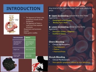
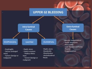
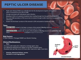
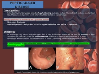
GASTROINTESTINA
L
BLEEDING
BY: UPARNA SAYURI HEENATIGALA
GROUP 10
INTRODUCTION Any kind of the haemorrhage from oral cavity to
anus
● Upper GI bleeding (proximal to the Treitz
ligament)
• Oesophageal varices bleeding
• Bleeding Peptic Ulcers
● Lower GI bleeding (distal to the Treitz
ligament)
• Diverticular Disease – Malignancy
• Small Bowel Disease
Overt Bleeding
• Clinical Manifestations:
• • Haematemesis
• Melena
• Haematochezia
Occult Bleeding
• Clinical Manifestations:
• Non-specific symptoms related to Iron deficiency
• The ligament of Treitz is the
suspensory muscle of the
duodenum.
• The ligament of Treitz is
made up of two separate
structures.
(Suspensory and accessory
muscle)
Helps gastric motility
. UPPER GI BLEEDING
Intra-luminal
Causes
Extra-luminal
Causes
DUODENAL
GASTRIC
ESOPHAGEAL
- Vascular malformations
- Aortic aneurysms
- Pancreatitis
- Liver diseases (cirrhosis)
- Trauma
- Coagulopathies
- Esophagitis
- Varices (enlarged
veins)
- Tumors (benign or
malignant)
- Peptic ulcers
- Gastric erosions
- Mallory-Weiss
tears
- Tumors (benign or
malignant)
-Peptic ulcers
-Duodenitis
-Diverticulum
-Aortoduodenal
fistula
• Esophageal varices are abnormal ,dilated veins that occur at the lower end of the oesophagus
• Varices are serious because they have weakened walls that can leak or break and bleed. Internal bleeding from a
ruptured vein can be sudden, severe and life-threatening.
• Esophageal varices occur in people with portal hypertension, which is high blood pressure in the portal vein that
runs through your liver and the other veins that branch off from it. Abnormal pressure causes the thin
esophageal veins to swell and enlarge. This most often occurs in people with liver disease.
SYMPTOMS
• Variceal bleed
• Alcoholism, exposure to blood-borne viruses
• Hematemesis, melena, or hematochezia
• Rapid upper GI bleed can present as rectal bleeding.
• Weight loss in patients with chronic liver disease
• Anorexia
• Abdominal discomfort
• Jaundice
• Pruritus
TREATMENT
Reduction of pressure in portal vein by drugs
• The high blood pressure in portal veins is brought down by beta blockers -nadolol and propranolol.
Endoscopy therapy
• Endoscopic therapy is the main treatment for managing the esophageal varices and liver cirrhosis. It can be used as both primary and
secondary prophylactic therapies
Blood transfusion
• Reinstating the blood is essential to maintain the standard volume of blood in an individual since there would be heavy loss in blood due
to variceal. Blood transfusion is done for a patients with high hemorrhage. Prophylatic antibiotics are used to prevent bacterial infection
that occurs during bleeding and early rebleeding.
OESOPHAGEAL VARICES
MALLORY WEISS TEAR
PEPTIC ULCER DISEASE
• Peptic ulcer disease (PUD) is an umbrella term for the development of gastric and duodenal
ulcers; it accounts for up to 50% of UGIB.
• Strongly associated with Helicobacter pylori infection.
• Bleeding in PUD is of a posterior duodenal ulcer eroding through into the gastroduodenal artery.
• But most UGIB secondary to PUD occurs due to erosions through smaller-sized blood vessels within the
submucosa.
• Erosion into the lesser curvature of the stomach is 20% and posterior duodenum is 40%.
Types
• Gastric Ulcers: Located in the lesser curvature of the stomach. Bleeding occurs to left gastric
artery/splenic artery most frequent among those aged 40–70 years
• Duodenal Ulcers: Located in the 1st part of the duodenum. Bleeding occurs to the posterior wall
where gastroduodenal artery is located. the age of 25 and 55 years.
Risk Factors:
H. Pylori Infection- Prolonged NSAID use- Alcohol -Smoking
Clinical Manifestations:
• Pain:
- if it occurs with meals and is relieved by vomiting= gastric ulcer
- if it relieved by eating food and radiates to the back= duodenal ulcers.
- Duodenal ulcer pain also occurs 2-5 hours after meals and at night (11pm-2am)
• bloating, abdominal fullness, nausea,
• heartburn or acid regurgitation
PEPTIC ULCER
DISEASE
Investigations
• Patients should undergo non-invasive H. pylori testing, such as a carbon-13 urea breath test or a stool antigen
test. Those who test positive for H. pylori should be started on triple therapy.
Red flag symptoms, as outlined by NICE guidance, include:
• New-onset dysphagia
• Aged >55 years with weight loss and either upper abdominal pain, reflux, or dyspepsia
Endoscopy
• At endoscopy, any peptic ulceration seen (Fig. 3) can be biopsied, which will be sent for histology (if looks
suspicious for malignancy) and for rapid urease test (CLO test) to determine the presence of H. pylori.
• Endoscopic therapy can also be utilised in cases of upper GI bleeding, secondary to peptic ulcer disease.
• If H.Pylori is present = H.pylori Eradication therapy (3X therapy): PPI (proton pump inhibitors) + Amoxycillin/Metronidazole +
Clarithromycin for 14 days.
EROSIVE GASTRITIS
• Gastritis refers to inflammation of
the lining of the stomach ;erosive
gastritis accounts for 15-20% of
UGIB.
• Characterized by ulceration of the
superficial epithelium.
• Associated with major stress and/or
trauma and a common source of
upper GI bleeding in acutely ill
patients.
• Gastritis can be classified based
upon location, timing or type of
pathology:
• Antral or pangastritis
• Acute or chronic gastritis •
Erosive or non-erosive
INVESTIGATIONS AND
DIAGNOSIS
• Upper GI endoscopy is the main investigation. It is both diagnostic and therapeutic.
• Upper GI endoscopy should be completed within 24 hours of presentation.
Blood tests
Full blood count (FBC) •Venous /
arterial blood gas •Urea &
electrolytes (U&Es) •Liver function
tests (LFTs) •Clotting
•Group & save with cross match
Imaging
Chest x-ray: An erect film should
be arranged if possible.
Allows evaluation for aspiration,
pneumomediastinum
(oesophageal perforation) and
free air under the diaphragm
(perforation of abdominal
viscus).
Initial Stabilization
-Continuous monitoring
of vital signs, including
blood pressure, heart
rate, and oxygen
saturation.
-Monitor urine output
MANAGEMENT- RESUSCITATION
DRUGS
• IV PPI for acid suppression
• IV Terlipressin/Vasopressin
• IV Ocreotide
• IV somatostatin
Following resuscitation, unstable patients should be transferred for an immediate endoscopy. It is
recommended that all other patients have endoscopy within 24
hours of admission.
According to Forest Classification, F1a, F1b and
F2a are indications for endoscopic therapy.
Usually the endoscopic therapies are combined;
Vasopressin infusion + clipping
Surgical Intervention If bleeding persists
• Under running of the ulcer: suture the ulcer
• Left Gastric artery/Gastro duodenal artery
Ligation
• Partial/Total Gastrectomy: if nothing works
Non-Variceal Bleeding
1. Pharmacological intervention
Terlipressin (IV injection)
• Analogue of vasopressin (ADH)
• Causes splanchnic vasoconstriction
• This reduces portal pressures
Prophylactic antibiotic therapy
• Reduces the risk of spontaneous bacterial peritonitis.
2. Endoscopic intervention
Variceal band ligation (VBL)
• Completed acutely. Patients then need to undergo variceal banding programme every 2-4 weeks until varices
have gone.
Endoscopic sclerotherapy
• Alternative option toVBL that involves injection of a sclerosing agent.
SURGICAL INTERVENTION.
Variceal Bleeding
TIPS (Transjugular Intra Hepatic
Portosystemic Shunt)
Oesophageal
Resection
Balloon tamponade
TIPS (Transjugular Intra Hepatic
Portosystemic Shunt)
is a procedure that involves inserting a stent (tube)
to connect the portal veins to adjacent blood vessels
that have lower pressure.
TIPS creates a new route for blood to flow through
the liver and into the major veins that lead back to
the heart, reducing back pressure in the veins that
flow into the liver.
Complications may include:
• Fever
• Muscle stiffness
• Bruising on the neck near the insertion point
• Stenosis (the narrowing of the shunt passageway
Variceal Bleeding
Balloon tamponade
- risk of oesophageal necrosis if left > 24 hours
- o control variceal bleeding; it may be used to
stabilize a patient with massive bleeding prior
to definitive therapy.
Variceal Bleeding
Sengstaken–Blakemore tube
.
Acute LGIB: presents with PR
bleeding or altered blood with or
without signs of shock.
Chronic LGIB: typically,
asymptomatic and picked up
incidentally following blood tests
indicating iron deficiency anaemia.
Signs or biochemistry of chronic
LGIB are indications for urgent
suspected cancer referral.
.
INVESTIGATIONS AND DIAGNOSIS • LGIB is a clinical diagnosis
with an aetiology that may be
revealed by CT scan or lower
GI endoscopy
(colonoscopy/flexible
sigmoidoscopy/enteroscopy)
• A full blood count (FBC)
and digital rectal
examination (DRE) are
important initial tests for
assessing LGIB.
BUN is high in UGIB because
blood will be digested and
metabolised into urea. Therefore
urea will be high unequal to
creatinine
Imaging
•CT abdomen/pelvis: CT may be ordered to
identify the underlying diagnosis, e.g if a large colonic
malignancy is suspected or to look for evidence of
ischaemic colitis.
•CT angiography: CT angiogram may allow
identification of a bleeding point - typically only in
patients with brisk active bleeding.
If you have a gastrointestinal condition that has already
been diagnosed, the endoscope can be used in many
cases to treat GI disorders. There are many different
types of endoscopic therapies.
• Dilation. Through the endoscope, a narrowed segment in
the esophagus, stomach, or duodenum can be stretched
with a balloon passed through the scope to enable easier
passage of food and liquids.
• Control of bleeding. Patients can present with internal
bleeding coming from a stomach ulcer or esophageal
varices (swollen veins in the esophagus). Through the
endoscope, thermal energy or other maneuvers can be
applied to stop the hemorrhage.
• Polypectomy. Some patients can grow polyps in their
stomach and duodenum that, if not removed, can pose a
risk for growing into cancer. Just like removing colon polyps
with a colonoscopy, these polyps can be cut out using
upper endoscopy.
• Botulinum toxin Injection. Some patients develop a
swallowing disorder called achalasia where the lower
esophagus sphincter (valve) fails to relax and open when
someone swallows food. Using the endoscope, botulinum
toxin can be injected into the lower esophageal sphincter,
thereby temporarily paralyzing and opening up the
muscular valve.
• Enteral stent placement. Cancers of the esophagus
and pancreas can cause blockage of the esophagus and
duodenum respectively. Through the endoscope, a hollow
metal tube called an enteral stent can be placed through
the narrowed segment of the tumor to open up the
blockage, allowing people to eat and digest their foods.
THERAPEUTIC ENDOSCOPY DIAGNOSTIC ENDOSCOPY
Diagnostic upper endoscopy can be used as a
screening tool for esophageal or gastric cancer.
Gastric ulcers, cancer, Crohn’s disease, and other
diseases of the upper digestive tract can be
diagnosed via upper endoscopy.
ASSESSING THE AMOUNT
OF BLOOD LOST
• Blood Urea Nitrogen Levels: Higher in cases of
upper GI bleeding.
• Haematocrit levels: Most patients with acute
upper gastrointestinal haemorrhage have a
haematocrit level less than 30%.
• Hb levels: The initial haemoglobin level in
patients with acute upper GI bleeding may be at
the patient's baseline because the patient is losing
whole blood.With time, the haemoglobin level will
decline as the blood is diluted by the influx of
extravascular fluid into the vascular space and by
fluid administered during resuscitation.The
haemoglobin level should initially be monitored
every two to eight hours, depending upon the
severity of the bleed.
• MCV: Acute bleeding does not alter the mean
corpuscular volume (MCV). If the MCV is low, it
may suggest iron deficiency, which could be caused
by chronic bleeding.
Hypovolemic shock is a dangerous condition in which your
heart can't get your body the blood (and oxygen) it needs to
function. This happens because you've lost a large amount
― more than 20% ― of your blood volume.
Stages of Shock
Initial Stage
Hypoperfusion hypoxia anaerobic respiration
→ → →
Krebs cycle is slowed →
lactic acidosis.
Compensatory Stage
• – Sympathetic Nervous System
• • Vasoconstriction, HR, contractility, UOP
↑ ↑ ↓
• – Renin-Angiotensin-Aldosterone
• • Response to BP/ blood volume with Na+
↓ ↓ ↑
• – Tissue Injury cytokines
→
• • Vasodilation, vasoconstriction, capillary
↑
permeability
Progressive Stage - Compensatory mechanisms begin
to fail.
Metabolic acidosis, precapillary sphincters relax to
↑ ↑ →
leakage of fluid and protein into the surrounding
tissues Hemoconcentration, viscosity increase
→ ↑ ↑ →
blockage of the microcirculation vital organs get
→
compromised. Bowel ischemia bacterial translocation
→
endotoxic shock.
→
Refractory Stage - vital organs have failed Brain
damage and cell death
ATP has been degraded into adenosine Adenosine
easily perfuses out of cellular membranes, there is no
adenosine to phosphorylate into ATP
Two large bore IVs (18 gauge or larger) or Central vein access
– Intraosseous Access if IV Access is unavailable
• Fluid Replacement
• – Crystalloid (LR or NS)
• – Packed Red Blood Cell transfusion as indicated (replace blood
loss with blood)
• Fluid Responsiveness Markers
• Vasopressors (refractory severe Hypotension)
• – Norepinephrine
• – Vasopressin
• – Epinephrine
• – Dopamine, dobutamine
• Respiratory support as needed
– Noninvasive Ventilation
- Intubation
THANK
YOU 
REFERENCES
• Upper Gi Bleeding Notes. Pulsenotes. (n. d. ). https: //app. pulsenotes. com/medicine/gastroenterology/notes/upper-gi-bleeding
• Approach to acute upper gastrointestinal bleeding in adults. UpToDate. (n.d.). https://www.uptodate.com/contents/approach-to-
acute-upper-gastrointestinal-bleeding- in-adults/print
• Acute lower gi bleed notes. Pulsenotes. (n. d. -a). https: //app. pulsenotes. com/medicine/gastroenterology/notes/acute-lower-gi-
bleed
• Lower gastrointestinal (GI) bleeding:ACG.American College of Gastroenterology. (2023,August 4). https://gi.org/topics/lower-gi-
• bleeding/#: ~: text=Lower%20GI%20bleeding%20refers%20to, with%20or%20without%20a%20stool.
• Gastrointestinal bleeding. Next. amboss. com. (n. d. -b). https: //next. amboss. com/us/article/ZS0Zy2# TJ16t30

GI bleeding Introduction & References.pptx

  • 1.
  • 2.
    INTRODUCTION Any kindof the haemorrhage from oral cavity to anus ● Upper GI bleeding (proximal to the Treitz ligament) • Oesophageal varices bleeding • Bleeding Peptic Ulcers ● Lower GI bleeding (distal to the Treitz ligament) • Diverticular Disease – Malignancy • Small Bowel Disease Overt Bleeding • Clinical Manifestations: • • Haematemesis • Melena • Haematochezia Occult Bleeding • Clinical Manifestations: • Non-specific symptoms related to Iron deficiency • The ligament of Treitz is the suspensory muscle of the duodenum. • The ligament of Treitz is made up of two separate structures. (Suspensory and accessory muscle) Helps gastric motility
  • 3.
    . UPPER GIBLEEDING Intra-luminal Causes Extra-luminal Causes DUODENAL GASTRIC ESOPHAGEAL - Vascular malformations - Aortic aneurysms - Pancreatitis - Liver diseases (cirrhosis) - Trauma - Coagulopathies - Esophagitis - Varices (enlarged veins) - Tumors (benign or malignant) - Peptic ulcers - Gastric erosions - Mallory-Weiss tears - Tumors (benign or malignant) -Peptic ulcers -Duodenitis -Diverticulum -Aortoduodenal fistula
  • 4.
    • Esophageal varicesare abnormal ,dilated veins that occur at the lower end of the oesophagus • Varices are serious because they have weakened walls that can leak or break and bleed. Internal bleeding from a ruptured vein can be sudden, severe and life-threatening. • Esophageal varices occur in people with portal hypertension, which is high blood pressure in the portal vein that runs through your liver and the other veins that branch off from it. Abnormal pressure causes the thin esophageal veins to swell and enlarge. This most often occurs in people with liver disease. SYMPTOMS • Variceal bleed • Alcoholism, exposure to blood-borne viruses • Hematemesis, melena, or hematochezia • Rapid upper GI bleed can present as rectal bleeding. • Weight loss in patients with chronic liver disease • Anorexia • Abdominal discomfort • Jaundice • Pruritus TREATMENT Reduction of pressure in portal vein by drugs • The high blood pressure in portal veins is brought down by beta blockers -nadolol and propranolol. Endoscopy therapy • Endoscopic therapy is the main treatment for managing the esophageal varices and liver cirrhosis. It can be used as both primary and secondary prophylactic therapies Blood transfusion • Reinstating the blood is essential to maintain the standard volume of blood in an individual since there would be heavy loss in blood due to variceal. Blood transfusion is done for a patients with high hemorrhage. Prophylatic antibiotics are used to prevent bacterial infection that occurs during bleeding and early rebleeding. OESOPHAGEAL VARICES
  • 5.
  • 6.
    PEPTIC ULCER DISEASE •Peptic ulcer disease (PUD) is an umbrella term for the development of gastric and duodenal ulcers; it accounts for up to 50% of UGIB. • Strongly associated with Helicobacter pylori infection. • Bleeding in PUD is of a posterior duodenal ulcer eroding through into the gastroduodenal artery. • But most UGIB secondary to PUD occurs due to erosions through smaller-sized blood vessels within the submucosa. • Erosion into the lesser curvature of the stomach is 20% and posterior duodenum is 40%. Types • Gastric Ulcers: Located in the lesser curvature of the stomach. Bleeding occurs to left gastric artery/splenic artery most frequent among those aged 40–70 years • Duodenal Ulcers: Located in the 1st part of the duodenum. Bleeding occurs to the posterior wall where gastroduodenal artery is located. the age of 25 and 55 years. Risk Factors: H. Pylori Infection- Prolonged NSAID use- Alcohol -Smoking Clinical Manifestations: • Pain: - if it occurs with meals and is relieved by vomiting= gastric ulcer - if it relieved by eating food and radiates to the back= duodenal ulcers. - Duodenal ulcer pain also occurs 2-5 hours after meals and at night (11pm-2am) • bloating, abdominal fullness, nausea, • heartburn or acid regurgitation
  • 7.
    PEPTIC ULCER DISEASE Investigations • Patientsshould undergo non-invasive H. pylori testing, such as a carbon-13 urea breath test or a stool antigen test. Those who test positive for H. pylori should be started on triple therapy. Red flag symptoms, as outlined by NICE guidance, include: • New-onset dysphagia • Aged >55 years with weight loss and either upper abdominal pain, reflux, or dyspepsia Endoscopy • At endoscopy, any peptic ulceration seen (Fig. 3) can be biopsied, which will be sent for histology (if looks suspicious for malignancy) and for rapid urease test (CLO test) to determine the presence of H. pylori. • Endoscopic therapy can also be utilised in cases of upper GI bleeding, secondary to peptic ulcer disease. • If H.Pylori is present = H.pylori Eradication therapy (3X therapy): PPI (proton pump inhibitors) + Amoxycillin/Metronidazole + Clarithromycin for 14 days.
  • 8.
    EROSIVE GASTRITIS • Gastritisrefers to inflammation of the lining of the stomach ;erosive gastritis accounts for 15-20% of UGIB. • Characterized by ulceration of the superficial epithelium. • Associated with major stress and/or trauma and a common source of upper GI bleeding in acutely ill patients. • Gastritis can be classified based upon location, timing or type of pathology: • Antral or pangastritis • Acute or chronic gastritis • Erosive or non-erosive
  • 9.
    INVESTIGATIONS AND DIAGNOSIS • UpperGI endoscopy is the main investigation. It is both diagnostic and therapeutic. • Upper GI endoscopy should be completed within 24 hours of presentation. Blood tests Full blood count (FBC) •Venous / arterial blood gas •Urea & electrolytes (U&Es) •Liver function tests (LFTs) •Clotting •Group & save with cross match Imaging Chest x-ray: An erect film should be arranged if possible. Allows evaluation for aspiration, pneumomediastinum (oesophageal perforation) and free air under the diaphragm (perforation of abdominal viscus). Initial Stabilization -Continuous monitoring of vital signs, including blood pressure, heart rate, and oxygen saturation. -Monitor urine output
  • 10.
    MANAGEMENT- RESUSCITATION DRUGS • IVPPI for acid suppression • IV Terlipressin/Vasopressin • IV Ocreotide • IV somatostatin Following resuscitation, unstable patients should be transferred for an immediate endoscopy. It is recommended that all other patients have endoscopy within 24 hours of admission.
  • 11.
    According to ForestClassification, F1a, F1b and F2a are indications for endoscopic therapy. Usually the endoscopic therapies are combined; Vasopressin infusion + clipping Surgical Intervention If bleeding persists • Under running of the ulcer: suture the ulcer • Left Gastric artery/Gastro duodenal artery Ligation • Partial/Total Gastrectomy: if nothing works Non-Variceal Bleeding
  • 12.
    1. Pharmacological intervention Terlipressin(IV injection) • Analogue of vasopressin (ADH) • Causes splanchnic vasoconstriction • This reduces portal pressures Prophylactic antibiotic therapy • Reduces the risk of spontaneous bacterial peritonitis. 2. Endoscopic intervention Variceal band ligation (VBL) • Completed acutely. Patients then need to undergo variceal banding programme every 2-4 weeks until varices have gone. Endoscopic sclerotherapy • Alternative option toVBL that involves injection of a sclerosing agent. SURGICAL INTERVENTION. Variceal Bleeding TIPS (Transjugular Intra Hepatic Portosystemic Shunt) Oesophageal Resection Balloon tamponade
  • 13.
    TIPS (Transjugular IntraHepatic Portosystemic Shunt) is a procedure that involves inserting a stent (tube) to connect the portal veins to adjacent blood vessels that have lower pressure. TIPS creates a new route for blood to flow through the liver and into the major veins that lead back to the heart, reducing back pressure in the veins that flow into the liver. Complications may include: • Fever • Muscle stiffness • Bruising on the neck near the insertion point • Stenosis (the narrowing of the shunt passageway Variceal Bleeding Balloon tamponade - risk of oesophageal necrosis if left > 24 hours - o control variceal bleeding; it may be used to stabilize a patient with massive bleeding prior to definitive therapy.
  • 14.
  • 15.
    . Acute LGIB: presentswith PR bleeding or altered blood with or without signs of shock. Chronic LGIB: typically, asymptomatic and picked up incidentally following blood tests indicating iron deficiency anaemia. Signs or biochemistry of chronic LGIB are indications for urgent suspected cancer referral.
  • 16.
    . INVESTIGATIONS AND DIAGNOSIS• LGIB is a clinical diagnosis with an aetiology that may be revealed by CT scan or lower GI endoscopy (colonoscopy/flexible sigmoidoscopy/enteroscopy) • A full blood count (FBC) and digital rectal examination (DRE) are important initial tests for assessing LGIB. BUN is high in UGIB because blood will be digested and metabolised into urea. Therefore urea will be high unequal to creatinine Imaging •CT abdomen/pelvis: CT may be ordered to identify the underlying diagnosis, e.g if a large colonic malignancy is suspected or to look for evidence of ischaemic colitis. •CT angiography: CT angiogram may allow identification of a bleeding point - typically only in patients with brisk active bleeding.
  • 17.
    If you havea gastrointestinal condition that has already been diagnosed, the endoscope can be used in many cases to treat GI disorders. There are many different types of endoscopic therapies. • Dilation. Through the endoscope, a narrowed segment in the esophagus, stomach, or duodenum can be stretched with a balloon passed through the scope to enable easier passage of food and liquids. • Control of bleeding. Patients can present with internal bleeding coming from a stomach ulcer or esophageal varices (swollen veins in the esophagus). Through the endoscope, thermal energy or other maneuvers can be applied to stop the hemorrhage. • Polypectomy. Some patients can grow polyps in their stomach and duodenum that, if not removed, can pose a risk for growing into cancer. Just like removing colon polyps with a colonoscopy, these polyps can be cut out using upper endoscopy. • Botulinum toxin Injection. Some patients develop a swallowing disorder called achalasia where the lower esophagus sphincter (valve) fails to relax and open when someone swallows food. Using the endoscope, botulinum toxin can be injected into the lower esophageal sphincter, thereby temporarily paralyzing and opening up the muscular valve. • Enteral stent placement. Cancers of the esophagus and pancreas can cause blockage of the esophagus and duodenum respectively. Through the endoscope, a hollow metal tube called an enteral stent can be placed through the narrowed segment of the tumor to open up the blockage, allowing people to eat and digest their foods. THERAPEUTIC ENDOSCOPY DIAGNOSTIC ENDOSCOPY Diagnostic upper endoscopy can be used as a screening tool for esophageal or gastric cancer. Gastric ulcers, cancer, Crohn’s disease, and other diseases of the upper digestive tract can be diagnosed via upper endoscopy.
  • 18.
    ASSESSING THE AMOUNT OFBLOOD LOST • Blood Urea Nitrogen Levels: Higher in cases of upper GI bleeding. • Haematocrit levels: Most patients with acute upper gastrointestinal haemorrhage have a haematocrit level less than 30%. • Hb levels: The initial haemoglobin level in patients with acute upper GI bleeding may be at the patient's baseline because the patient is losing whole blood.With time, the haemoglobin level will decline as the blood is diluted by the influx of extravascular fluid into the vascular space and by fluid administered during resuscitation.The haemoglobin level should initially be monitored every two to eight hours, depending upon the severity of the bleed. • MCV: Acute bleeding does not alter the mean corpuscular volume (MCV). If the MCV is low, it may suggest iron deficiency, which could be caused by chronic bleeding. Hypovolemic shock is a dangerous condition in which your heart can't get your body the blood (and oxygen) it needs to function. This happens because you've lost a large amount ― more than 20% ― of your blood volume. Stages of Shock Initial Stage Hypoperfusion hypoxia anaerobic respiration → → → Krebs cycle is slowed → lactic acidosis. Compensatory Stage • – Sympathetic Nervous System • • Vasoconstriction, HR, contractility, UOP ↑ ↑ ↓ • – Renin-Angiotensin-Aldosterone • • Response to BP/ blood volume with Na+ ↓ ↓ ↑ • – Tissue Injury cytokines → • • Vasodilation, vasoconstriction, capillary ↑ permeability Progressive Stage - Compensatory mechanisms begin to fail. Metabolic acidosis, precapillary sphincters relax to ↑ ↑ → leakage of fluid and protein into the surrounding tissues Hemoconcentration, viscosity increase → ↑ ↑ → blockage of the microcirculation vital organs get → compromised. Bowel ischemia bacterial translocation → endotoxic shock. → Refractory Stage - vital organs have failed Brain damage and cell death ATP has been degraded into adenosine Adenosine easily perfuses out of cellular membranes, there is no adenosine to phosphorylate into ATP
  • 19.
    Two large boreIVs (18 gauge or larger) or Central vein access – Intraosseous Access if IV Access is unavailable • Fluid Replacement • – Crystalloid (LR or NS) • – Packed Red Blood Cell transfusion as indicated (replace blood loss with blood) • Fluid Responsiveness Markers • Vasopressors (refractory severe Hypotension) • – Norepinephrine • – Vasopressin • – Epinephrine • – Dopamine, dobutamine • Respiratory support as needed – Noninvasive Ventilation - Intubation
  • 20.
    THANK YOU  REFERENCES • UpperGi Bleeding Notes. Pulsenotes. (n. d. ). https: //app. pulsenotes. com/medicine/gastroenterology/notes/upper-gi-bleeding • Approach to acute upper gastrointestinal bleeding in adults. UpToDate. (n.d.). https://www.uptodate.com/contents/approach-to- acute-upper-gastrointestinal-bleeding- in-adults/print • Acute lower gi bleed notes. Pulsenotes. (n. d. -a). https: //app. pulsenotes. com/medicine/gastroenterology/notes/acute-lower-gi- bleed • Lower gastrointestinal (GI) bleeding:ACG.American College of Gastroenterology. (2023,August 4). https://gi.org/topics/lower-gi- • bleeding/#: ~: text=Lower%20GI%20bleeding%20refers%20to, with%20or%20without%20a%20stool. • Gastrointestinal bleeding. Next. amboss. com. (n. d. -b). https: //next. amboss. com/us/article/ZS0Zy2# TJ16t30