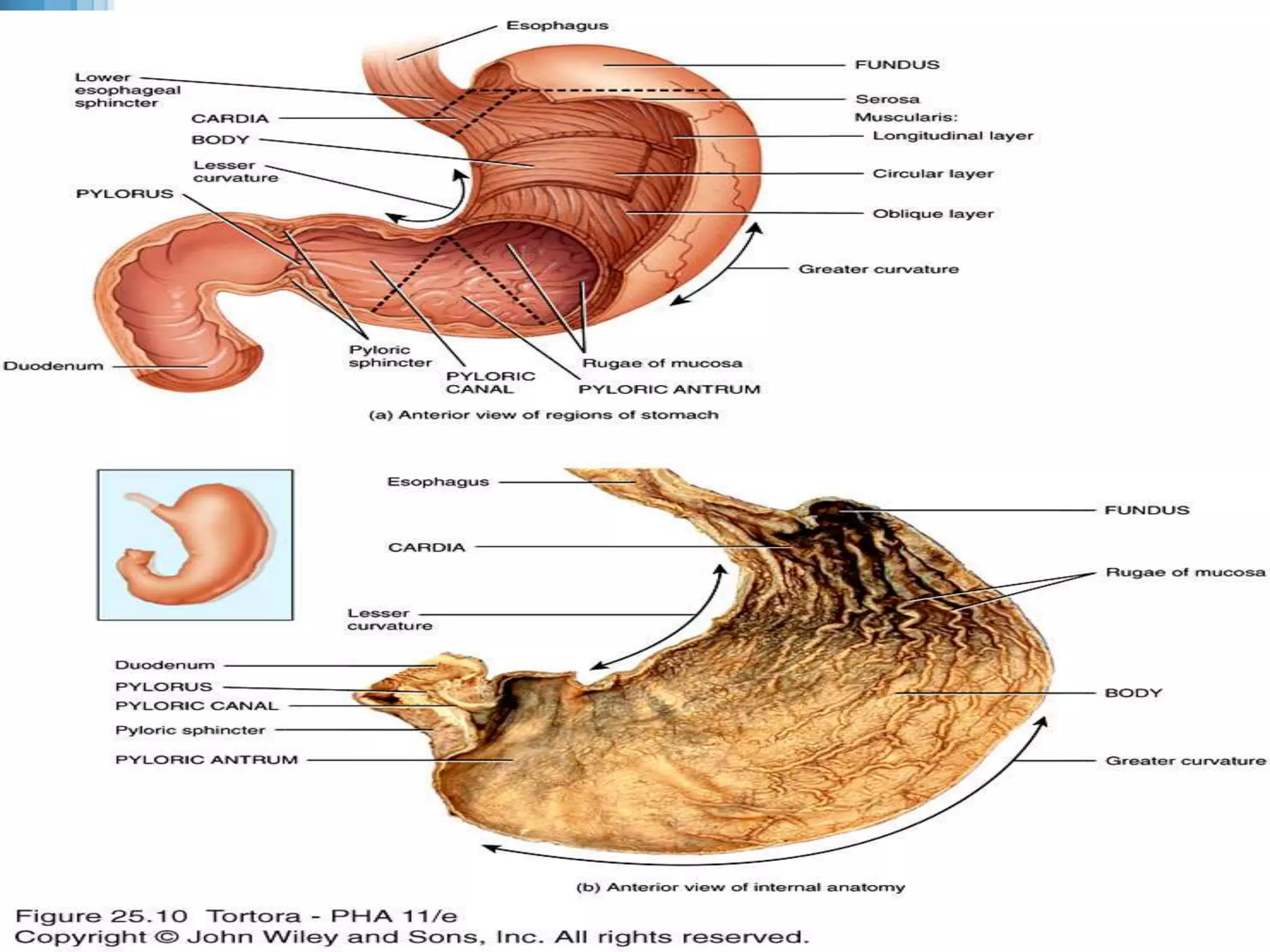
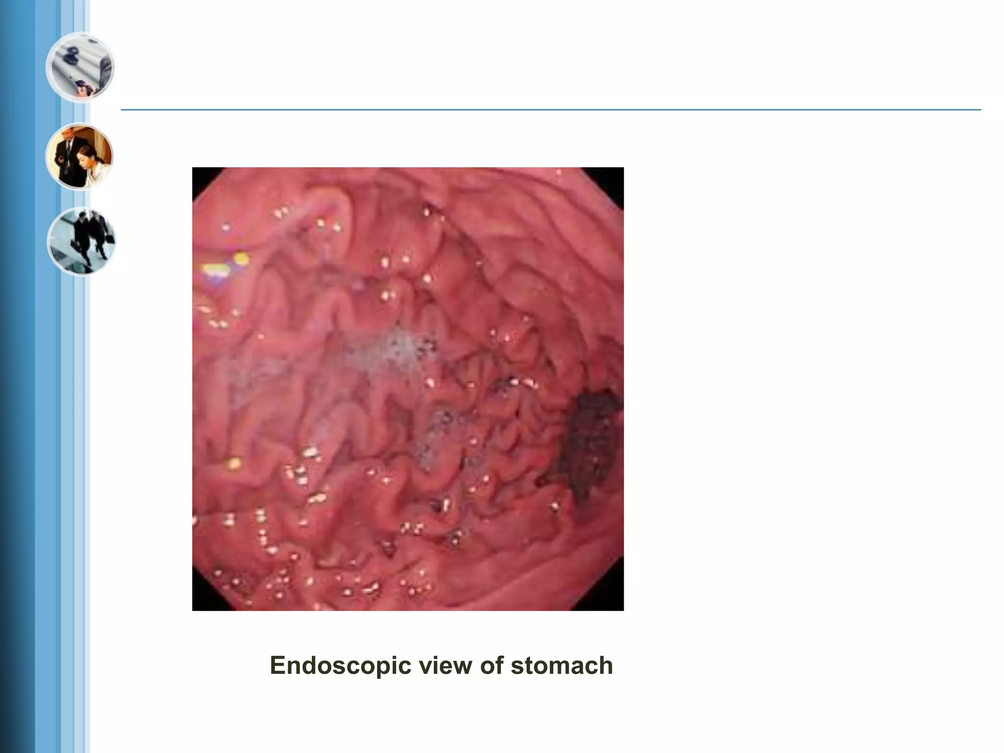
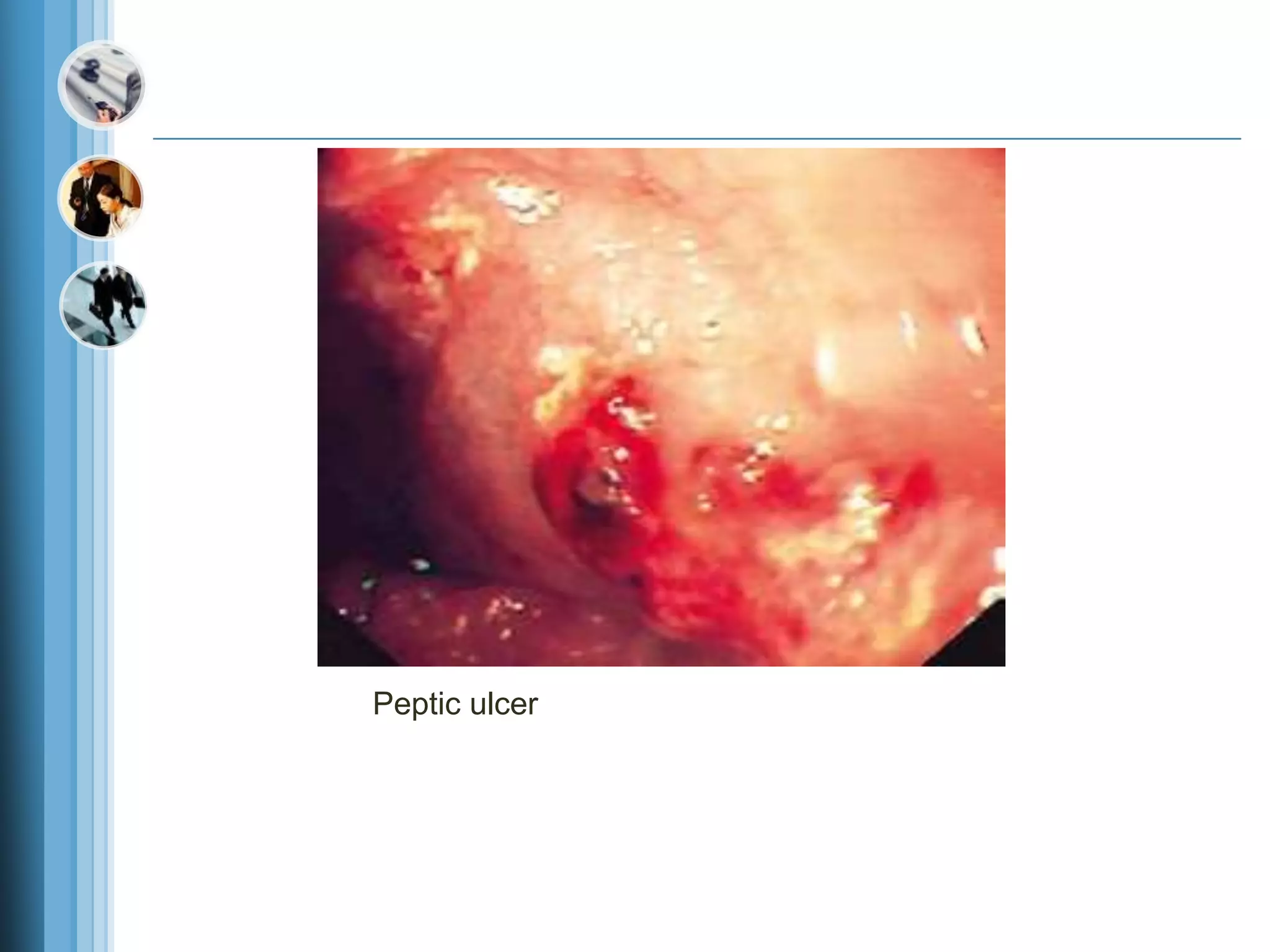
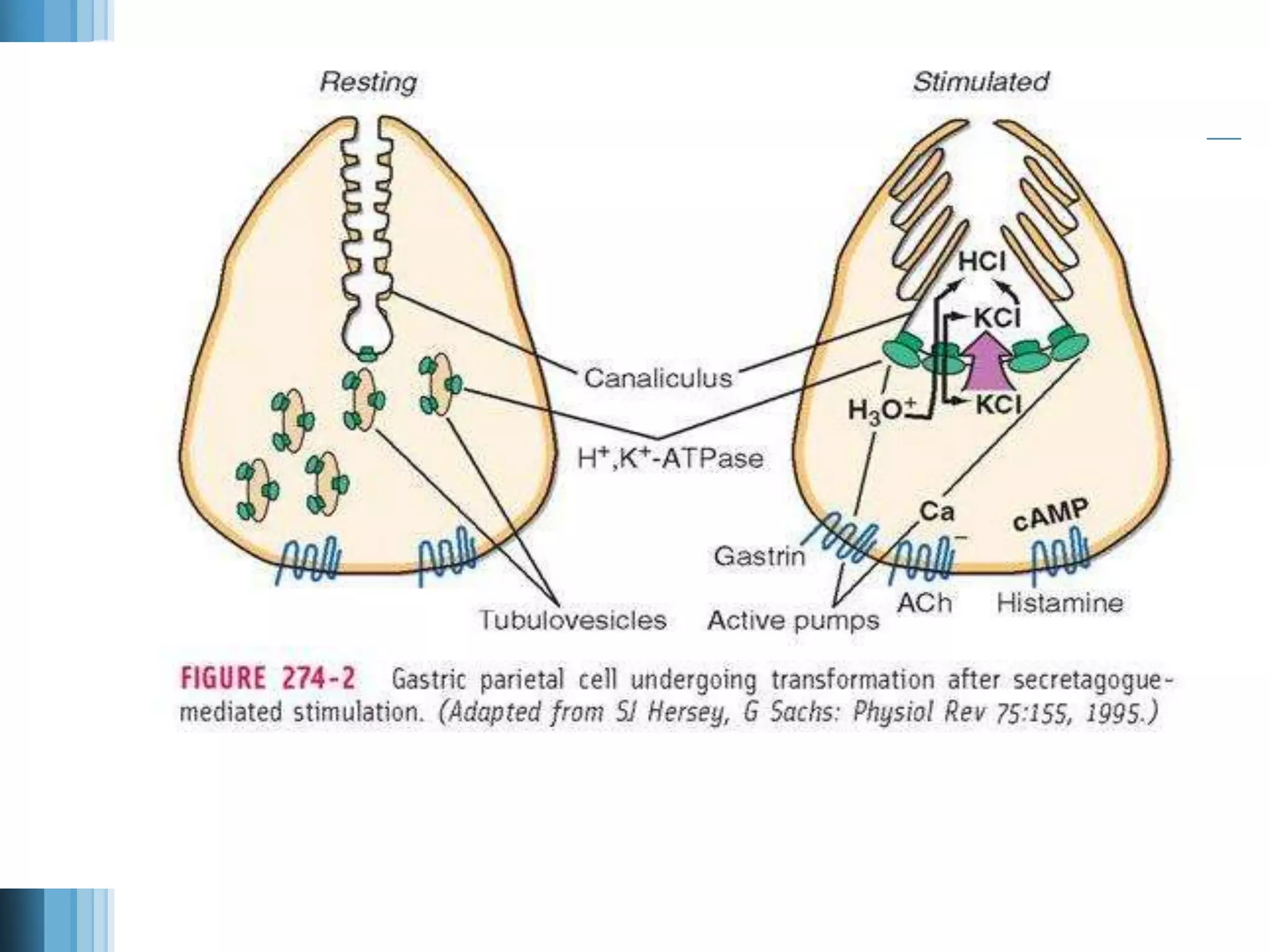
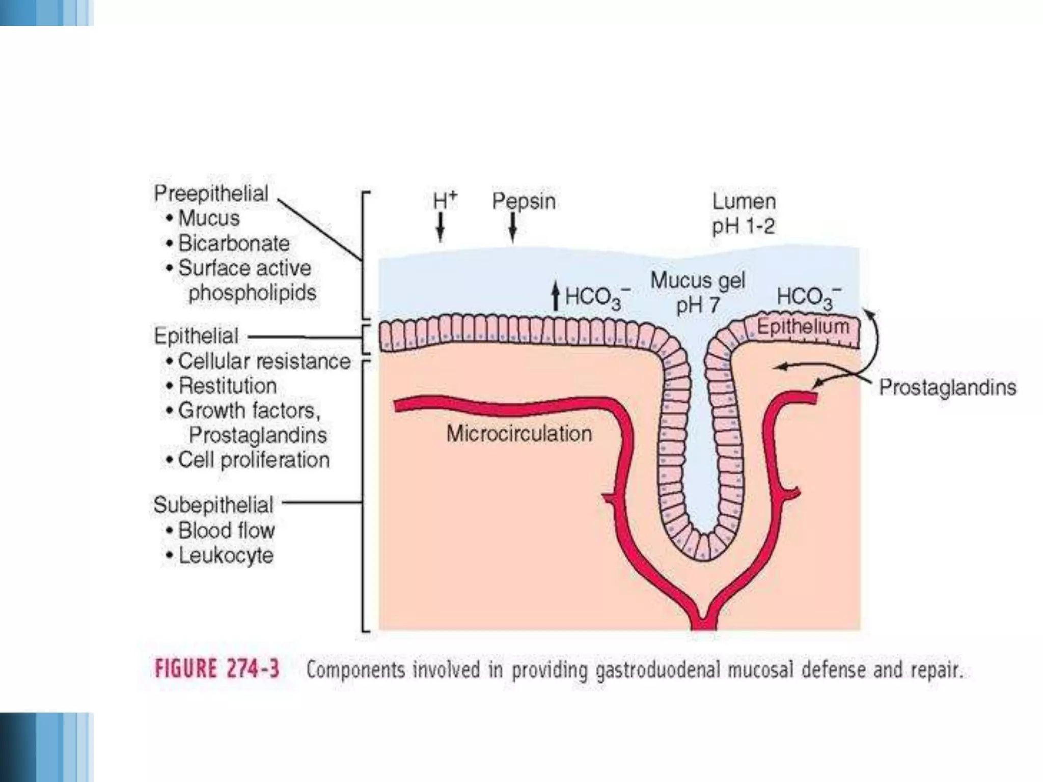
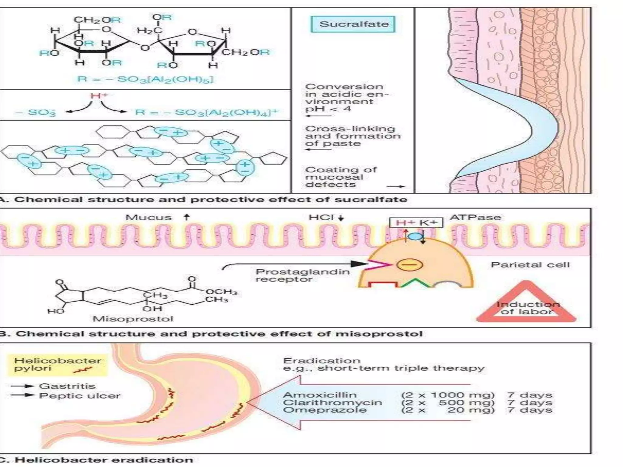
www.themegallery.com
LOGO
PEPTIC ULCER
Copyright 2009, John Wiley & Sons,
Endoscopic view of stomach
Peptic ulcer
Histology of the Stomach
 The submucosa of the stomach is composed of aereolar connective tissue.
 The muscularis has three layers of smooth muscle (rather than the two found in the
lower esophagus and small and large intestines):
 an outer longitudinal layer,
 a middle circular layer, and
 an inner oblique layer.
 The oblique layer is limited primarily to the body of the stomach, allowing the
stomach to more effectively churn and mix the food.
 The serosa is composed of simple squamous epithelium (mesothelium), the
visceralperitoneum, and areolar connective tissue.
Three dimensional view of layers of the stomach
Gastric Glands
 The gastric glands contain three types of exocrine gland cells that secrete their
products into the stomach lumen: mucous neck cells, chief cells, and parietal cells.
 Both mucous surface cells and mucous neck cells secrete mucus.
 Parietal cells produce intrinsic factor (needed for absorption of vitamin B12) and
hydrochloric acid.
 The chief (zymogenic) cells secrete pepsinogen and gastric lipase.
 The secretions of the mucous, parietal, and chief cells form gastric juice, which
totals 2000–3000 mL (roughly 2–3 qt) per day.
 In addition, a type of enteroendocrine cell, the G cell, located mainly in the pyloric
antrum, secretes the hormone gastrin into the bloodstream.
 Gastrin stimulates growth of the gastric glands and secretion of large amounts of
gastric juice. It also strengthens contraction of the lower esophageal sphincter,
increases motility of the stomach, and relaxes the pyloric and ileocecal sphincters.
Gastric gland
The parietal cell
 The parietal cell, also known as the oxyntic cell, is usually found in the
neck, or isthmus, or the oxyntic gland. The resting, or
unstimulated,parietal cell has prominent cytoplasmic tubulovesicles and
intracellular canaliculi containing short microvilli along its apical
surface H, K-ATPase is expressed in the tubulovesicle membrane; upon
cell stimulation, this membrane, along with apical membranes,
transforms into a dense network of apical intracellular canaliculi
containing long microvilli.
 Acid secretion, a process requiring high energy, occurs at the apical
canalicular surface. Numerous mitochondria (30 to 40% of total cell
volume) generate the energy required for secretion.
Gastroduodenal Mucosal Defense
 The gastric epithelium is under a constant assault by a series of
endogenous noxious factors including HCl, pepsinogen/pepsin,
and bile salts. In addition, a steady flow of exogenous substances
such as medications, alcohol, and bacteria encounter the gastric
mucosa.
 A highly intricate biologic system is in place to provide defense
from mucosal injury and to repair any injury that may occur.
 The mucosal defense system can be envisioned as a three-level
barrier, composed of pre-epithelial, epithelial, and sub-epithelial
elements
Mucus is secreted in a regulated fashion by gastroduodenal surface epithelial cells.
 It consists primarily of water (95%) and a mixture of lipids and glycoproteins.
 Mucin is the constituent glycoprotein that, in combination with phospholipids (also
secreted by gastric mucous cells), forms a hydrophobic surface with fatty acids that
extend into the lumen from the cell membrane.
 The mucous gel functions as a nonstirred water layer impeding diffusion of ions and
molecules such as pepsin.
Bicarbonate, secreted by surface epithelial cells of the gastroduodenal mucosa into the
mucous gel, forms a pH gradient ranging from 1 to 2 at the gastric luminal surface and
reaching 6 to 7 along the epithelial cell surface.
Bicarbonate secretion is stimulated by calcium, prostaglandins, cholinergic input, and
luminal acidification.
 Surface epithelial cells provide the next line of defense through several factors,
including mucus production, epithelial cell ionic transporters that maintain
intracellular pH and bicarbonate production, and intracellular tight junctions.
 If the preepithelial barrier were breached, gastric epithelial cells bordering a site of
injury can migrate to restore a damaged region (restitution). This process occurs
independent of cell division and requires uninterrupted blood flow and an alkaline
pH in the surrounding environment.
 Several growth factors including epidermal growth factor (EGF), transforming
growth factor (TGF) , and basic fibroblast growth factor (FGF) modulate the
process of restitution. Larger defects that are not effectively repaired by restitution
require cell proliferation. Epithelial cell regeneration is regulated by prostaglandins
and growth factors such as EGF and TGF-. In tandem with epithelial cell renewal,
formation of new vessels (angiogenesis) within the injured microvascular bed
occurs. Both FGF and vascular endothelial growth factor (VEGF) are important in
regulating angiogenesis in the gastric mucosa.
 Prostaglandins play a central role in gastric epithelial defense/repair
Physiology of Gastric Secretion
PATHOPHYSIOLOGIC BASIS OF
PEPTIC ULCER DISEASE
PUD encompasses both gastric and duodenal
ulcers.
Ulcers are defined as a break in the mucosal
surface 5 mm in size, with depth to the sub-
mucosa.
Duodenal ulcers (DUs) and gastric ulcers
(GUs); share many commonm features in
terms of pathogenesis, diagnosis, and
treatment, but several factors distinguish
them from one another.
Epidemiology
 DUODENAL ULCERS
DUs are estimated to occur in 6 to 15% of the western
population. The incidence of DUs declined steadily
from 1960 to 1980 and has remained stable since
then. The death rates, need for surgery, and physician
visits have decreased by 50% over the past 30 years.
The reason for the reduction in the frequency ofDUs is
likely related to the decreasing frequency of
Helicobacter pylori.
 Before the discovery of H. pylori, the natural history
of DUs was typified by frequent recurrences after
initial therapy.
 Eradication of H. pylori has greatly reduced these
recurrence rates.
GASTRIC ULCERS
GUs tend to occur later in life than duodenal lesions, with
a peak incidence reported in the sixth decade. More
than half of GUs occur in males and are less common
than DUs, perhaps due to the higher likelihood of GUs
being silent and presenting only after a complication
develops. Autopsy studies suggest a similar incidence
of DUs and GUs.
Pathology
 DUODENAL ULCERS
 DUs occur most often in the first portion of duodenum (95%), with 90%
located within 3 cm of the pylorus.
 They are usually 1 cm in diameter but can occasionally reach 3 to 6 cm
(giant ulcer). Ulcers are sharply demarcated, with depth at times
reaching the muscularis propria. The base of the ulcer often consists of a
zone of eosinophilic necrosis with surrounding fibrosis. Malignant
duodenal ulcers are extremely rare.
 GASTRIC ULCERS
 In contrast to DUs, GUs can represent a malignancy.
 Benign GUs are most often found distal to the junction between the
antrum and the acid secretory mucosa. This junction is variable, but in
general the antral mucosa extends about two-thirds of the distance of the
lesser curvature and one-third the way up the greater curvature.
 Benign GUs are quite rare in the gastric fundus and are histologically
similar to DUs. Benign GUs associated with H. pylori are associated
with antral gastritis. In contrast, NSAID-related GUs are not
accompanied by chronic active gastritis but may instead have evidence
of a chemical gastropathy.
PATHOPHYSIOLOGY
It is now clear that H. pylori and NSAID-induced injury
account for the majority of DUs. Gastric acid contributes to
mucosalinjury but does not play a primary role.
H. PYLORI
 is a gram-negative microaerophilic rod found most commonly in the deeper portions of the
mucous gel coating the gastric mucosa or between the mucous layer and the gastric
epithelium.
 It may attach to gastric epithelium but under normal circumstances does not appear to
invade cells. It is strategically designed to live within the aggressive environment of the
stomach. It is S-shaped (0.5 3 m in size) and contains multiple sheathed flagella.
 Initially, H. pylori resides in the antrum but, over time, migrates toward the more proximal
segments of the stomach. The organism is capable of transforming into a coccoid form,
which represents a dormant state that may facilitate survival in adverse conditions.
 The genome of H. pylori has been sequenced (1.65 million base pairs) and encodes 1500
proteins.
 Amongst this multitude of proteins there are factors that are essential determinants of H.
pylori–mediated pathogenesis and colonization such as the outer membrane protein (Hop
proteins), urease, and the vacuolating cytotoxin (Vac A).
 Moreover, the majority of H. pylori strains contain a genomic fragment, which encodes the
cag pathogenicity island (cag-PAI). Several of the genes that make up cag-PAI encode
components of a type IV secretion island that translocates Cag A into host cells. Once in
the cell, Cag A activates a series of cellular events important in cell growth and cytokine
production.
 The first step in infection by H. pylori is dependent on the bacteria’s motility and its ability
to produce urease. Urease produces ammonia from urea, an essential step in alkalinizing
the surrounding pH. Additional bacterial factors include catalase, lipase, adhesins, platelet-
activating factor, and pic B (induces cytokines)
 The prevalence of H. pylori varies throughout the
world and depends to a great extent on the overall
standard of living in the region.
 In developing parts of the world, 80% of the
population may be infected by the age of 20, whereas
the prevalence is 20 to 50% in industrialized
countries
 Transmission of H. pylori occurs from person to
person, following an oral-oral or fecal-oral route.
 The risk of H. pylori infection is declining in
developing countries. The rate of infection in the
United States has fallen by 50% when compared to
30 years ago.
 The particular end result of H. pylori infection (gastritis, PUD, gastric
MALT lymphoma, gastric cancer) is determined by a complex interplay
between bacterial and host factors .
 1. Bacterial factors:
 H. pylori is able to facilitate gastric residence,
 induce mucosal injury, and avoid host defense.
 The virulence factors Cag A and pic B.
 Vac A also contributes to pathogenicity,
 These virulence factors, in conjunction with additional bacterial
constituents, can cause mucosal damage.
 Urease, which allows the bacteria to reside in the acidic stomach,
generates NH3, which can damage epithelial cells.
 The bacteria produce surface factors that are chemotactic for neutrophils
and monocytes, which in turn contribute to epithelial cell injury
 H. pylori makes proteases and phospholipases that break down the
glycoprotein lipid complex of the mucous gel, thus reducing the efficacy
of this first line of mucosal defense.
 H. pylori expresses adhesins, which facilitate attachment of the bacteria
to gastric epithelial cells.
2. Host factors:
The inflammatory response to H. pylori includes
 recruitment of neutrophils, lymphocytes (T and B), macrophages, and plasma
cells.
 The pathogen leads to local injury by binding to class II MHC molecules
expressed on gastric epithelial cells leading to cell death (apoptosis).
 Moreover, bacterial strains that encode cag-PAI can introduce Cag A into the
host cells, leading to further cell injury and activation of cellular pathways
involved in cytokine production.
 Elevated concentrations of multiple cytokines are found in the gastric
epithelium of H. pylori–infected individuals, including interleukin (IL) 1/, IL-2,
IL-6, IL-8, tumor necrosis factor (TNF) and interferon (IFN) . H. pylori
infection also leads to both a mucosal and systemic humoral response, which
does not lead to eradication of the bacteria but further compounds epithelial cell
injury.
NSAIDS-INDUCED DISEASE
TREATMENT
Acid Neutralizing/Inhibitory Drugs
1.ANTACIDS
 often used by patients for symptomatic relief of dyspepsia.
 The most commonly used agents are mixtures of aluminum hydroxide and magnesium
hydroxide.
 Aluminum hydroxide can produce constipation and phosphate depletion;
 Magnesium hydroxide may cause loose stools.
 Many of the commonly used antacids (e.g: gelusil MPS,Digene) have a combination of both
aluminum and magnesium hydroxide in order to avoid these side effects.
 The magnesium-containing preparation should not be used in chronic renal failure patients
because of possible hypermagnesemia,
 Aluminum may cause chronic neurotoxicity in these patients.
 Calcium carbonate and sodium bicarbonate are potent antacids with varying levels of potential
problems. The long-term use of calcium carbonate (converts to calcium chloride in the stomach)
can lead to milk-alkali syndrome (hypercalcemia, hyperphosphatemia with possible renal
calcinosis and progression to renal insufficiency).
 Sodium bicarbonate may induce systemic alkalosis.
H2 RECEPTOR ANTAGONISTS
 Four of these agents are presently available (cimetidine, ranitidine, famotidine, and nizatidine), and their
structures share homology with histamine. Although each has different potency, all will significantly
inhibit basal and stimulated acid secretion to comparable levels when used at therapeutic doses. Moreover,
similar ulcerhealing rates are achieved with each drug when used at the correct dosage. Presently, this
class of drug is often used for treatment of active ulcers (4 to 6 weeks) in combination with antibiotics
directed at eradicating H. pylori .
 Cimetidine was the first H2 receptor antagonist used for the treatment of acid peptic disorders. The initial
recommended dosing profile for cimetidine was 300 mg four times per day. Subsequent studies have
documented the efficacy of using 800 mg at bedtime for treatment
 of active ulcer, with healing rates approaching 80% at 4 weeks. Cimetidine may have weak antiandrogenic
side effects resulting in reversible
 gynecomastia and impotence, primarily in patients receiving high doses for prolonged periods of time
(months to years, as in ZES).
 In view of cimetidine’s ability to inhibit cytochrome P450, careful monitoring of drugs such as warfarin,
phenytoin, and theophylline is indicated with long-term usage. Other rare reversible adverse effects
reported with cimetidine include confusion and elevated levels of serum aminotransferases, creatinine, and
serum prolactin.
 Ranitidine, famotidine, and nizatidine are more potent H2 receptor antagonists than
cimetidine.
 Each can be used once a day at bedtime. Comparable nighttime dosing regimens are
ranitidine, 300 mg, famotidine, 40 mg, and
 nizatidine, 300 mg. Additional rare, reversible systemic toxicities reported with H2
receptor antagonists include pancytopenia, neutropenia, anemia, and thrombocytopenia,
with a prevalence rate varying from 0.01 to 0.2%. Cimetidine and rantidine (to a lesser
extent) can bind to hepatic cytochromeP450; famotidine and nizatidine do not.
PROTON PUMP (H,K-ATPASE) INHIBITORS
 Omeprazole, esomeprazole, lansoprazole, rabeprazole, and pantoprazole are substituted
benzimidazole derivatives that covalently bind and irreversibly inhibit H,K-ATPase.
 Esomeprazole, the newest member of this drug class, is the S-enantiomer of omeprazole, which
is a racemic mixture of both S- and R-optical isomers. These are the most potent acid inhibitory
agents available.
 Omeprazole and lansoprazole are the proton pump inhibitors (PPIs) that have been used for the
longest time. Both are acid labile and are administered as enteric-coated granules in a sustained-
release capsule that dissolves within the small intestine at a pH of 6.
 Pantoprazole and rabeprazole are available as enteric-coated tablets.
 Pantoprazole is also available as a parenteral formulation for intravenous use. These agents are
lipophilic compounds; upon entering the parietal cell, they are protonated and trapped within the
acid environment of the tubulovesicular and canalicular system.
 These agents potently inhibit all phases of gastric acid secretion. Onset of action is rapid, with a
maximum acid inhibitory effect between 2 and 6 h after administration and duration of inhibition
lasting up to 72 to 96 h. With repeated daily dosing, progressive acid inhibitory effects are
observed, with basal and secretagogue-stimulated acid production being inhibited by 95% after 1
week of therapy.
 Because the pumps need to be activated for these agents to be effective, their efficacy is
maximized if they are administered before a meal (e.g., in the morning before breakfast).
 Standard dosing for omeprazole and lansoprazole is 20 mg and 30 mg once per day,
respectively.
 As with any agent that leads to significant hypochlorhydria, PPIs may interfere with
absorption of drugs such as ketoconazole, ampicillin, iron, and digoxin.
 Hepatic cytochrome P450 can be inhibited by the earlier PPIs (omeprazole, lansoprazole).
 Rabeprazole, pantoprazole, and esomeprazole do not appear to interact significantly with
drugs metabolized by the cytochrome P450 system. The overall clinical significance of
this observation is not definitely established.
 Caution should be taken when using warfarin, diazepam, and phenytoin concomitantly
with PPIs
Cytoprotective Agents
 SUCRALFATE Sucralfate is a complex sucrose salt in which the hydroxyl groups have been
substituted by aluminum hydroxide and sulfate.
 This compound is insoluble in water and becomes a viscous paste within the stomach and
duodenum, binding primarily to sites of active ulceration.
 Sucralfate may act by several mechanisms. In the gastric environment, aluminum hydroxide
dissociates, leaving the polar sulfate anion, which can bind to positively charged tissue
 proteins found within the ulcer bed, and providing a physicochemical
 barrier impeding further tissue injury by acid and pepsin. Sucralfate may also induce a trophic
effect by binding growth factors such as EGF, enhance prostaglandin synthesis, stimulate
mucous and bicarbonate secretion, and enhance mucosal defense and repair.
 Toxicity from this drug is rare, with constipation being the most common one reported (2 to 3%).
It should be avoided in patients with chronic renalinsufficienc y to prevent aluminum-induced
neurotoxicity. Hypophosphatemia and gastric bezoar formation have also been rarely reported.
 Standard dosing of sucralfate is 1 g four times per day.
BISMUTH-CONTAINING PREPARATIONS
 Sir William Osler considered bismuth- containing compounds the drug of choice for
treating PUD.
 The resurgence in the use of these agents is due to their effect against H. pylori.
 Colloidal bismuth subcitrate (CBS) and bismuth subsalicylate (BSS, Pepto-Bismol) are
the most widely used preparations.
 The mechanism by which these agents induce ulcer healing is unclear.
 Potential mechanisms include ulcer coating; prevention of further pepsin/ HCl-induced
damage; binding of pepsin; and stimulation of prostaglandins, bicarbonate, and mucous
secretion.
 Adverse effects with short-term usage are rare with bismuth compounds. Long-term usage
with high doses, especially with the avidly absorbed CBS, may lead to neurotoxicity.
These compounds are commonly used as one of the
 agents in an anti-H. pylori regimen
PROSTAGLANDIN ANALOGUES
 In view of their central role in maintaining mucosal integrity and repair, stable
prostaglandin analogues were developed for the treatment of PUD.
 The prostaglandin E1 derivative misoprostal is the only agent of this class approved by the
U.S. Food and Drug Administration for clinical use in the prevention of NSAIDinduced
 gastroduodenal mucosal injury
 The mechanism by which this rapidly absorbed drug provides its therapeutic effect is
through enhancement of mucosal defense and repair.
 Prostaglandin analogues enhance mucous bicarbonate secretion, stimulate mucosal blood
flow, and decrease mucosal cell turnover.
 The most common toxicity noted with this drug is diarrhea (10 to 30% incidence).
 Other major toxicities include uterine bleeding and contractions; misoprostal is
contraindicated in women who may be pregnant, and women of childbearing age must be
made clearly aware of this potential drug toxicity. The standard therapeutic dose is 200 g
four times per day.
MISCELLANEOUS DRUGS
 A number of drugs aimed at treating acid peptic disorders have been developed over the
years. In view of their limited utilization in the United States, if any, they will only be
listed briefly.
 Anticholinergics, designed to inhibit activation of the muscarinic receptor in parietal cells,
met with limited success due to their relatively weak acid-inhibiting effect and significant
side effects (dry eyes, dry mouth, urinary retention)
 Tricyclic antidepressants have been suggested by some, but again the toxicity of these
agents in comparison to the safe, effective drugs already described precludes their utility.
 Finally, the licorice extract carbenoxolone has aldosterone-like side effects with fluid
retention and hypokalemia, making it an undesirable therapeutic option.
Therapy of H. pylori Extensive effort has been placed into determining
who of the many individuals with H. pylori infection should be treated.
The common conclusion arrived at by multiple consensus conferences
(National Institutes of Health Consensus Development, American Digestive
Health Foundation International Update Conference, European
Maastricht Consensus, and Asia Pacific Consensus Conference) is that
H. pylori should be eradicated in patients with documented PUD.
This holds true independent of time of presentation (first episode or not),
severity of symptoms, presence of confounding factors such as ingestion
of NSAIDs, or whether the ulcer is in remission. Some have advocated
treating patients with a history of documented PUD who are
found to be H. pylori–positive by serology or breath testing.
 Side effects have been reported in up to 20 to 30% of patients on triple therapy.
 Bismuth may cause black stools, constipation, or darkening of the tongue.
 The most feared complication with amoxicillin is pseudomembranous colitis, but this
occurs in 1 to 2% of patients.
 Amoxicillin can also lead to antibiotic-associated diarrhea, nausea, vomiting, skin rash,
and allergic reaction.
 Tetracycline has been reported to cause rashes and very rarely hepatotoxicity and
anaphylaxis.
Surgical procedures
 Specific Operations for Duodenal Ulcers Surgical treatment is designed to
decrease gastric acid secretion. Operations most commonly performed
 include (1) vagotomy and drainage (by pyloroplasty, gastroduodenostomy,or
gastrojejunostomy),
 (2) highly selective vagotomy (which does not require a drainage procedure),
and
 (3) vagotomy with antrectomy.
 The specific procedure performed is dictated by the underlying circumstances:
elective vs. emergency, the degree and extent of duodenal ulceration, and the
expertise of the surgeon.
 Specific Operations for Gastric Ulcers The location and the presence of a
concomitant DU dictate the operative procedure performed for a GU.
 Antrectomy (including the ulcer) with a Billroth I anastomosis is the treatment of
choice for an antral ulcer.
Over view of ulcer
 General
Mild epigastric pain or acute life-threatening upper gastrointestinal complications
 Symptoms
Abdominal pain that is often epigastric and described as burning, but may present as
vague discomfort, abdominal fullness, or cramping A typical nocturnal pain that awakens
the patient from sleep (especially between 12 AM and 3 AM) The severity of ulcer pain
varies from patient to patient, and may be seasonal, occurring more frequently in the
spring or fall; episodes of discomfort usually occur in clusters, lasting up to a few weeks
and followed by a pain-free period or remission lasting from weeks to year Changes in the
character of the pain may suggest the presence of complications
 • Heartburn, belching, and bloating often accompany the pain , Nausea, vomiting, and
anorexia, are more common in patients with gastric
 ulcer than with duodenal ulcer, but may also be signs of an ulcer-related complication
 Signs
 • Weight loss associated with nausea, vomiting, and anorexia
 • Complications, including ulcer bleeding, perforation, penetration, or obstruction
 Laboratory tests
 • Gastric acid secretory studies
 • Fasting serum gastrin concentrations are only recommended for patients who
are unresponsive to therapy, or for those in whom hypersecretory diseases are
suspected
 The hematocrit and hemoglobin are low with bleeding, and stool hemoccult
tests are positive
 • Tests for Helicobacter pylori (see Table )
 Other diagnostic tests
 • Fiberoptic upper endoscopy (esophagogastroduodenoscopy) detects more than
90% of peptic ulcers and permits direct inspection, biopsy, visualization of
superficial erosions, and sites of active bleeding
 • Routine single-barium contrast techniques detect 30% of peptic ulcers; optimal
 double-contrast radiography detects 60% to 80% of ulcers
peptic ulcer

peptic ulcer

  • 1.
  • 2.
    Copyright 2009, JohnWiley & Sons,
  • 3.
  • 4.
  • 5.
    Histology of theStomach  The submucosa of the stomach is composed of aereolar connective tissue.  The muscularis has three layers of smooth muscle (rather than the two found in the lower esophagus and small and large intestines):  an outer longitudinal layer,  a middle circular layer, and  an inner oblique layer.  The oblique layer is limited primarily to the body of the stomach, allowing the stomach to more effectively churn and mix the food.  The serosa is composed of simple squamous epithelium (mesothelium), the visceralperitoneum, and areolar connective tissue.
  • 6.
    Three dimensional viewof layers of the stomach
  • 7.
    Gastric Glands  Thegastric glands contain three types of exocrine gland cells that secrete their products into the stomach lumen: mucous neck cells, chief cells, and parietal cells.  Both mucous surface cells and mucous neck cells secrete mucus.  Parietal cells produce intrinsic factor (needed for absorption of vitamin B12) and hydrochloric acid.  The chief (zymogenic) cells secrete pepsinogen and gastric lipase.  The secretions of the mucous, parietal, and chief cells form gastric juice, which totals 2000–3000 mL (roughly 2–3 qt) per day.  In addition, a type of enteroendocrine cell, the G cell, located mainly in the pyloric antrum, secretes the hormone gastrin into the bloodstream.  Gastrin stimulates growth of the gastric glands and secretion of large amounts of gastric juice. It also strengthens contraction of the lower esophageal sphincter, increases motility of the stomach, and relaxes the pyloric and ileocecal sphincters.
  • 8.
  • 9.
    The parietal cell The parietal cell, also known as the oxyntic cell, is usually found in the neck, or isthmus, or the oxyntic gland. The resting, or unstimulated,parietal cell has prominent cytoplasmic tubulovesicles and intracellular canaliculi containing short microvilli along its apical surface H, K-ATPase is expressed in the tubulovesicle membrane; upon cell stimulation, this membrane, along with apical membranes, transforms into a dense network of apical intracellular canaliculi containing long microvilli.  Acid secretion, a process requiring high energy, occurs at the apical canalicular surface. Numerous mitochondria (30 to 40% of total cell volume) generate the energy required for secretion.
  • 11.
    Gastroduodenal Mucosal Defense The gastric epithelium is under a constant assault by a series of endogenous noxious factors including HCl, pepsinogen/pepsin, and bile salts. In addition, a steady flow of exogenous substances such as medications, alcohol, and bacteria encounter the gastric mucosa.  A highly intricate biologic system is in place to provide defense from mucosal injury and to repair any injury that may occur.  The mucosal defense system can be envisioned as a three-level barrier, composed of pre-epithelial, epithelial, and sub-epithelial elements
  • 13.
    Mucus is secretedin a regulated fashion by gastroduodenal surface epithelial cells.  It consists primarily of water (95%) and a mixture of lipids and glycoproteins.  Mucin is the constituent glycoprotein that, in combination with phospholipids (also secreted by gastric mucous cells), forms a hydrophobic surface with fatty acids that extend into the lumen from the cell membrane.  The mucous gel functions as a nonstirred water layer impeding diffusion of ions and molecules such as pepsin. Bicarbonate, secreted by surface epithelial cells of the gastroduodenal mucosa into the mucous gel, forms a pH gradient ranging from 1 to 2 at the gastric luminal surface and reaching 6 to 7 along the epithelial cell surface. Bicarbonate secretion is stimulated by calcium, prostaglandins, cholinergic input, and luminal acidification.
  • 14.
     Surface epithelialcells provide the next line of defense through several factors, including mucus production, epithelial cell ionic transporters that maintain intracellular pH and bicarbonate production, and intracellular tight junctions.  If the preepithelial barrier were breached, gastric epithelial cells bordering a site of injury can migrate to restore a damaged region (restitution). This process occurs independent of cell division and requires uninterrupted blood flow and an alkaline pH in the surrounding environment.  Several growth factors including epidermal growth factor (EGF), transforming growth factor (TGF) , and basic fibroblast growth factor (FGF) modulate the process of restitution. Larger defects that are not effectively repaired by restitution require cell proliferation. Epithelial cell regeneration is regulated by prostaglandins and growth factors such as EGF and TGF-. In tandem with epithelial cell renewal, formation of new vessels (angiogenesis) within the injured microvascular bed occurs. Both FGF and vascular endothelial growth factor (VEGF) are important in regulating angiogenesis in the gastric mucosa.  Prostaglandins play a central role in gastric epithelial defense/repair
  • 16.
  • 17.
    PATHOPHYSIOLOGIC BASIS OF PEPTICULCER DISEASE PUD encompasses both gastric and duodenal ulcers. Ulcers are defined as a break in the mucosal surface 5 mm in size, with depth to the sub- mucosa. Duodenal ulcers (DUs) and gastric ulcers (GUs); share many commonm features in terms of pathogenesis, diagnosis, and treatment, but several factors distinguish them from one another.
  • 18.
    Epidemiology  DUODENAL ULCERS DUsare estimated to occur in 6 to 15% of the western population. The incidence of DUs declined steadily from 1960 to 1980 and has remained stable since then. The death rates, need for surgery, and physician visits have decreased by 50% over the past 30 years. The reason for the reduction in the frequency ofDUs is likely related to the decreasing frequency of Helicobacter pylori.  Before the discovery of H. pylori, the natural history of DUs was typified by frequent recurrences after initial therapy.  Eradication of H. pylori has greatly reduced these recurrence rates.
  • 19.
    GASTRIC ULCERS GUs tendto occur later in life than duodenal lesions, with a peak incidence reported in the sixth decade. More than half of GUs occur in males and are less common than DUs, perhaps due to the higher likelihood of GUs being silent and presenting only after a complication develops. Autopsy studies suggest a similar incidence of DUs and GUs.
  • 20.
    Pathology  DUODENAL ULCERS DUs occur most often in the first portion of duodenum (95%), with 90% located within 3 cm of the pylorus.  They are usually 1 cm in diameter but can occasionally reach 3 to 6 cm (giant ulcer). Ulcers are sharply demarcated, with depth at times reaching the muscularis propria. The base of the ulcer often consists of a zone of eosinophilic necrosis with surrounding fibrosis. Malignant duodenal ulcers are extremely rare.  GASTRIC ULCERS  In contrast to DUs, GUs can represent a malignancy.  Benign GUs are most often found distal to the junction between the antrum and the acid secretory mucosa. This junction is variable, but in general the antral mucosa extends about two-thirds of the distance of the lesser curvature and one-third the way up the greater curvature.  Benign GUs are quite rare in the gastric fundus and are histologically similar to DUs. Benign GUs associated with H. pylori are associated with antral gastritis. In contrast, NSAID-related GUs are not accompanied by chronic active gastritis but may instead have evidence of a chemical gastropathy.
  • 21.
    PATHOPHYSIOLOGY It is nowclear that H. pylori and NSAID-induced injury account for the majority of DUs. Gastric acid contributes to mucosalinjury but does not play a primary role.
  • 22.
    H. PYLORI  isa gram-negative microaerophilic rod found most commonly in the deeper portions of the mucous gel coating the gastric mucosa or between the mucous layer and the gastric epithelium.  It may attach to gastric epithelium but under normal circumstances does not appear to invade cells. It is strategically designed to live within the aggressive environment of the stomach. It is S-shaped (0.5 3 m in size) and contains multiple sheathed flagella.  Initially, H. pylori resides in the antrum but, over time, migrates toward the more proximal segments of the stomach. The organism is capable of transforming into a coccoid form, which represents a dormant state that may facilitate survival in adverse conditions.  The genome of H. pylori has been sequenced (1.65 million base pairs) and encodes 1500 proteins.  Amongst this multitude of proteins there are factors that are essential determinants of H. pylori–mediated pathogenesis and colonization such as the outer membrane protein (Hop proteins), urease, and the vacuolating cytotoxin (Vac A).  Moreover, the majority of H. pylori strains contain a genomic fragment, which encodes the cag pathogenicity island (cag-PAI). Several of the genes that make up cag-PAI encode components of a type IV secretion island that translocates Cag A into host cells. Once in the cell, Cag A activates a series of cellular events important in cell growth and cytokine production.  The first step in infection by H. pylori is dependent on the bacteria’s motility and its ability to produce urease. Urease produces ammonia from urea, an essential step in alkalinizing the surrounding pH. Additional bacterial factors include catalase, lipase, adhesins, platelet- activating factor, and pic B (induces cytokines)
  • 23.
     The prevalenceof H. pylori varies throughout the world and depends to a great extent on the overall standard of living in the region.  In developing parts of the world, 80% of the population may be infected by the age of 20, whereas the prevalence is 20 to 50% in industrialized countries  Transmission of H. pylori occurs from person to person, following an oral-oral or fecal-oral route.  The risk of H. pylori infection is declining in developing countries. The rate of infection in the United States has fallen by 50% when compared to 30 years ago.
  • 24.
     The particularend result of H. pylori infection (gastritis, PUD, gastric MALT lymphoma, gastric cancer) is determined by a complex interplay between bacterial and host factors .  1. Bacterial factors:  H. pylori is able to facilitate gastric residence,  induce mucosal injury, and avoid host defense.  The virulence factors Cag A and pic B.  Vac A also contributes to pathogenicity,  These virulence factors, in conjunction with additional bacterial constituents, can cause mucosal damage.  Urease, which allows the bacteria to reside in the acidic stomach, generates NH3, which can damage epithelial cells.  The bacteria produce surface factors that are chemotactic for neutrophils and monocytes, which in turn contribute to epithelial cell injury  H. pylori makes proteases and phospholipases that break down the glycoprotein lipid complex of the mucous gel, thus reducing the efficacy of this first line of mucosal defense.  H. pylori expresses adhesins, which facilitate attachment of the bacteria to gastric epithelial cells.
  • 25.
    2. Host factors: Theinflammatory response to H. pylori includes  recruitment of neutrophils, lymphocytes (T and B), macrophages, and plasma cells.  The pathogen leads to local injury by binding to class II MHC molecules expressed on gastric epithelial cells leading to cell death (apoptosis).  Moreover, bacterial strains that encode cag-PAI can introduce Cag A into the host cells, leading to further cell injury and activation of cellular pathways involved in cytokine production.  Elevated concentrations of multiple cytokines are found in the gastric epithelium of H. pylori–infected individuals, including interleukin (IL) 1/, IL-2, IL-6, IL-8, tumor necrosis factor (TNF) and interferon (IFN) . H. pylori infection also leads to both a mucosal and systemic humoral response, which does not lead to eradication of the bacteria but further compounds epithelial cell injury.
  • 29.
  • 31.
  • 34.
    Acid Neutralizing/Inhibitory Drugs 1.ANTACIDS often used by patients for symptomatic relief of dyspepsia.  The most commonly used agents are mixtures of aluminum hydroxide and magnesium hydroxide.  Aluminum hydroxide can produce constipation and phosphate depletion;  Magnesium hydroxide may cause loose stools.  Many of the commonly used antacids (e.g: gelusil MPS,Digene) have a combination of both aluminum and magnesium hydroxide in order to avoid these side effects.  The magnesium-containing preparation should not be used in chronic renal failure patients because of possible hypermagnesemia,  Aluminum may cause chronic neurotoxicity in these patients.  Calcium carbonate and sodium bicarbonate are potent antacids with varying levels of potential problems. The long-term use of calcium carbonate (converts to calcium chloride in the stomach) can lead to milk-alkali syndrome (hypercalcemia, hyperphosphatemia with possible renal calcinosis and progression to renal insufficiency).  Sodium bicarbonate may induce systemic alkalosis.
  • 35.
    H2 RECEPTOR ANTAGONISTS Four of these agents are presently available (cimetidine, ranitidine, famotidine, and nizatidine), and their structures share homology with histamine. Although each has different potency, all will significantly inhibit basal and stimulated acid secretion to comparable levels when used at therapeutic doses. Moreover, similar ulcerhealing rates are achieved with each drug when used at the correct dosage. Presently, this class of drug is often used for treatment of active ulcers (4 to 6 weeks) in combination with antibiotics directed at eradicating H. pylori .  Cimetidine was the first H2 receptor antagonist used for the treatment of acid peptic disorders. The initial recommended dosing profile for cimetidine was 300 mg four times per day. Subsequent studies have documented the efficacy of using 800 mg at bedtime for treatment  of active ulcer, with healing rates approaching 80% at 4 weeks. Cimetidine may have weak antiandrogenic side effects resulting in reversible  gynecomastia and impotence, primarily in patients receiving high doses for prolonged periods of time (months to years, as in ZES).  In view of cimetidine’s ability to inhibit cytochrome P450, careful monitoring of drugs such as warfarin, phenytoin, and theophylline is indicated with long-term usage. Other rare reversible adverse effects reported with cimetidine include confusion and elevated levels of serum aminotransferases, creatinine, and serum prolactin.
  • 36.
     Ranitidine, famotidine,and nizatidine are more potent H2 receptor antagonists than cimetidine.  Each can be used once a day at bedtime. Comparable nighttime dosing regimens are ranitidine, 300 mg, famotidine, 40 mg, and  nizatidine, 300 mg. Additional rare, reversible systemic toxicities reported with H2 receptor antagonists include pancytopenia, neutropenia, anemia, and thrombocytopenia, with a prevalence rate varying from 0.01 to 0.2%. Cimetidine and rantidine (to a lesser extent) can bind to hepatic cytochromeP450; famotidine and nizatidine do not.
  • 37.
    PROTON PUMP (H,K-ATPASE)INHIBITORS  Omeprazole, esomeprazole, lansoprazole, rabeprazole, and pantoprazole are substituted benzimidazole derivatives that covalently bind and irreversibly inhibit H,K-ATPase.  Esomeprazole, the newest member of this drug class, is the S-enantiomer of omeprazole, which is a racemic mixture of both S- and R-optical isomers. These are the most potent acid inhibitory agents available.  Omeprazole and lansoprazole are the proton pump inhibitors (PPIs) that have been used for the longest time. Both are acid labile and are administered as enteric-coated granules in a sustained- release capsule that dissolves within the small intestine at a pH of 6.  Pantoprazole and rabeprazole are available as enteric-coated tablets.  Pantoprazole is also available as a parenteral formulation for intravenous use. These agents are lipophilic compounds; upon entering the parietal cell, they are protonated and trapped within the acid environment of the tubulovesicular and canalicular system.  These agents potently inhibit all phases of gastric acid secretion. Onset of action is rapid, with a maximum acid inhibitory effect between 2 and 6 h after administration and duration of inhibition lasting up to 72 to 96 h. With repeated daily dosing, progressive acid inhibitory effects are observed, with basal and secretagogue-stimulated acid production being inhibited by 95% after 1 week of therapy.
  • 38.
     Because thepumps need to be activated for these agents to be effective, their efficacy is maximized if they are administered before a meal (e.g., in the morning before breakfast).  Standard dosing for omeprazole and lansoprazole is 20 mg and 30 mg once per day, respectively.  As with any agent that leads to significant hypochlorhydria, PPIs may interfere with absorption of drugs such as ketoconazole, ampicillin, iron, and digoxin.  Hepatic cytochrome P450 can be inhibited by the earlier PPIs (omeprazole, lansoprazole).  Rabeprazole, pantoprazole, and esomeprazole do not appear to interact significantly with drugs metabolized by the cytochrome P450 system. The overall clinical significance of this observation is not definitely established.  Caution should be taken when using warfarin, diazepam, and phenytoin concomitantly with PPIs
  • 39.
    Cytoprotective Agents  SUCRALFATESucralfate is a complex sucrose salt in which the hydroxyl groups have been substituted by aluminum hydroxide and sulfate.  This compound is insoluble in water and becomes a viscous paste within the stomach and duodenum, binding primarily to sites of active ulceration.  Sucralfate may act by several mechanisms. In the gastric environment, aluminum hydroxide dissociates, leaving the polar sulfate anion, which can bind to positively charged tissue  proteins found within the ulcer bed, and providing a physicochemical  barrier impeding further tissue injury by acid and pepsin. Sucralfate may also induce a trophic effect by binding growth factors such as EGF, enhance prostaglandin synthesis, stimulate mucous and bicarbonate secretion, and enhance mucosal defense and repair.  Toxicity from this drug is rare, with constipation being the most common one reported (2 to 3%). It should be avoided in patients with chronic renalinsufficienc y to prevent aluminum-induced neurotoxicity. Hypophosphatemia and gastric bezoar formation have also been rarely reported.  Standard dosing of sucralfate is 1 g four times per day.
  • 40.
    BISMUTH-CONTAINING PREPARATIONS  SirWilliam Osler considered bismuth- containing compounds the drug of choice for treating PUD.  The resurgence in the use of these agents is due to their effect against H. pylori.  Colloidal bismuth subcitrate (CBS) and bismuth subsalicylate (BSS, Pepto-Bismol) are the most widely used preparations.  The mechanism by which these agents induce ulcer healing is unclear.  Potential mechanisms include ulcer coating; prevention of further pepsin/ HCl-induced damage; binding of pepsin; and stimulation of prostaglandins, bicarbonate, and mucous secretion.  Adverse effects with short-term usage are rare with bismuth compounds. Long-term usage with high doses, especially with the avidly absorbed CBS, may lead to neurotoxicity. These compounds are commonly used as one of the  agents in an anti-H. pylori regimen
  • 41.
    PROSTAGLANDIN ANALOGUES  Inview of their central role in maintaining mucosal integrity and repair, stable prostaglandin analogues were developed for the treatment of PUD.  The prostaglandin E1 derivative misoprostal is the only agent of this class approved by the U.S. Food and Drug Administration for clinical use in the prevention of NSAIDinduced  gastroduodenal mucosal injury  The mechanism by which this rapidly absorbed drug provides its therapeutic effect is through enhancement of mucosal defense and repair.  Prostaglandin analogues enhance mucous bicarbonate secretion, stimulate mucosal blood flow, and decrease mucosal cell turnover.  The most common toxicity noted with this drug is diarrhea (10 to 30% incidence).  Other major toxicities include uterine bleeding and contractions; misoprostal is contraindicated in women who may be pregnant, and women of childbearing age must be made clearly aware of this potential drug toxicity. The standard therapeutic dose is 200 g four times per day.
  • 42.
    MISCELLANEOUS DRUGS  Anumber of drugs aimed at treating acid peptic disorders have been developed over the years. In view of their limited utilization in the United States, if any, they will only be listed briefly.  Anticholinergics, designed to inhibit activation of the muscarinic receptor in parietal cells, met with limited success due to their relatively weak acid-inhibiting effect and significant side effects (dry eyes, dry mouth, urinary retention)  Tricyclic antidepressants have been suggested by some, but again the toxicity of these agents in comparison to the safe, effective drugs already described precludes their utility.  Finally, the licorice extract carbenoxolone has aldosterone-like side effects with fluid retention and hypokalemia, making it an undesirable therapeutic option.
  • 44.
    Therapy of H.pylori Extensive effort has been placed into determining who of the many individuals with H. pylori infection should be treated. The common conclusion arrived at by multiple consensus conferences (National Institutes of Health Consensus Development, American Digestive Health Foundation International Update Conference, European Maastricht Consensus, and Asia Pacific Consensus Conference) is that H. pylori should be eradicated in patients with documented PUD. This holds true independent of time of presentation (first episode or not), severity of symptoms, presence of confounding factors such as ingestion of NSAIDs, or whether the ulcer is in remission. Some have advocated treating patients with a history of documented PUD who are found to be H. pylori–positive by serology or breath testing.
  • 45.
     Side effectshave been reported in up to 20 to 30% of patients on triple therapy.  Bismuth may cause black stools, constipation, or darkening of the tongue.  The most feared complication with amoxicillin is pseudomembranous colitis, but this occurs in 1 to 2% of patients.  Amoxicillin can also lead to antibiotic-associated diarrhea, nausea, vomiting, skin rash, and allergic reaction.  Tetracycline has been reported to cause rashes and very rarely hepatotoxicity and anaphylaxis.
  • 47.
    Surgical procedures  SpecificOperations for Duodenal Ulcers Surgical treatment is designed to decrease gastric acid secretion. Operations most commonly performed  include (1) vagotomy and drainage (by pyloroplasty, gastroduodenostomy,or gastrojejunostomy),  (2) highly selective vagotomy (which does not require a drainage procedure), and  (3) vagotomy with antrectomy.  The specific procedure performed is dictated by the underlying circumstances: elective vs. emergency, the degree and extent of duodenal ulceration, and the expertise of the surgeon.
  • 48.
     Specific Operationsfor Gastric Ulcers The location and the presence of a concomitant DU dictate the operative procedure performed for a GU.  Antrectomy (including the ulcer) with a Billroth I anastomosis is the treatment of choice for an antral ulcer.
  • 49.
    Over view ofulcer  General Mild epigastric pain or acute life-threatening upper gastrointestinal complications  Symptoms Abdominal pain that is often epigastric and described as burning, but may present as vague discomfort, abdominal fullness, or cramping A typical nocturnal pain that awakens the patient from sleep (especially between 12 AM and 3 AM) The severity of ulcer pain varies from patient to patient, and may be seasonal, occurring more frequently in the spring or fall; episodes of discomfort usually occur in clusters, lasting up to a few weeks and followed by a pain-free period or remission lasting from weeks to year Changes in the character of the pain may suggest the presence of complications  • Heartburn, belching, and bloating often accompany the pain , Nausea, vomiting, and anorexia, are more common in patients with gastric  ulcer than with duodenal ulcer, but may also be signs of an ulcer-related complication  Signs  • Weight loss associated with nausea, vomiting, and anorexia  • Complications, including ulcer bleeding, perforation, penetration, or obstruction
  • 50.
     Laboratory tests • Gastric acid secretory studies  • Fasting serum gastrin concentrations are only recommended for patients who are unresponsive to therapy, or for those in whom hypersecretory diseases are suspected  The hematocrit and hemoglobin are low with bleeding, and stool hemoccult tests are positive  • Tests for Helicobacter pylori (see Table )  Other diagnostic tests  • Fiberoptic upper endoscopy (esophagogastroduodenoscopy) detects more than 90% of peptic ulcers and permits direct inspection, biopsy, visualization of superficial erosions, and sites of active bleeding  • Routine single-barium contrast techniques detect 30% of peptic ulcers; optimal  double-contrast radiography detects 60% to 80% of ulcers