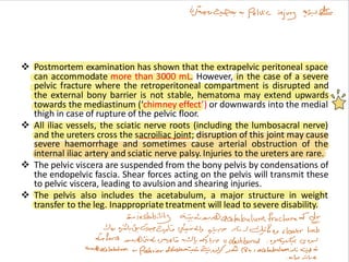
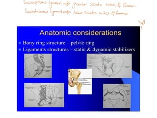
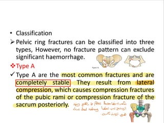
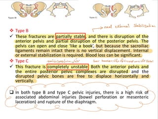
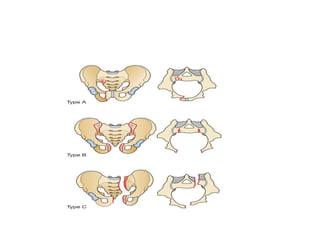
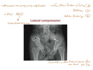
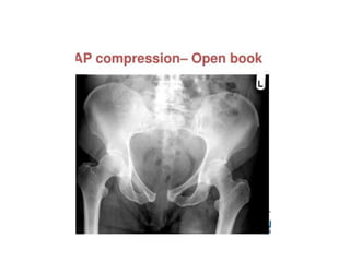
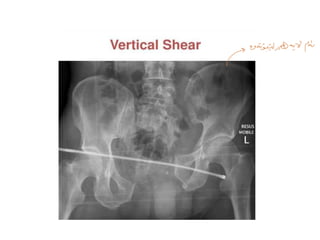
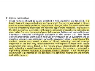
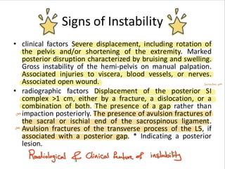
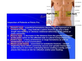
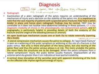
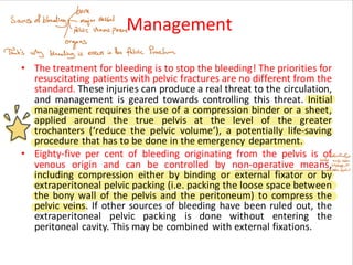
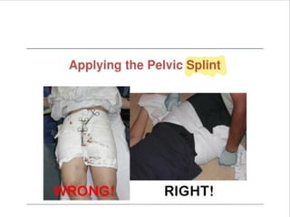
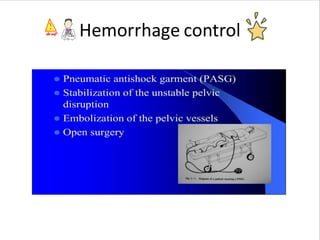
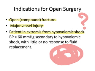
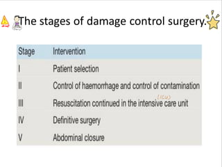
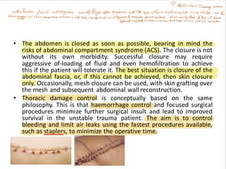
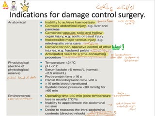
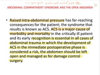
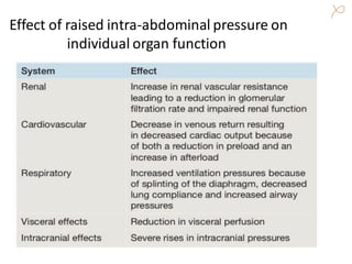
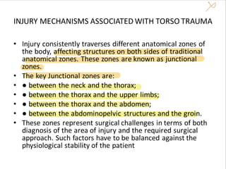
abdominal trauma
dr.hassan a. ahmed
m.b.ch.b, fkbms
general surgery
2022-2023
THE PELVIS
• Although mortality following severe pelvic fractures has
decreased dramatically with better methods of controlling
haemorrhage, these patients still represent a significant
challenge to every link of the treatment chain. Mortality
rates exceeding 40% have recently been reported. Further,
pelvic bleeding as one of the ‘hidden bleeding sources’ is
still underestimated or missed. Extreme force is required to
disrupt the pelvic ring, and associated injuries and extra
pelvic bleeding sources are common (up to 50% of cases).
The haemodynamically unstable patient with severe pelvic
fracture has a 90% risk of associated injuries and a 30% risk
of intra-abdominal bleeding. To save these patients, three
questions need to be addressed:
● Is the patient at high risk of massive bleeding?
● Where is the source of the bleeding?
● How to stop the bleeding?
➢ Anatomy
• The surgical anatomy of the pelvis is key to the understanding of pelvic
injuries.
❖ The pelvic inlet is circular. It is a structure that is immensely strong, but
routinely gives way at more than one point should sufficient force be
applied to it. Therefore, isolated fractures of the anterior or posterior
pelvic ring are uncommon.
❖ The forces required to fracture the pelvic ring do not respect the
surrounding organ systems.
❖ The pelvis has a rich collateral blood supply, especially across the sacrum
and posterior part of the ileum. The cancellous bone of the pelvis also has
an excellent blood supply. Most pelvic haemorrhage emanates from
venous injury and fracture sites. However, in the haemodynamically
unstable patient with severe pelvic injury, arterial bleeding is more
frequent. Important for the treatment is that the surgeon has to deal with
both arterial and venous bleeding.
❖ Postmortem examination has shown that the extrapelvic peritoneal space
can accommodate more than 3000 mL. However, in the case of a severe
pelvic fracture where the retroperitoneal compartment is disrupted and
the external bony barrier is not stable, hematoma may extend upwards
towards the mediastinum (‘chimney effect’) or downwards into the medial
thigh in case of rupture of the pelvic floor.
❖ All iliac vessels, the sciatic nerve roots (including the lumbosacral nerve)
and the ureters cross the sacroiliac joint; disruption of this joint may cause
severe haemorrhage and sometimes cause arterial obstruction of the
internal iliac artery and sciatic nerve palsy. Injuries to the ureters are rare.
❖ The pelvic viscera are suspended from the bony pelvis by condensations of
the endopelvic fascia. Shear forces acting on the pelvis will transmit these
to pelvic viscera, leading to avulsion and shearing injuries.
❖ The pelvis also includes the acetabulum, a major structure in weight
transfer to the leg. Inappropriate treatment will lead to severe disability.
• Classification
➢Pelvic ring fractures can be classified into three
types, However, no fracture pattern can exclude
significant haemorrhage.
❖Type A
✓Type A are the most common fractures and are
completely stable. They result from lateral
compression, which causes compression fractures
of the pubic rami or compression fracture of the
sacrum posteriorly.
❖ Type B
✓ These fractures are partially stable, and there is disruption of the
anterior pelvis and partial disruption of the posterior pelvis. The
pelvis can open and close ‘like a book’, but because the sacroiliac
ligaments remain intact there is no vertical displacement. Internal
or external stabilization is required. Blood loss can be significant.
❖ Type C
✓ This fracture is completely unstable. Both the anterior pelvis and
the entire posterior pelvic complexes are disrupted and the
disrupted pelvic bones are free to displace horizontally and
vertically.
❑ In both type B and type C pelvic injuries, there is a high risk of
associated abdominal injuries (bowel perforation or mesenteric
laceration) and rupture of the diaphragm.
❖ Clinical examination
• Pelvic fractures should be easily identified if ATLS guidelines are followed.. If a
binder has not been applied and an ‘open book’ fracture is suspected, a binder
must be immediately applied as the presence of major pelvic fracture is associated
with life-threatening blood loss and requires appropriate measures. Inspection of
the skin may reveal lacerations in the groin, perineum or sacral area, indicating an
open pelvic fracture, the result of gross deformation. Evidence of perineal injury or
haematuria mandates radiological evaluation of the urinary tract from below
upwards (retrograde urethrogram followed by cystogram or CT cystogram and an
excretory urogram, as appropriate) when the physiology allows. Inspection of the
urethral meatus may reveal a drop of blood, indicating urethral damage.
Inspection of the anus may reveal lacerations to the sphincter mechanism. Rectal
examination may reveal blood in the rectum and/or discontinuity of the rectal
wall, indicating a rectal laceration. In male patients, the prostate is palpated; a
high-riding prostate indicates a complete urethral avulsion. A full neurological
examination is performed of the perineal area, sphincter mechanism and femoral
and sciatic nerves.
Signs of Instability
• clinical factors Severe displacement, including rotation of
the pelvis and/or shortening of the extremity. Marked
posterior disruption characterized by bruising and swelling.
Gross instability of the hemi-pelvis on manual palpation.
Associated injuries to viscera, blood vessels, or nerves.
Associated open wound.
• radiographic factors Displacement of the posterior SI
complex >1 cm, either by a fracture, a dislocation, or a
combination of both. The presence of a gap rather than
impaction posteriorly. The presence of avulsion fractures of
the sacral or ischial end of the sacrospinous ligament.
Avulsion fractures of the transverse process of the L5, if
associated with a posterior gap. * Indicating a posterior
lesion.
•Leg-length discrepancy or rotational deformity without
fracture suggests unstable pelvis fracture
• Motion may be detected by grasping iliac crests and
pushing inward, then outward
• Once pelvic instability is demonstrated, no further
maneuvers are necessary
• Inspect flank, scrotum, and perianal area for bleeding
including urethral meatus
• Inspect for lacerations in perineum, vagina, rectum, or
buttocks for open pelvic fractures
• genital exam to OR
• Palpate for high-riding prostate gland in males.
Diagnosis
➢ Radiograph
• Examination of a plain radiograph of the pelvis requires an understanding of the
mechanism of injury and a decision on the stability of the pelvic rim. It is important to
note that the vast majority of patients with suspected pelvic fractures may have a pelvic
binder in place and hence plain radiograph findings may be normal. FAST may be
unreliable as it does not localize intra-abdominal bleeding in these patients. CT is the
diagnostic modality of choice in the physiologically non-compromised patient, and CT
angiography is particularly helpful in providing details of both the anatomy of the
fracture and the origin of the bleeding (venous or arterial).
• An open book-type mechanism causes one or both ilia to rotate externally (opening,
like a book).
• A lateral compression mechanism causes the pelvis to collapse. An ‘open book fracture’
is seen as a widening of the pubic symphysis or widening at the site of a fracture in the
pubic ramus. Not only is there disruption of the bony pelvis, but also tearing of the
pelvic floor and thus the pelvic venous plexus is at risk. The more unstable the pelvis,
the more likely the structures are to be damaged. When the pelvis collapses from a
lateral compression injury, the pubic bones usually fracture. Displacement of the
anterior pelvis by greater than 2 cm indicatesat least partialinstability.
• A vertical shear disruption of the sacroiliac joint with apparent shortening of the limb
on the affected side implies significant energy of injury.
Management
• The treatment for bleeding is to stop the bleeding! The priorities for
resuscitating patients with pelvic fractures are no different from the
standard. These injuries can produce a real threat to the circulation,
and management is geared towards controlling this threat. Initial
management requires the use of a compression binder or a sheet,
applied around the true pelvis at the level of the greater
trochanters (‘reduce the pelvic volume’), a potentially life-saving
procedure that has to be done in the emergency department.
• Eighty-five per cent of bleeding originating from the pelvis is of
venous origin and can be controlled by non-operative means,
including compression either by binding or external fixator or by
extraperitoneal pelvic packing (i.e. packing the loose space between
the bony wall of the pelvis and the peritoneum) to compress the
pelvic veins. If other sources of bleeding have been ruled out, the
extraperitoneal pelvic packing is done without entering the
peritoneal cavity. This may be combined with external fixations.
• If the bleeding is of arterial origin, interventional angioembolisation is
the next choice for bleeding control. The techniques for bleeding
control (compression, packing, fixation and angioembolisation) do not
exclude each other but rather may complement each other. Persistent
bleeding after packing may require angioembolisation and vice versa.
• Severe pelvic injuries require a multidisciplinary team approach. If
adequate orthopedic experience is unavailable, consideration should
be given towards early transfer of this patient to an institution with
the necessary expertise.
• If the source of the bleeding is in doubt or FAST/CT results are positive,
showing a significant amount of blood in the peritoneal cavity,
concurrent intra-abdominal injury cannot be excluded and it is wise to
perform an exploratory laparotomy to treat or rule out intra-
abdominal bleeding
Hemorrhage control
Indications for Open Surgery
• Open (compound) fracture.
• Major vessel injury.
• Patient in extremis from hypovolemic shock.
BP < 60 mmHg secondaryto hypovolemic
shock, with little or no response to fluid
replacement.
Pelvic injury
● Associated injuries can only be managed once the
patient is physiologically non-compromised
● Decision on the stability is of paramount
importance
● Procedures for damage control may be the only
available option
● External stabilisation of the pelvic ring is the basis
of all treatment
● If necessary, further bleeding control can be
achieved either by angioembolisation or by
extraperitoneal packing
DAMAGE CONTROL
• Following major injury, protracted surgery in the physiologically unstable
patient can in itself prove fatal. Patients with the ‘deadly triad’
(hypothermia, acidosis and coagulopathy) are those at highest risk.
Damagecontrol or damage limitation surgery is a concept that originated
from a naval shipbuilding strategy, whereby ships were designed so that
the damage was kept ‘local’ and only minimal repairs were needed to
prevent the ship from sinking while definitive repairs waited until it had
reached port. The technique has been adopted following major trauma
and includes initial care and resuscitation (damage control resuscitation)
and the surgical correction of the injury (damage control surgery).
• The minimum amount of surgery needed to stabilize the patient’s
condition may be the safest course until the physiological derangement
can be corrected. Damagecontrol surgery is restricted to only three goals:
● stopping any active surgical bleeding;
● controlling any contamination;
● restoring normal physiology.
• Once the first two have been achieved then the
operation is suspended and the abdomen
temporarily closed to allow for restoration of
physiology to occur. The patient’s resuscitation
then continues in the ICU, where other
therapeutic interventions can take place. Once
the physiology has been corrected, the patient
warmed and the coagulopathy corrected, the
patient is returned to the operating theatre for
any definitive surgery
Damage control resuscitation
• The concept of damage control has been broadened to include the
techniques used in resuscitation as well as in surgery. The time in
the emergency department is minimized and the majority of
resuscitation of the patient is carried out in the operating theatre
and not in the resuscitation bay. Resuscitation is individualized
through repeated point-of-care testing of haemoglobin, acidosis
(pH and lactate) and clotting, and is therefore directed towards the
early delivery of biologically active colloids, clotting products and
whole blood in order to buy time. The physiological disturbances
that are associated with the downward spiral of acidosis,
coagulopathy and hypothermia in these serious injuries are
predicted and attempts are made to avoid them rather than react
to them.
Damage control surgery
• This is a key component of damage control resuscitation. The decision of whether
damage control surgery is the appropriate course should be made early and allows
the whole surgical and anesthetic team to work together to limit the time in surgery
and achieve the earliest possible admission of the patient to the ICU. Damage control
is a staged process.
• The initial focus is haemorrhage control, followed by control and limitation of
contamination, which are achieved using a range of abbreviated techniques including
simple ligation of bleeding vessels, shunting of major arteries and veins, drainage,
temporary stapling of bowel and therapeutic packing. Following the above, the
abdomen is closed in a temporary fashion either by using commercially available
products or by using a sheet of plastic (e.g. OPSITE or similar product) over the bowel,
an intermediate pack to allow suction and a further sheet of adherent plastic drape to
the skin to form a watertight and airtight seal. Suction is applied to the intermediate
pack area to collect abdominal fluid. This technique is known as the ‘Vac-Pac’ or
‘OPSITE sandwich’. As soon as control has been achieved the patient is transferred to
the ICU, where resuscitation is continued.
• The next stage following damage control surgery and physiological stabilization is
definitive surgery. The team should aim to perform definitive anastomoses, vascular
reconstruction and closure of the body cavity within 24–72 hours of injury. However,
this must be individualized to the patient, the response to critical care resuscitation
and the progression of injury.
The stages of damage control surgery.
• The abdomen is closed as soon as possible, bearing in mind the
risks of abdominal compartment syndrome (ACS). The closure is not
without its own morbidity. Successful closure may require
aggressive of-loading of fluid and even hemofiltration to achieve
this if the patient will tolerate it. The best situation is closure of the
abdominal fascia, or, if this cannot be achieved, then skin closure
only. Occasionally, mesh closure can be used, with skin grafting over
the mesh and subsequent abdominal wall reconstruction.
• Thoracic damage control is conceptually based on the same
philosophy. This is that haemorrhage control and focused surgical
procedures minimize further surgical insult and lead to improved
survival in the unstable trauma patient. The aim is to control
bleeding and limit air leaks using the fastest procedures available,
such as staplers, to minimize the operative time.
• The indications and techniques for emergency
thoracic surgery have already been described.
• Damage control applies equally to the
extremities. In this case, it is shunting of blood
vessels, identifying and marking damaged
structures such as nerves, fasciotomy and
removal of contaminated tissue that are the
main tasks. Subsequent definitive
management can be carried out at a later
Indications for damage control surgery.
ABDOMINAL COMPARTMENT SYNDROME AND THE OPEN ABDOMEN
• Raised intra-abdominal pressure has far-reaching
consequences for the patient; the syndrome that
results is known as ACS. ACS is a major cause of
morbidity and mortality in the critically ill patient
and its early recognition is essential In all cases of
abdominal trauma in which the development of
ACS in the immediate postoperative phase is
considered a risk, the abdomen should be left
open and managed as for damage control
surgery.
Effect of raised intra-abdominal pressure on
individual organ function
INJURY MECHANISMS ASSOCIATED WITH TORSO TRAUMA
• Injury consistently traverses different anatomical zones of
the body, affecting structures on both sides of traditional
anatomical zones. These zones are known as junctional
zones.
• The key Junctional zones are:
• ● between the neck and the thorax;
• ● between the thorax and the upper limbs;
• ● between the thorax and the abdomen;
• ● between the abdominopelvic structures and the groin.
• These zones represent surgical challenges in terms of both
diagnosis of the area of injury and the required surgical
approach. Such factors have to be balanced against the
physiological stability of the patient
thanks

pelvis trauma & damage control surgery and its management

  • 1.
    abdominal trauma dr.hassan a.ahmed m.b.ch.b, fkbms general surgery 2022-2023
  • 3.
    THE PELVIS • Althoughmortality following severe pelvic fractures has decreased dramatically with better methods of controlling haemorrhage, these patients still represent a significant challenge to every link of the treatment chain. Mortality rates exceeding 40% have recently been reported. Further, pelvic bleeding as one of the ‘hidden bleeding sources’ is still underestimated or missed. Extreme force is required to disrupt the pelvic ring, and associated injuries and extra pelvic bleeding sources are common (up to 50% of cases). The haemodynamically unstable patient with severe pelvic fracture has a 90% risk of associated injuries and a 30% risk of intra-abdominal bleeding. To save these patients, three questions need to be addressed: ● Is the patient at high risk of massive bleeding? ● Where is the source of the bleeding? ● How to stop the bleeding?
  • 4.
    ➢ Anatomy • Thesurgical anatomy of the pelvis is key to the understanding of pelvic injuries. ❖ The pelvic inlet is circular. It is a structure that is immensely strong, but routinely gives way at more than one point should sufficient force be applied to it. Therefore, isolated fractures of the anterior or posterior pelvic ring are uncommon. ❖ The forces required to fracture the pelvic ring do not respect the surrounding organ systems. ❖ The pelvis has a rich collateral blood supply, especially across the sacrum and posterior part of the ileum. The cancellous bone of the pelvis also has an excellent blood supply. Most pelvic haemorrhage emanates from venous injury and fracture sites. However, in the haemodynamically unstable patient with severe pelvic injury, arterial bleeding is more frequent. Important for the treatment is that the surgeon has to deal with both arterial and venous bleeding.
  • 5.
    ❖ Postmortem examinationhas shown that the extrapelvic peritoneal space can accommodate more than 3000 mL. However, in the case of a severe pelvic fracture where the retroperitoneal compartment is disrupted and the external bony barrier is not stable, hematoma may extend upwards towards the mediastinum (‘chimney effect’) or downwards into the medial thigh in case of rupture of the pelvic floor. ❖ All iliac vessels, the sciatic nerve roots (including the lumbosacral nerve) and the ureters cross the sacroiliac joint; disruption of this joint may cause severe haemorrhage and sometimes cause arterial obstruction of the internal iliac artery and sciatic nerve palsy. Injuries to the ureters are rare. ❖ The pelvic viscera are suspended from the bony pelvis by condensations of the endopelvic fascia. Shear forces acting on the pelvis will transmit these to pelvic viscera, leading to avulsion and shearing injuries. ❖ The pelvis also includes the acetabulum, a major structure in weight transfer to the leg. Inappropriate treatment will lead to severe disability.
  • 8.
    • Classification ➢Pelvic ringfractures can be classified into three types, However, no fracture pattern can exclude significant haemorrhage. ❖Type A ✓Type A are the most common fractures and are completely stable. They result from lateral compression, which causes compression fractures of the pubic rami or compression fracture of the sacrum posteriorly.
  • 9.
    ❖ Type B ✓These fractures are partially stable, and there is disruption of the anterior pelvis and partial disruption of the posterior pelvis. The pelvis can open and close ‘like a book’, but because the sacroiliac ligaments remain intact there is no vertical displacement. Internal or external stabilization is required. Blood loss can be significant. ❖ Type C ✓ This fracture is completely unstable. Both the anterior pelvis and the entire posterior pelvic complexes are disrupted and the disrupted pelvic bones are free to displace horizontally and vertically. ❑ In both type B and type C pelvic injuries, there is a high risk of associated abdominal injuries (bowel perforation or mesenteric laceration) and rupture of the diaphragm.
  • 14.
    ❖ Clinical examination •Pelvic fractures should be easily identified if ATLS guidelines are followed.. If a binder has not been applied and an ‘open book’ fracture is suspected, a binder must be immediately applied as the presence of major pelvic fracture is associated with life-threatening blood loss and requires appropriate measures. Inspection of the skin may reveal lacerations in the groin, perineum or sacral area, indicating an open pelvic fracture, the result of gross deformation. Evidence of perineal injury or haematuria mandates radiological evaluation of the urinary tract from below upwards (retrograde urethrogram followed by cystogram or CT cystogram and an excretory urogram, as appropriate) when the physiology allows. Inspection of the urethral meatus may reveal a drop of blood, indicating urethral damage. Inspection of the anus may reveal lacerations to the sphincter mechanism. Rectal examination may reveal blood in the rectum and/or discontinuity of the rectal wall, indicating a rectal laceration. In male patients, the prostate is palpated; a high-riding prostate indicates a complete urethral avulsion. A full neurological examination is performed of the perineal area, sphincter mechanism and femoral and sciatic nerves.
  • 15.
    Signs of Instability •clinical factors Severe displacement, including rotation of the pelvis and/or shortening of the extremity. Marked posterior disruption characterized by bruising and swelling. Gross instability of the hemi-pelvis on manual palpation. Associated injuries to viscera, blood vessels, or nerves. Associated open wound. • radiographic factors Displacement of the posterior SI complex >1 cm, either by a fracture, a dislocation, or a combination of both. The presence of a gap rather than impaction posteriorly. The presence of avulsion fractures of the sacral or ischial end of the sacrospinous ligament. Avulsion fractures of the transverse process of the L5, if associated with a posterior gap. * Indicating a posterior lesion.
  • 16.
    •Leg-length discrepancy orrotational deformity without fracture suggests unstable pelvis fracture • Motion may be detected by grasping iliac crests and pushing inward, then outward • Once pelvic instability is demonstrated, no further maneuvers are necessary • Inspect flank, scrotum, and perianal area for bleeding including urethral meatus • Inspect for lacerations in perineum, vagina, rectum, or buttocks for open pelvic fractures • genital exam to OR • Palpate for high-riding prostate gland in males.
  • 18.
    Diagnosis ➢ Radiograph • Examinationof a plain radiograph of the pelvis requires an understanding of the mechanism of injury and a decision on the stability of the pelvic rim. It is important to note that the vast majority of patients with suspected pelvic fractures may have a pelvic binder in place and hence plain radiograph findings may be normal. FAST may be unreliable as it does not localize intra-abdominal bleeding in these patients. CT is the diagnostic modality of choice in the physiologically non-compromised patient, and CT angiography is particularly helpful in providing details of both the anatomy of the fracture and the origin of the bleeding (venous or arterial). • An open book-type mechanism causes one or both ilia to rotate externally (opening, like a book). • A lateral compression mechanism causes the pelvis to collapse. An ‘open book fracture’ is seen as a widening of the pubic symphysis or widening at the site of a fracture in the pubic ramus. Not only is there disruption of the bony pelvis, but also tearing of the pelvic floor and thus the pelvic venous plexus is at risk. The more unstable the pelvis, the more likely the structures are to be damaged. When the pelvis collapses from a lateral compression injury, the pubic bones usually fracture. Displacement of the anterior pelvis by greater than 2 cm indicatesat least partialinstability. • A vertical shear disruption of the sacroiliac joint with apparent shortening of the limb on the affected side implies significant energy of injury.
  • 19.
    Management • The treatmentfor bleeding is to stop the bleeding! The priorities for resuscitating patients with pelvic fractures are no different from the standard. These injuries can produce a real threat to the circulation, and management is geared towards controlling this threat. Initial management requires the use of a compression binder or a sheet, applied around the true pelvis at the level of the greater trochanters (‘reduce the pelvic volume’), a potentially life-saving procedure that has to be done in the emergency department. • Eighty-five per cent of bleeding originating from the pelvis is of venous origin and can be controlled by non-operative means, including compression either by binding or external fixator or by extraperitoneal pelvic packing (i.e. packing the loose space between the bony wall of the pelvis and the peritoneum) to compress the pelvic veins. If other sources of bleeding have been ruled out, the extraperitoneal pelvic packing is done without entering the peritoneal cavity. This may be combined with external fixations.
  • 21.
    • If thebleeding is of arterial origin, interventional angioembolisation is the next choice for bleeding control. The techniques for bleeding control (compression, packing, fixation and angioembolisation) do not exclude each other but rather may complement each other. Persistent bleeding after packing may require angioembolisation and vice versa. • Severe pelvic injuries require a multidisciplinary team approach. If adequate orthopedic experience is unavailable, consideration should be given towards early transfer of this patient to an institution with the necessary expertise. • If the source of the bleeding is in doubt or FAST/CT results are positive, showing a significant amount of blood in the peritoneal cavity, concurrent intra-abdominal injury cannot be excluded and it is wise to perform an exploratory laparotomy to treat or rule out intra- abdominal bleeding
  • 22.
  • 23.
    Indications for OpenSurgery • Open (compound) fracture. • Major vessel injury. • Patient in extremis from hypovolemic shock. BP < 60 mmHg secondaryto hypovolemic shock, with little or no response to fluid replacement.
  • 24.
    Pelvic injury ● Associatedinjuries can only be managed once the patient is physiologically non-compromised ● Decision on the stability is of paramount importance ● Procedures for damage control may be the only available option ● External stabilisation of the pelvic ring is the basis of all treatment ● If necessary, further bleeding control can be achieved either by angioembolisation or by extraperitoneal packing
  • 25.
    DAMAGE CONTROL • Followingmajor injury, protracted surgery in the physiologically unstable patient can in itself prove fatal. Patients with the ‘deadly triad’ (hypothermia, acidosis and coagulopathy) are those at highest risk. Damagecontrol or damage limitation surgery is a concept that originated from a naval shipbuilding strategy, whereby ships were designed so that the damage was kept ‘local’ and only minimal repairs were needed to prevent the ship from sinking while definitive repairs waited until it had reached port. The technique has been adopted following major trauma and includes initial care and resuscitation (damage control resuscitation) and the surgical correction of the injury (damage control surgery). • The minimum amount of surgery needed to stabilize the patient’s condition may be the safest course until the physiological derangement can be corrected. Damagecontrol surgery is restricted to only three goals: ● stopping any active surgical bleeding; ● controlling any contamination; ● restoring normal physiology.
  • 26.
    • Once thefirst two have been achieved then the operation is suspended and the abdomen temporarily closed to allow for restoration of physiology to occur. The patient’s resuscitation then continues in the ICU, where other therapeutic interventions can take place. Once the physiology has been corrected, the patient warmed and the coagulopathy corrected, the patient is returned to the operating theatre for any definitive surgery
  • 27.
    Damage control resuscitation •The concept of damage control has been broadened to include the techniques used in resuscitation as well as in surgery. The time in the emergency department is minimized and the majority of resuscitation of the patient is carried out in the operating theatre and not in the resuscitation bay. Resuscitation is individualized through repeated point-of-care testing of haemoglobin, acidosis (pH and lactate) and clotting, and is therefore directed towards the early delivery of biologically active colloids, clotting products and whole blood in order to buy time. The physiological disturbances that are associated with the downward spiral of acidosis, coagulopathy and hypothermia in these serious injuries are predicted and attempts are made to avoid them rather than react to them.
  • 28.
    Damage control surgery •This is a key component of damage control resuscitation. The decision of whether damage control surgery is the appropriate course should be made early and allows the whole surgical and anesthetic team to work together to limit the time in surgery and achieve the earliest possible admission of the patient to the ICU. Damage control is a staged process. • The initial focus is haemorrhage control, followed by control and limitation of contamination, which are achieved using a range of abbreviated techniques including simple ligation of bleeding vessels, shunting of major arteries and veins, drainage, temporary stapling of bowel and therapeutic packing. Following the above, the abdomen is closed in a temporary fashion either by using commercially available products or by using a sheet of plastic (e.g. OPSITE or similar product) over the bowel, an intermediate pack to allow suction and a further sheet of adherent plastic drape to the skin to form a watertight and airtight seal. Suction is applied to the intermediate pack area to collect abdominal fluid. This technique is known as the ‘Vac-Pac’ or ‘OPSITE sandwich’. As soon as control has been achieved the patient is transferred to the ICU, where resuscitation is continued. • The next stage following damage control surgery and physiological stabilization is definitive surgery. The team should aim to perform definitive anastomoses, vascular reconstruction and closure of the body cavity within 24–72 hours of injury. However, this must be individualized to the patient, the response to critical care resuscitation and the progression of injury.
  • 29.
    The stages ofdamage control surgery.
  • 30.
    • The abdomenis closed as soon as possible, bearing in mind the risks of abdominal compartment syndrome (ACS). The closure is not without its own morbidity. Successful closure may require aggressive of-loading of fluid and even hemofiltration to achieve this if the patient will tolerate it. The best situation is closure of the abdominal fascia, or, if this cannot be achieved, then skin closure only. Occasionally, mesh closure can be used, with skin grafting over the mesh and subsequent abdominal wall reconstruction. • Thoracic damage control is conceptually based on the same philosophy. This is that haemorrhage control and focused surgical procedures minimize further surgical insult and lead to improved survival in the unstable trauma patient. The aim is to control bleeding and limit air leaks using the fastest procedures available, such as staplers, to minimize the operative time.
  • 31.
    • The indicationsand techniques for emergency thoracic surgery have already been described. • Damage control applies equally to the extremities. In this case, it is shunting of blood vessels, identifying and marking damaged structures such as nerves, fasciotomy and removal of contaminated tissue that are the main tasks. Subsequent definitive management can be carried out at a later
  • 32.
    Indications for damagecontrol surgery.
  • 33.
    ABDOMINAL COMPARTMENT SYNDROMEAND THE OPEN ABDOMEN • Raised intra-abdominal pressure has far-reaching consequences for the patient; the syndrome that results is known as ACS. ACS is a major cause of morbidity and mortality in the critically ill patient and its early recognition is essential In all cases of abdominal trauma in which the development of ACS in the immediate postoperative phase is considered a risk, the abdomen should be left open and managed as for damage control surgery.
  • 34.
    Effect of raisedintra-abdominal pressure on individual organ function
  • 35.
    INJURY MECHANISMS ASSOCIATEDWITH TORSO TRAUMA • Injury consistently traverses different anatomical zones of the body, affecting structures on both sides of traditional anatomical zones. These zones are known as junctional zones. • The key Junctional zones are: • ● between the neck and the thorax; • ● between the thorax and the upper limbs; • ● between the thorax and the abdomen; • ● between the abdominopelvic structures and the groin. • These zones represent surgical challenges in terms of both diagnosis of the area of injury and the required surgical approach. Such factors have to be balanced against the physiological stability of the patient
  • 36.