
Recommended
More Related Content
Similar to PELVIC FRACTURE ppt by dr.bharti pawar.ppt
Similar to PELVIC FRACTURE ppt by dr.bharti pawar.ppt (20)
More from bharti pawar
More from bharti pawar (17)
Recently uploaded
Recently uploaded (20)
PELVIC FRACTURE ppt by dr.bharti pawar.ppt
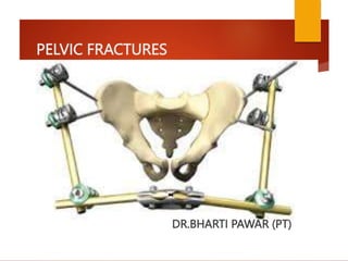
- 1. PELVIC FRACTURES DR.BHARTI PAWAR (PT)
- 2. PELVIC FRACTURES Fractures of the pelvis account for less than 5% of all skeletal injuries, but it is important because it associated with:- 1. Soft tissue injuries and blood loss. 2. Shock. 3. Sepsis. 4. ARDS. Because of those mortality rate exceeds 10%.
- 5. PELVIC FRACTURES Fractures of the adult pelvis, exclusive of the acetabulum, generally are either stable fractures resulting from low- energy trauma, such as falls in elderly patients, or fractures caused by high-energy trauma that result in significant morbidity and mortality.
- 6. Mechanisms of injury The basic mechanisms of pelvic ring injury are: 1. Anteroposterior compression (APC). 2. Lateral compression (LC). 3. Vertical shear (VS). 4. Combinations of these.
- 7. Anteroposterior compression (APC) Usually caused by a frontal collision between pedestrian and a car. This injury may lead to: 1. Fracture of the rami. 2. The innominate bones are sprung apart and externally rotated with disruption of the symphysis. 3. The anterior sacroiliac joint is partially torn. 4. Fracture of the posterior part of the ilium. This is called open book injury.
- 9. Lateral compression (LC) Side to side compression of the pelvis causes the ring to buckle and break. This is due to a side –on impact in a road accident or a fall from a height. This injury may lead to 1. Anteriorly the pubic rami on one side or both sides are fractured. 2. Posteriorly there is severe sacroiliac strain or fracture of the sacrum or ilium, either on the same side of the pubic fracture or on the opposite side.
- 11. Vertical shear (VS) The innominate bone on one side is displaced vertically, fracturing the pubic rami and disrupting the sacroiliac region on the same side. This is typically occurs when falls from a height on one leg. These are severe unstable injuries with gross tearing of the soft tissues and associated with retroperitoneal hemorrhage.
- 13. Combination injuries In severe pelvic injuries there may be a combination of the above.
- 14. CLASSIFICATIONS
- 19. Clinical features and clinical assessment 1. Fracture of the pelvis should be suspected in every patient with serious abdominal injury or lower limb injury. 2. HO road traffic accident, fall from a height or crush injury. 3. Severe pain, swelling and bruises in the lower abdomen, perineum, thighs, scrotum or valva. 4. Extravasations of urine. 5. Symptoms and signs of bleeding and hemorrhagic shock.
- 21. Clinical features and clinical assessment 6. Tenderness all over the pelvic bone especially when attempt to compress or distract the pelvis. 7. Tender abdomen due to bleeding or intrapelvic structure injuries. 8. Rectal examination should be done in every case.
- 23. Clinical features and clinical assessment 9. Bleeding in external meatus indicates urethral injury. If no bleeding ask the patient to void and give direct look to the urine, if the patient able to void this indicates either no urethral injury or there is only minimal damage to the urethra. Note no attempt should be made to pass a catheter, as this could convert the partial injury to complete injury. 10. Neurological examination should be done to exclude sacral and lumber plexus injury.
- 24. Radiography 1. plain radiography: 5 views are necessary 1. Anteroposterior view. 2. Pelvic inlet view in which the tube is cephalad to the pelvis and tilted 45° downwards. 3. Pelvic outlet view in which the tube is caudad to the pelvis and tilted 45° upwards. 4. Right oblique view. 5. Left oblique view.
- 25. RADIOGRAPH POSITIONING : AP VIEW Patient lies supine with the x ray beam centered over the pelvis
- 26. RADIOGRAPH POSITIONING : INLET VIEW X Ray beam is directed 45 degrees caudally. Simulates a direct view of pelvis from above along its longitudinal axis.
- 27. RADIOGRAPH POSITIONING : OUTLET VIEW X Ray beam is directed 45 degrees cephalad. Simulates looking at sacrum and SI joint en face.
- 28. X RAY PELVIS : AP VIEW
- 29. X RAY PELVIS : AP VIEW PUBIC SYMPHYSIS SHOULD BE COLINEAR WITH THE SACRAL SPINOUS PROCESS
- 30. X RAY PELVIS : AP VIEW The Iliopectineal line should be traced back to its intersection with lateral margin of ala. It should be at the same level ( usually at superior margin of S2 foramen) bilaterally.
- 31. X RAY PELVIS : AP VIEW Asymmetry of SI joint or foramina : possibility of SI joint dislocation or sacral fracture.
- 32. X ray Pelvis : AP view Fracture of L5 transverse process : Vertical shear injury due to avulsion of the processes via Iliolumbar ligament.
- 33. Symphyseal diastasis or pubic rami fracture : may have additional injuries in the posterior ring.
- 34. X RAY PELVIS : INLET VIEW • AP translation of hemi pelvis. • External/ Internal rotation of hemi pelvis. • Opening of SI joint. • Impaction # of sacral ala.
- 35. X RAY PELVIS : OUTLET VIEW • Vertical shift of hemi pelvis. • Sacral fractures relative to foramina. • Flexion or Extension deformity of pelvic ring
- 36. CT SCAN CT scan gives accurate details and much information about the injury. CT scanning is imperative in any suspected pelvic injury or in suspected sacral fractures. 2mm to 3mm axial sections are recommended.
- 37. CT SCAN LATERAL X RAY OF SACRUM and CT SCAN of the same patient showing SACRAL FRACTURE DISLOCATION WITH SPINAL PELVIC DISASSOCIATION.
- 38. Urethrographyfor diagnosis of urethral injury
- 40. Management 1. Early management Treatment should not await full and detailed diagnosis. Doctor should move according to the priority of life saving measures with the already available information.Six questions must be asked and the answers acting upon as they emerge:
- 41. Management 1. Is there a clear airway? 2. Are the lungs adequately ventilated? 3. Is the patient losing blood? 4. Is there an intra abdominal injury? 5. Is there a bladder or urethral injury? 6. Is the pelvic fracture stable or not?
- 42. Management After exclusion of the above, the doctor now has a good idea about the patient general condition and the associated injuries so further investigation can be done.
- 43. Management 2. Management of severe bleeding Treatment of shock – Rapid fluid resuscitation,blood transfusion. Wrapping of pelvis with sheets with internal rotation & slight flexion of the knees. Anterior external fixation,pelvic C- clamp,pneumatic antishock garments. Pelvic packing & angiographic embolisation if required. 3. Management of urethral and bladder injury.
- 44. Management 4.Control of contamination Repair of genitourinary and rectal injuries. Debridement of necrotic tissue in case of open injury. 5.Laparotomy if required
- 45. Management 5. Treatment of the fracture 1. Isolated fractures and minimally displaced fractures(LC 1 & APC 1): need only bed rest with lower limb traction.
- 46. Management 2. More severe pelvic fractures (LC 2 & APC2) with pubic symphysis diastasis of more than 2.5 cm,pubic rami fractures with more than 2 cm displacement or other rotationally unstable fractures with limb length discrepancy of more than 1.5 cm require surgical intervention either by external fixation or by closed reduction and internal fixation.
- 51. Management 4. AP-III and VC are the most dangerous and the most difficult to treat. These are unstable fractures and needs reduction and fixation by either external fixation or plate and screws.
- 53. Secondary complications 1. Sciatic nerve injury. 2. Urogenital problem like stricture, incontinence and impotence. 3. Persistent sacroiliac pain due to unstable pelvis.
- 54. ACKNOWLEDGEMENT