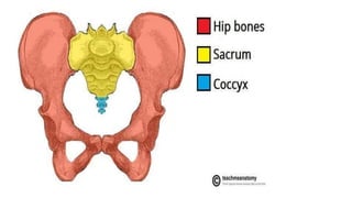
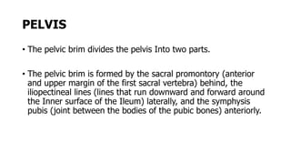
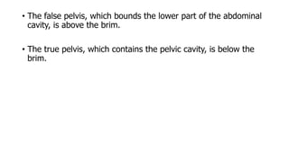
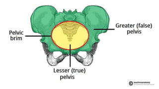
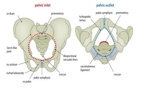
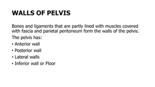
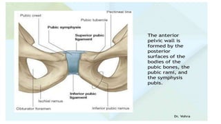
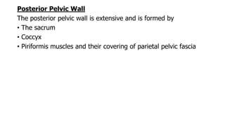
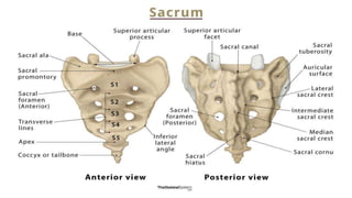
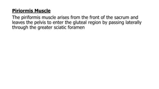
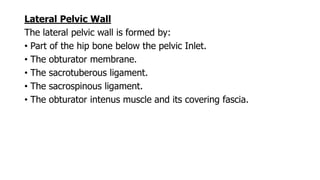
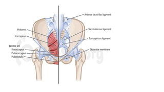
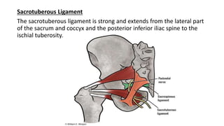
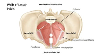
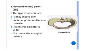
3. INTRODUCTION
• The term pelvis is loosely used to describe the region where the
trunk and lower limbs meet.
• The word pelvis means “basin” and is more correctly applied to
the skeleton of the region, that is, the pelvic girdle or bony
pelvis.
• Thus, the pelvis is a bowl-shaped bony structure.
• it contains, supports, and protects the lower parts of the
gastrointestinal and urinary tracts and the male and female
internal organs of reproduction.
4. • It also contains important nerves, blood vessels, and lymphatic
tissues and provides attachment for trunk, lower limb and
muscles.
5. Four bones make up the bony pelvis:
• The two hip bones, which form the lateral and anterior walls.
• The sacrum.
• The coccyx, which are part of the vertebral column and form
the posterior wall.
The two hip bones articulate with each other anteriorly at the
symphysis pubis and posteriorly with the sacrum at the
sacroillac Joints.
The bony pelvis thus forms a strong basin-shaped structure.
6.
7. PELVIS
• The pelvic brim divides the pelvis Into two parts.
• The pelvic brim is formed by the sacral promontory (anterior
and upper margin of the first sacral vertebra) behind, the
iliopectineal lines (lines that run downward and forward around
the Inner surface of the Ileum) laterally, and the symphysis
pubis (joint between the bodies of the pubic bones) anteriorly.
8. • The false pelvis, which bounds the lower part of the abdominal
cavity, is above the brim.
• The true pelvis, which contains the pelvic cavity, is below the
brim.
9.
10. True Pelvis
The anatomy of the true pelvis in females is important for
obstetrics because it forms the bony canal through which the
child passes during birth.
The true pelvis in both sexes has an inlet, an outlet, and a cavity.
Pelvic lnlet (pelvic brim):
its boundaries are the
Sacral promontory posteriorly.
The illopectineal lines laterally.
The symphysls pubis anteriorly.
11. Pelvic outlet
Its boundaries are:
The coccyx posteriorly
The ischial tuberosities laterally
The pubic arch anteriory.
12.
13. • The pelvic outlet has three wide notches.
• The pubic arch lies anteriorly, between the ischiopubic rami.
• The sciatic notches lie laterally and are divided into the greater
and lesser sciatic foramina by the sacrotuberous and
sacrospinous ligaments
14.
15. Pelvic cavity:
this short, curved canal, with a shallow anterior wall and a much
deeper posterior wall, lies between the inlet and the outlet.
16. WALLS OF PELVIS
Bones and ligaments that are partly lined with muscles covered
with fascia and parietal peritoneum form the walls of the pelvis.
The pelvis has:
• Anterior wall
• Posterior wall
• Lateral walls
• Inferior wall or Floor
17. Anterior Wall
The anterior pelvic wall is the shallowest wall and is
formed by
• The bodies of the pubic bones.
• The pubic rami.
• The symphysis pubis.
18.
19.
20. Posterior Pelvic Wall
The posterior pelvic wall is extensive and is formed by
• The sacrum
• Coccyx
• Piriformis muscles and their covering of parietal pelvic fascia
21.
22. Piriormis Muscle
The piriformis muscle arises from the front of the sacrum and
leaves the pelvis to enter the gluteal region by passing laterally
through the greater sciatic foramen
23.
24. Lateral Pelvic Wall
The lateral pelvic wall is formed by:
• Part of the hip bone below the pelvic Inlet.
• The obturator membrane.
• The sacrotuberous ligament.
• The sacrospinous ligament.
• The obturator intenus muscle and its covering fascia.
25.
26. Sacrotuberous Ligament
The sacrotuberous ligament is strong and extends from the lateral part
of the sacrum and coccyx and the posterior inferior iliac spine to the
ischial tuberosity.
27.
28. Sacrospinous Ligament
• The sacrospinous ligament is strong and triangle shaped.
• It is attached by its base to the lateral part of the sacrum and
coccyx and by its apex to the spine of the ischium.
• The sacrotuberous and sacrospinous ligaments prevent the
lower end of the sacrum and the coccyx from being rotated
upward at the sacroiliac joint by the weight of the body.
• The two ligaments also convert the greater and lesser sciatic
notches into the greater and laser sciatic foramina.
29.
30. Inferior Pelvic Wall or Pelvic Floor
The floor of the pelvis supports the pelvic viscera and is formed
by the pelvic diaphragm and associated fascia.