SlideShare a Scribd company logo
1 of 81
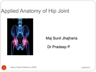
Applied Anatomy of Hip Joint
2/22/20151 Dept of Sports Medicine, AFMC
Maj Sunil Jhajharia
Dr Pradeep P
Applied Anatomy of Hip Joint
 Introduction
 Bones
 Ligaments
 Muscles & Movement
 Blood and Nerve
Supply
 Applied Radiology
 Applied Anatomy
2/22/2015Dept of Sports Medicine, AFMC2
Introduction
 It is the largest joint of
the human body
 It is the 2nd largest
weight bearing joint of
the human body
 Type:- Ball & Socket
variety of Synovial
joint
2/22/20153 Dept of Sports Medicine, AFMC
Head of Femur
 Globular, more than a
hemisphere
 Directed upward,
medially, and a little
forward
 Fovea capatis
femoris:- an ovoid
depression
2/22/2015Dept of Sports Medicine, AFMC4
Head of Femur
 Covered with hyaline
cartilage, except over
fovea capitis femoris
 The fovea capatis
gives attachment to
the ligament of the
head of femur
2/22/2015Dept of Sports Medicine, AFMC5
Acetabulum
 Horseshoe shaped
 Lunate articular
surface
 Acetabular fossa
 Acetabular notch
 Acetabular labrum
2/22/2015Dept of Sports Medicine, AFMC6
Acetabulum
2/22/2015Dept of Sports Medicine, AFMC7
Horse-shoe shaped
articular surface
Deepened by
fibro-cartilaginous
rim called
acetabular
labrum
Nonarticular
part, acetabular
fossa, lodges
pad of fat
Deficient
inferiorly as the
acetabular
notch that is
bridged up by
transverse
acetabular
ligament
Articulation
 Hemispherical head of
Femur
 Horseshoe-shaped
acetabulum of the hip
bone
 Articular surfaces
covered with hyaline
cartilage
2/22/20158 Dept of Sports Medicine, AFMC
Stability
 Unique in having a high degree of stability as well
as mobility
 Stability depends upon:
 Depth of Acetabulum and the narrowing of its mouth
by the Acetabular labrum
 Tension and strength of ligaments
 Strength of surrounding muscles
 Length and obliquity of the neck of the Femur
2/22/20159 Dept of Sports Medicine, AFMC
Ligaments
 Fibrous capsule
 Iliofemoral ligament
 Pubofemoral ligament
 Ischiofemoral
ligament
 Ligament of the head
of the Femur
 Acetabular labrum
 Transverse acetabular
ligament
2/22/2015Dept of Sports Medicine, AFMC10
Joint capsule
 Encloses the joint
 Anterosuperiorly-
 Thick & firmly
attached
 Posteroinferiorly-
 Thin & loosely
attached
 Two types of fibers
 Outer-longitudinal
 Inner-circular-zona
orbicularis
2/22/2015Dept of Sports Medicine, AFMC11
Iliofemoral ligament
 Strong, Y-shaped
 Covers the joint
anteriorly
 Prevents over-extension
during standing posture
 Attachment:
• Base to anterior inferior
iliac spine
• Two limbs to upper &
lower ends of
intertrochanteric line
2/22/201512 Dept of Sports Medicine, AFMC
Pubofemoral ligament
 Support the joint
inferomedially
 Triangular in shape
 Attachment:-
 Superiorly, attached to the
iliopubic eminence,the
obturator creast
 Inferiorly, merges with the
capsule and lower band of
iliofemoral ligament
 It limits extension &
abduction
2/22/201513 Dept of Sports Medicine, AFMC
Ischiofemoral Ligament
 Comparatively weak
and twisted & spiral
in shape
 Supports the joint
posteriorly
 Extend from the
ischium to the
acetabulum
 Limits the extension
of joint
2/22/2015Dept of Sports Medicine, AFMC14
Ligament of head of the Femur
 Round ligament or
ligamentum teres
 Flat and triangular in
shape
 Attachment:-
 Apex to the fovea
capatis
 Base to the
transverse ligament
and margins of the
acetabular notch
2/22/2015Dept of Sports Medicine, AFMC15
Ligament of head of the Femur
 It transmits arteries to
the head of Femur,
from the acetabular
branches of the
obturator and medial
circumflex femoral
arteries
2/22/2015Dept of Sports Medicine, AFMC16
Acetabular labrum
 Fibrocartilaginous rim
attached to the
margins of the
acetabulum
 Narrows the mouth of
the acetabulum
 Helps in holding the
head of femur in
position
2/22/2015Dept of Sports Medicine, AFMC17
Transverse acetabular ligament
 Formed by
acetabular labrum
 Bridges acetabular
notch, converting it
into tunnel through
which vessels and
nerves enter the
joint
2/22/201518 Dept of Sports Medicine, AFMC
Gluteus maximus
 Origin:-
 lower posterior iliac crest and
posterior surface of the
sacrum
 Insertion:-
 gluteal tuberosity (upper,
posterior aspect of the
femur) & IT band
 Actions:
 Extension of the hip
 External rotation of the hip
 Lower fibers (below the center
of motion) assist in adduction
2/22/2015Dept of Sports Medicine, AFMC19
Gluteus maximus
 Produces hip
extension beyond 15
degrees; not used
extensively during
walking
 Strongly used during
running, hopping,
skipping, and jumping
 Best isolated with the
knee flexed to reduce
hip extension from the
hamstrings
2/22/2015Dept of Sports Medicine, AFMC20
Gluteus medius
 Origin:-
 outer surface of the
ilium just below the
crest
 Insertion:-
 greater trochanter
 Actions:
 Abduction of the hip
 Anterior fibers:
Internal rotation,
 Posterior fibers:
External rotation
2/22/2015Dept of Sports Medicine, AFMC21
Gluteus minimus
 Origin:-
 Outer surface of the
ilium beneath the
gluteus medius
 Insertion:-
 Greater trochanter of
the femur
 Actions:-
 Abduction of the hip
 Internal rotation
2/22/2015Dept of Sports Medicine, AFMC22
Gluteus medius & minimus
 During walking these
muscles abduct (or
hold up) the free leg,
preventing it from
sagging.
 Both are important in
transferring weight
from one leg to the
other (e.g. running,
hopping, skipping,
etc.)
 Their effectiveness
decreases with age.
2/22/2015Dept of Sports Medicine, AFMC23
Tensor fasciae latae
 Origin:- iliac crest
 Insertion:- Iliotibial
band
 Actions:-
 Flexion of the hip
 Internal rotation
 Abduction of the hip
2/22/2015Dept of Sports Medicine, AFMC24
Iliopsoas
 Iliacus:-
• takes origin from upper 2/3rd
of iliac fossa
• Inner lip of the Iliac creast
• Upper surface of lateral part
of the Sacrum
• Psoas major:-
 From aterior surface and
lower borders of transverse
process of all lumber
vertebrae
2/22/2015Dept of Sports Medicine, AFMC25
Iliopsoas
 Insertion:-
 Iliacus and Psoas
Major both are
inserted into Lesser
trochanter of the
femur
 Actions:-
• Chief flexor of Hip
joint
• External rotation
2/22/2015Dept of Sports Medicine, AFMC26
Biceps femoris
 Origin:
 Long head:- ischial
tuberosity
 Short head:- lower half
of the linea aspera
 Insertion:-
 Head of the fibula
 Action:
 Extension of hip
 External rotation of the hip
(and knee)
 (Flexion of knee)
2/22/2015Dept of Sports Medicine, AFMC27
Semitendinosus
 Origin: Ischial
tuberosity
 Insertion: Medial
surface of proximal
end of the tibia
 Action:
 Extension of the
hip
 Internal rotation of
the hip (and knee)
 Flexion of the knee
2/22/2015Dept of Sports Medicine, AFMC28
Semimembranosus
 Origin: Ischial
tuberosity
 Insertion: Medial
surface of the tibia
 Action:-
 Flexion of the knee
 Extension of the hip
 Internal rotation of the
hip
2/22/2015Dept of Sports Medicine, AFMC29
2/22/2015Dept of Sports Medicine, AFMC30
Hamstrings
Rectus femoris
 Two joint muscle; most
superficial
 Origin: anterior-inferior
iliac spine of the ilium
 Insertion: top of the
patella and patellar
ligament to the tibial
tuberosity
 Actions:
 Flexion of the hip
 Extension of the knee
2/22/2015Dept of Sports Medicine, AFMC31
Sartorius
 Origin:-
 Anterior-Superior Iliac Spine
 Insertion:-
 Upper part of the medial
surface of shaft of Tibia
 Action:-
 Abduction and Lateral
rotation of the hip
 Flexion of the leg at knee
joint
2/22/2015Dept of Sports Medicine, AFMC32
Gracilis
 Origin:-
 Pubic crest
 Insertion:-
 Medial condyle of
tibia
 Actions:-
 Adduction at the hip
 Internal rotation
 Flexion (weak)
2/22/2015Dept of Sports Medicine, AFMC33
Pectineous
 Origin:
 pubic crest or ramus
 Insertion:-
 below the linea
aspera
 Actions
 Flexion
 Adduction
 External rotation
2/22/2015Dept of Sports Medicine, AFMC34
Adductor Brevis
 Origin:-
 Inferior ramus of
pubis
 Insertion:
 Pectineal line (linea
aspera)
 Actions:
 Adduction
 External rotation
 Flexion (weak)
2/22/2015Dept of Sports Medicine, AFMC35
Adductor Longus
 Below the adductor brevis
 Origin:-
 front of the pubis just
below its crest
 Insertion:-
 middle third of the linea
aspera
 Actions:-
 Adduction
 Flexion (weak)
2/22/2015Dept of Sports Medicine, AFMC36
Adductor Magnus
 Located posterior to the longus
 Origin:
 edge of the pubic crest and
ischial tuberosity
 Insertion:
 linea aspera
 Actions:
 Adduction
 External rotation
 Extension
2/22/2015Dept of Sports Medicine, AFMC37
Blood Supply
 Obturator artery
 Medial and lateral
circumflex femoral
Arteries
 Gluteal arteries
2/22/2015Dept of Sports Medicine, AFMC38
Nerve Supply
 Femoral nerve
 Superior Gluteal
nerve
 Anterior division of
Obturator nerve
 Nerve to Rectus
femoris
 Nerve to Quadratus
femoris
2/22/2015Dept of Sports Medicine, AFMC39
X RAY PELVIS
2/22/2015Dept of Sports Medicine, AFMC40
 Two standard views are taken to visualize hip
joint
 1. AP view
 2.lateral view
X RAY PELVIS
2/22/2015Dept of Sports Medicine, AFMC41
 Most trauma to the pelvis and hips can be
evaluated with an AP view of the pelvis and hips.
 Symptoms from fractures of the hip, acetabulum
and pelvis may be quite similar, thus, a full AP
pelvis radiograph including the hip must be
obtained if any of the above fractures are
expected.
 The femurs should be internally rotated when
obtaining an AP pelvis film so that the femoral
necks can be appropriately assessed for
fractures.
X RAY PELVIS
2/22/2015Dept of Sports Medicine, AFMC42
X Ray Of PELVIS
2/22/2015Dept of Sports Medicine, AFMC43
 Hip X-ray anatomy - Normal AP
 The five bones that comprise the pelvis are the
ilium, ischium, pubis, sacrum, coccyx, acetabulum
and head and neck of femur.
 Shenton's line is formed by the medial edge of
the femoral neck and the inferior edge of the
superior pubic ramus
 Loss of contour of Shenton's line is a sign of a
fractured neck of femur
Lateral X Ray Of Hip Joint
2/22/2015Dept of Sports Medicine, AFMC44
 Lateral x ray of hip joint is not routinely taken
 The Lateral view is often not so clear because
those with hip pain find the positioning required
difficult
 Taken to confirm the displacement of fracture
fragment and
 To confirm type of dislocation
Lateral X Ray Of Hip Joint
2/22/2015Dept of Sports Medicine, AFMC45
Arthritis Of Hip Joint
2/22/2015Dept of Sports Medicine, AFMC46
Avulsion Fracture Of ASIS
2/22/2015Dept of Sports Medicine, AFMC47
USG
2/22/2015Dept of Sports Medicine, AFMC48
 Usg is safe and painless procedure
 Is used to evaluate abnormalities of muscle, fluid
collection, benign and malignant tumors
 To visualize each structure position of probe and
patient differs
2/22/2015Dept of Sports Medicine, AFMC49
USG
2/22/2015Dept of Sports Medicine, AFMC50
STRUCTURES VISUALIZED
MUSCLE AND FAT HYPOECHOIC
TENDON HYPERECHOIC
LIGAMENTS HYPERECHOIC
BONE (CORTEX) HYPERECHOIC
USG
2/22/2015Dept of Sports Medicine, AFMC51
 Ultrasound of the hip is divided into anterior,
medial, lateral, and posterior approaches.
USG
2/22/2015Dept of Sports Medicine, AFMC52
 To visualize iliopsoas tendon:
Patient : supine position
Transducer : longitudinal or vertical
over the joint space
USG
2/22/2015Dept of Sports Medicine, AFMC53
To visualize Rectus femoris :
Patient : supine
Transducer : longitudinally or transversely
over AIIS
USG – RECTUS FEMORIS
2/22/2015Dept of Sports Medicine, AFMC54
USG – ADDUCTOR MUSCLE
2/22/2015Dept of Sports Medicine, AFMC55
 To visualize adductors muscle
Patient : thigh abducted and externally rotated
the knee joint
Transducer : longitudinally over medial aspect
of thigh
 Three layer are recognized on axial plane
superficial – Adductor longus and Gracilis
intermediate – Adductor brevis
deep – Adductor magnus
USG – ADDUCTOR MUSCLE
2/22/2015Dept of Sports Medicine, AFMC56
USG – ABDUCTOR MUSCLE
2/22/2015Dept of Sports Medicine, AFMC57
 To visualize abductors muscle
Patient lie on the opposite hip assuming an
oblique lateral or true lateral position
Transducer : Transverse and longitudinal US
planes obtained cranial to the greater trochanter
show the gluteus medius (superficial) and gluteus
minimus (deep) muscles.
USG – ABDUCTOR MUSCLE
2/22/2015Dept of Sports Medicine, AFMC58
USG - HAMSTRING
2/22/2015Dept of Sports Medicine, AFMC59
 To visualize hamstring muscles
The patient lies prone with the feet hanging out
of the bed.
Proximal origin of the semimembranosus,
semitendinosus and long head of the biceps
femoris muscles is visualized
 The ischial tuberosity is the main landmark
USG - HAMSTRING
2/22/2015Dept of Sports Medicine, AFMC60
USG - HAMSTRING
2/22/2015Dept of Sports Medicine, AFMC61
USG
2/22/2015Dept of Sports Medicine, AFMC62
Mri Of Hip Joint
2/22/2015Dept of Sports Medicine, AFMC63
MRI OF KNEE JOINT
2/22/2015Dept of Sports Medicine, AFMC64
STRUCTURE T1W1 T2W1
Bone, Tendon
and Muscles
Dark Dark
Fat Bright Less bright
Fluid Dark Bright
MRI OF KNEE JOINT
2/22/2015Dept of Sports Medicine, AFMC65
Three standard views are
1.Coranal views
2.Axial views
3.Saggital
Coronal View
2/22/2015Dept of Sports Medicine, AFMC66
1.Iliac
2.Iliac Muscle
3.Gluteus med
4.Tensor fascia
lata
5.Pectineus
6.Urinary
bladder
7.Sym pubis
8. Gluteus mini
Axial View
2/22/2015Dept of Sports Medicine, AFMC67
1.Gluteus medius
2.Rectus femoris
3.Femoral vessels
4.Urinary bladder
5.Ilio psoas
6.Sartorius
7.TFL
8.Femoral head
9.Obturator
internus and
10. Gluteus maxi
Applied Anatomy
2/22/2015Dept of Sports Medicine, AFMC68
Avulsion Injuries Of Pelvis
2/22/2015Dept of Sports Medicine, AFMC69
 Avulsion injuries commonly seen in the
skeletally immature patient
 Avulsion injuries in adults involve tendinous
origins.
 The most common site of avulsion fractures in
the skeletally immature athlete are ischial
tuberosity, AIIS, and ASIS avulsions
 Most injuries occur from an eccentric muscle
contraction.
 Most injuries may be managed
nonoperatively.
Avulsion Injuries Of AIIS
2/22/2015Dept of Sports Medicine, AFMC70
Snapping Hip Syndrome
2/22/2015Dept of Sports Medicine, AFMC71
 Snapping hip syndrome, sometimes called
dancer's hip
 In most cases, snapping is caused by the
movement of a muscle or tendon over a bony
structure in the hip.
 The most common site is
1.when the iliotibial band passes over the
greater trochanter.
2.Iliopsoas tendon moves over the Iliopectineal
eminence
 Commonly seen in sports like
1.Atheletes
2/22/2015Dept of Sports Medicine, AFMC72
Snapping Hip Syndrome
2/22/2015Dept of Sports Medicine, AFMC73
 Fabers test positive
 MRI and ultrasound can confirm the diagnosis.
 Treatment : Avoidance of precipitating activities,
particularly hip flexion greater than 90°,
 NSAIDs and physiotherapy are the mainstay of
treatment
Adductor Muscle Strain
2/22/2015Dept of Sports Medicine, AFMC74
 Commonly seen in sports that involve sudden change
in direction
 1.Football
 2.Rugby
 3.Batminton etc..
 Treatment : 0-48 hrs
1.RICE
2.Active pain free ROM
• More than 48hrs
1.Strengthning exercise and
2. Sports specific skills
Iliopsoas Strain
2/22/2015Dept of Sports Medicine, AFMC75
 Iliopsoas is the strongest flexor of hip joint
 Iliopsoas problem occurs due to over use of hip
flexion
 Commonly injured in sports like
1.Football
2.Sprinters
Treatment :1. Avoid the aggravating
Factor
2. Stretching and strengthening of
Iliopsoas
Trochantric Bursitis
2/22/2015Dept of Sports Medicine, AFMC76
Bursa around Hip
Joint
Trochantric Bursitis
2/22/2015Dept of Sports Medicine, AFMC77
 Commonly seen in long distance runners
 c/o lateral hip pain , aggrevated by hip movments
 Releived after rest
 Pain can be reproduced by stretching
of Gluteus medius
Treatment :1. Avoid the aggravating
Factor
2. Stretching and strengthening of
Gluteus medius
Trochantric Bursitis
2/22/2015Dept of Sports Medicine, AFMC78
Conclusion
2/22/2015Dept of Sports Medicine, AFMC79
 It is essential to understand the basic anatomy of
hip joint
 Understanding the anatomy will be helpful to
come to final diagnosis and we can start early
treatment without depending upon the any
radiological investigation
 Which can also prevent the patient from any kind
of radiological exposure
References
22 February 2015Dept of Sports Medicine80
 Grays anatomy 40th edition
 Clinical sports medicine by Peter brukner and Karim
kahn 3 edition
 Current diagnosis and treatment by Patrick mc Mahon
1st edition
 http://www.rad.washington.edu/academics/academic-
sections/msk/teaching-materials/radiology-anatomy-
teaching-modules/basic-knee-anatomy
 Musculoskeletal Ultrasound Anatomy and Technique
by John O’Neill, MD
 Ultasound of Muscle Sports injuries by Jarret MD
Radiology
Thank you
2/22/2015Dept of Sports Medicine, AFMC81

More Related Content

What's hot (20)

Vertebral Column
Vertebral ColumnVertebral Column
Vertebral Column
 
The patella
The patellaThe patella
The patella
 
The shoulder joint
The shoulder jointThe shoulder joint
The shoulder joint
 
Hip joint anatomy
Hip joint anatomyHip joint anatomy
Hip joint anatomy
 
Knee Joint Anatomy
Knee Joint AnatomyKnee Joint Anatomy
Knee Joint Anatomy
 
Dr. kamal kant applied anatomy of hip
Dr. kamal kant   applied anatomy of hipDr. kamal kant   applied anatomy of hip
Dr. kamal kant applied anatomy of hip
 
Shoulder joint, sterno clavicular joint, acromio-clavicular joint (2)
Shoulder joint,  sterno clavicular joint, acromio-clavicular joint (2)Shoulder joint,  sterno clavicular joint, acromio-clavicular joint (2)
Shoulder joint, sterno clavicular joint, acromio-clavicular joint (2)
 
Radioulnar joints
Radioulnar jointsRadioulnar joints
Radioulnar joints
 
The clavicle
The clavicleThe clavicle
The clavicle
 
Knee joint
Knee jointKnee joint
Knee joint
 
Anatomy of Humerus
Anatomy of HumerusAnatomy of Humerus
Anatomy of Humerus
 
Cubital fossa
Cubital fossaCubital fossa
Cubital fossa
 
Sacrum
SacrumSacrum
Sacrum
 
Arches of foot
Arches of footArches of foot
Arches of foot
 
5.posterior compartment of the thigh
5.posterior compartment of the thigh5.posterior compartment of the thigh
5.posterior compartment of the thigh
 
Slideshow: Tibia
Slideshow: TibiaSlideshow: Tibia
Slideshow: Tibia
 
Popliteal fossa
Popliteal fossa   Popliteal fossa
Popliteal fossa
 
Anatomy of ankle joint
Anatomy of ankle jointAnatomy of ankle joint
Anatomy of ankle joint
 
3.medial side of the thigh
3.medial side of the thigh3.medial side of the thigh
3.medial side of the thigh
 
Elbow joint
Elbow jointElbow joint
Elbow joint
 

Viewers also liked

Viewers also liked (10)

hip joint
hip jointhip joint
hip joint
 
Anatomy of Hip joint
Anatomy of Hip joint Anatomy of Hip joint
Anatomy of Hip joint
 
Hydrotherapy by pradeep
Hydrotherapy by pradeepHydrotherapy by pradeep
Hydrotherapy by pradeep
 
Respiratory response to exercise
Respiratory response to exerciseRespiratory response to exercise
Respiratory response to exercise
 
Cryotherapy
CryotherapyCryotherapy
Cryotherapy
 
The Hips
The HipsThe Hips
The Hips
 
Hip joint
Hip jointHip joint
Hip joint
 
Hip joint
Hip jointHip joint
Hip joint
 
the gross antomy of the hip hoint and applied anatomy focused for undergradua...
the gross antomy of the hip hoint and applied anatomy focused for undergradua...the gross antomy of the hip hoint and applied anatomy focused for undergradua...
the gross antomy of the hip hoint and applied anatomy focused for undergradua...
 
Hip joint anatomy and biomechanics
Hip joint anatomy and biomechanicsHip joint anatomy and biomechanics
Hip joint anatomy and biomechanics
 

Similar to Applied Anatomy of Hip Joint

Kin191 A.Ch.6.Knee.Patellofemoral.Anatomy
Kin191 A.Ch.6.Knee.Patellofemoral.AnatomyKin191 A.Ch.6.Knee.Patellofemoral.Anatomy
Kin191 A.Ch.6.Knee.Patellofemoral.AnatomyJLS10
 
OSTEOLOGY OF THE HIP JOINT presentattion.pptx
OSTEOLOGY OF THE HIP JOINT presentattion.pptxOSTEOLOGY OF THE HIP JOINT presentattion.pptx
OSTEOLOGY OF THE HIP JOINT presentattion.pptxOluseyi7
 
Anatomy and biomechanics of hip joint
Anatomy and biomechanics of hip joint Anatomy and biomechanics of hip joint
Anatomy and biomechanics of hip joint BipulBorthakur
 
Anatomy and biomechanics of hip joint [autosaved]
Anatomy and biomechanics of hip joint [autosaved]Anatomy and biomechanics of hip joint [autosaved]
Anatomy and biomechanics of hip joint [autosaved]ujjalrajbangshi
 
The Pelvis and Hip: Function and Anatomy
The Pelvis and Hip: Function and Anatomy The Pelvis and Hip: Function and Anatomy
The Pelvis and Hip: Function and Anatomy Jill Costley
 
Vertebral column anatomy . radiology training resource ncchanji nkeh keneth
Vertebral column anatomy . radiology training resource ncchanji nkeh kenethVertebral column anatomy . radiology training resource ncchanji nkeh keneth
Vertebral column anatomy . radiology training resource ncchanji nkeh kenethNchanji Nkeh Keneth
 
TOTAL KNEE REPLACEMENT
TOTAL KNEE REPLACEMENTTOTAL KNEE REPLACEMENT
TOTAL KNEE REPLACEMENTManoj Kumar R
 
fractureofneckofthefemur-121016113941-phpapp02.pdf
fractureofneckofthefemur-121016113941-phpapp02.pdffractureofneckofthefemur-121016113941-phpapp02.pdf
fractureofneckofthefemur-121016113941-phpapp02.pdfSbusisomtungwa
 
The knee and related structures f09
The knee and related structures f09The knee and related structures f09
The knee and related structures f09guest0dae325
 
The knee and related structures f09
The knee and related structures f09The knee and related structures f09
The knee and related structures f09guest6e3b98
 
Fracture neck of femur
Fracture neck of  femurFracture neck of  femur
Fracture neck of femurPrateek Singh
 
Surgical anatomy of knee joint
Surgical anatomy of knee jointSurgical anatomy of knee joint
Surgical anatomy of knee jointTausifahmed786
 
Soft tissue injury of the knee
Soft tissue injury of the kneeSoft tissue injury of the knee
Soft tissue injury of the kneeMONTHER ALKHAWLANY
 

Similar to Applied Anatomy of Hip Joint (20)

Kin191 A.Ch.6.Knee.Patellofemoral.Anatomy
Kin191 A.Ch.6.Knee.Patellofemoral.AnatomyKin191 A.Ch.6.Knee.Patellofemoral.Anatomy
Kin191 A.Ch.6.Knee.Patellofemoral.Anatomy
 
OSTEOLOGY OF THE HIP JOINT presentattion.pptx
OSTEOLOGY OF THE HIP JOINT presentattion.pptxOSTEOLOGY OF THE HIP JOINT presentattion.pptx
OSTEOLOGY OF THE HIP JOINT presentattion.pptx
 
Knee biomechanic
Knee biomechanicKnee biomechanic
Knee biomechanic
 
Anatomy and biomechanics of hip joint
Anatomy and biomechanics of hip joint Anatomy and biomechanics of hip joint
Anatomy and biomechanics of hip joint
 
Anatomy and biomechanics of hip joint [autosaved]
Anatomy and biomechanics of hip joint [autosaved]Anatomy and biomechanics of hip joint [autosaved]
Anatomy and biomechanics of hip joint [autosaved]
 
The Pelvis and Hip: Function and Anatomy
The Pelvis and Hip: Function and Anatomy The Pelvis and Hip: Function and Anatomy
The Pelvis and Hip: Function and Anatomy
 
Knee complex
Knee complexKnee complex
Knee complex
 
Vertebral column anatomy . radiology training resource ncchanji nkeh keneth
Vertebral column anatomy . radiology training resource ncchanji nkeh kenethVertebral column anatomy . radiology training resource ncchanji nkeh keneth
Vertebral column anatomy . radiology training resource ncchanji nkeh keneth
 
TOTAL KNEE REPLACEMENT
TOTAL KNEE REPLACEMENTTOTAL KNEE REPLACEMENT
TOTAL KNEE REPLACEMENT
 
fractureofneckofthefemur-121016113941-phpapp02.pdf
fractureofneckofthefemur-121016113941-phpapp02.pdffractureofneckofthefemur-121016113941-phpapp02.pdf
fractureofneckofthefemur-121016113941-phpapp02.pdf
 
Clinical Examination of Hip
Clinical Examination of HipClinical Examination of Hip
Clinical Examination of Hip
 
The knee and related structures f09
The knee and related structures f09The knee and related structures f09
The knee and related structures f09
 
The knee and related structures f09
The knee and related structures f09The knee and related structures f09
The knee and related structures f09
 
Fracture neck of femur
Fracture neck of  femurFracture neck of  femur
Fracture neck of femur
 
Hip pain treatment
Hip pain treatmentHip pain treatment
Hip pain treatment
 
The knee joint
The knee jointThe knee joint
The knee joint
 
Surgical anatomy of knee joint
Surgical anatomy of knee jointSurgical anatomy of knee joint
Surgical anatomy of knee joint
 
Soft tissue injury of the knee
Soft tissue injury of the kneeSoft tissue injury of the knee
Soft tissue injury of the knee
 
Ch8 Le 5
Ch8 Le 5Ch8 Le 5
Ch8 Le 5
 
PS SESSION : EXAMINATION OF HIP
PS SESSION : EXAMINATION OF HIPPS SESSION : EXAMINATION OF HIP
PS SESSION : EXAMINATION OF HIP
 

Recently uploaded

Crayon Activity Handout For the Crayon A
Crayon Activity Handout For the Crayon ACrayon Activity Handout For the Crayon A
Crayon Activity Handout For the Crayon AUnboundStockton
 
call girls in Kamla Market (DELHI) 🔝 >༒9953330565🔝 genuine Escort Service 🔝✔️✔️
call girls in Kamla Market (DELHI) 🔝 >༒9953330565🔝 genuine Escort Service 🔝✔️✔️call girls in Kamla Market (DELHI) 🔝 >༒9953330565🔝 genuine Escort Service 🔝✔️✔️
call girls in Kamla Market (DELHI) 🔝 >༒9953330565🔝 genuine Escort Service 🔝✔️✔️9953056974 Low Rate Call Girls In Saket, Delhi NCR
 
microwave assisted reaction. General introduction
microwave assisted reaction. General introductionmicrowave assisted reaction. General introduction
microwave assisted reaction. General introductionMaksud Ahmed
 
Separation of Lanthanides/ Lanthanides and Actinides
Separation of Lanthanides/ Lanthanides and ActinidesSeparation of Lanthanides/ Lanthanides and Actinides
Separation of Lanthanides/ Lanthanides and ActinidesFatimaKhan178732
 
Employee wellbeing at the workplace.pptx
Employee wellbeing at the workplace.pptxEmployee wellbeing at the workplace.pptx
Employee wellbeing at the workplace.pptxNirmalaLoungPoorunde1
 
Mastering the Unannounced Regulatory Inspection
Mastering the Unannounced Regulatory InspectionMastering the Unannounced Regulatory Inspection
Mastering the Unannounced Regulatory InspectionSafetyChain Software
 
Concept of Vouching. B.Com(Hons) /B.Compdf
Concept of Vouching. B.Com(Hons) /B.CompdfConcept of Vouching. B.Com(Hons) /B.Compdf
Concept of Vouching. B.Com(Hons) /B.CompdfUmakantAnnand
 
BASLIQ CURRENT LOOKBOOK LOOKBOOK(1) (1).pdf
BASLIQ CURRENT LOOKBOOK  LOOKBOOK(1) (1).pdfBASLIQ CURRENT LOOKBOOK  LOOKBOOK(1) (1).pdf
BASLIQ CURRENT LOOKBOOK LOOKBOOK(1) (1).pdfSoniaTolstoy
 
18-04-UA_REPORT_MEDIALITERAСY_INDEX-DM_23-1-final-eng.pdf
18-04-UA_REPORT_MEDIALITERAСY_INDEX-DM_23-1-final-eng.pdf18-04-UA_REPORT_MEDIALITERAСY_INDEX-DM_23-1-final-eng.pdf
18-04-UA_REPORT_MEDIALITERAСY_INDEX-DM_23-1-final-eng.pdfssuser54595a
 
Contemporary philippine arts from the regions_PPT_Module_12 [Autosaved] (1).pptx
Contemporary philippine arts from the regions_PPT_Module_12 [Autosaved] (1).pptxContemporary philippine arts from the regions_PPT_Module_12 [Autosaved] (1).pptx
Contemporary philippine arts from the regions_PPT_Module_12 [Autosaved] (1).pptxRoyAbrique
 
“Oh GOSH! Reflecting on Hackteria's Collaborative Practices in a Global Do-It...
“Oh GOSH! Reflecting on Hackteria's Collaborative Practices in a Global Do-It...“Oh GOSH! Reflecting on Hackteria's Collaborative Practices in a Global Do-It...
“Oh GOSH! Reflecting on Hackteria's Collaborative Practices in a Global Do-It...Marc Dusseiller Dusjagr
 
POINT- BIOCHEMISTRY SEM 2 ENZYMES UNIT 5.pptx
POINT- BIOCHEMISTRY SEM 2 ENZYMES UNIT 5.pptxPOINT- BIOCHEMISTRY SEM 2 ENZYMES UNIT 5.pptx
POINT- BIOCHEMISTRY SEM 2 ENZYMES UNIT 5.pptxSayali Powar
 
_Math 4-Q4 Week 5.pptx Steps in Collecting Data
_Math 4-Q4 Week 5.pptx Steps in Collecting Data_Math 4-Q4 Week 5.pptx Steps in Collecting Data
_Math 4-Q4 Week 5.pptx Steps in Collecting DataJhengPantaleon
 
Organic Name Reactions for the students and aspirants of Chemistry12th.pptx
Organic Name Reactions  for the students and aspirants of Chemistry12th.pptxOrganic Name Reactions  for the students and aspirants of Chemistry12th.pptx
Organic Name Reactions for the students and aspirants of Chemistry12th.pptxVS Mahajan Coaching Centre
 
Presentation by Andreas Schleicher Tackling the School Absenteeism Crisis 30 ...
Presentation by Andreas Schleicher Tackling the School Absenteeism Crisis 30 ...Presentation by Andreas Schleicher Tackling the School Absenteeism Crisis 30 ...
Presentation by Andreas Schleicher Tackling the School Absenteeism Crisis 30 ...EduSkills OECD
 
Introduction to ArtificiaI Intelligence in Higher Education
Introduction to ArtificiaI Intelligence in Higher EducationIntroduction to ArtificiaI Intelligence in Higher Education
Introduction to ArtificiaI Intelligence in Higher Educationpboyjonauth
 
Alper Gobel In Media Res Media Component
Alper Gobel In Media Res Media ComponentAlper Gobel In Media Res Media Component
Alper Gobel In Media Res Media ComponentInMediaRes1
 

Recently uploaded (20)

Crayon Activity Handout For the Crayon A
Crayon Activity Handout For the Crayon ACrayon Activity Handout For the Crayon A
Crayon Activity Handout For the Crayon A
 
call girls in Kamla Market (DELHI) 🔝 >༒9953330565🔝 genuine Escort Service 🔝✔️✔️
call girls in Kamla Market (DELHI) 🔝 >༒9953330565🔝 genuine Escort Service 🔝✔️✔️call girls in Kamla Market (DELHI) 🔝 >༒9953330565🔝 genuine Escort Service 🔝✔️✔️
call girls in Kamla Market (DELHI) 🔝 >༒9953330565🔝 genuine Escort Service 🔝✔️✔️
 
microwave assisted reaction. General introduction
microwave assisted reaction. General introductionmicrowave assisted reaction. General introduction
microwave assisted reaction. General introduction
 
Separation of Lanthanides/ Lanthanides and Actinides
Separation of Lanthanides/ Lanthanides and ActinidesSeparation of Lanthanides/ Lanthanides and Actinides
Separation of Lanthanides/ Lanthanides and Actinides
 
Employee wellbeing at the workplace.pptx
Employee wellbeing at the workplace.pptxEmployee wellbeing at the workplace.pptx
Employee wellbeing at the workplace.pptx
 
Mastering the Unannounced Regulatory Inspection
Mastering the Unannounced Regulatory InspectionMastering the Unannounced Regulatory Inspection
Mastering the Unannounced Regulatory Inspection
 
Concept of Vouching. B.Com(Hons) /B.Compdf
Concept of Vouching. B.Com(Hons) /B.CompdfConcept of Vouching. B.Com(Hons) /B.Compdf
Concept of Vouching. B.Com(Hons) /B.Compdf
 
BASLIQ CURRENT LOOKBOOK LOOKBOOK(1) (1).pdf
BASLIQ CURRENT LOOKBOOK  LOOKBOOK(1) (1).pdfBASLIQ CURRENT LOOKBOOK  LOOKBOOK(1) (1).pdf
BASLIQ CURRENT LOOKBOOK LOOKBOOK(1) (1).pdf
 
18-04-UA_REPORT_MEDIALITERAСY_INDEX-DM_23-1-final-eng.pdf
18-04-UA_REPORT_MEDIALITERAСY_INDEX-DM_23-1-final-eng.pdf18-04-UA_REPORT_MEDIALITERAСY_INDEX-DM_23-1-final-eng.pdf
18-04-UA_REPORT_MEDIALITERAСY_INDEX-DM_23-1-final-eng.pdf
 
Contemporary philippine arts from the regions_PPT_Module_12 [Autosaved] (1).pptx
Contemporary philippine arts from the regions_PPT_Module_12 [Autosaved] (1).pptxContemporary philippine arts from the regions_PPT_Module_12 [Autosaved] (1).pptx
Contemporary philippine arts from the regions_PPT_Module_12 [Autosaved] (1).pptx
 
“Oh GOSH! Reflecting on Hackteria's Collaborative Practices in a Global Do-It...
“Oh GOSH! Reflecting on Hackteria's Collaborative Practices in a Global Do-It...“Oh GOSH! Reflecting on Hackteria's Collaborative Practices in a Global Do-It...
“Oh GOSH! Reflecting on Hackteria's Collaborative Practices in a Global Do-It...
 
POINT- BIOCHEMISTRY SEM 2 ENZYMES UNIT 5.pptx
POINT- BIOCHEMISTRY SEM 2 ENZYMES UNIT 5.pptxPOINT- BIOCHEMISTRY SEM 2 ENZYMES UNIT 5.pptx
POINT- BIOCHEMISTRY SEM 2 ENZYMES UNIT 5.pptx
 
Código Creativo y Arte de Software | Unidad 1
Código Creativo y Arte de Software | Unidad 1Código Creativo y Arte de Software | Unidad 1
Código Creativo y Arte de Software | Unidad 1
 
Model Call Girl in Tilak Nagar Delhi reach out to us at 🔝9953056974🔝
Model Call Girl in Tilak Nagar Delhi reach out to us at 🔝9953056974🔝Model Call Girl in Tilak Nagar Delhi reach out to us at 🔝9953056974🔝
Model Call Girl in Tilak Nagar Delhi reach out to us at 🔝9953056974🔝
 
_Math 4-Q4 Week 5.pptx Steps in Collecting Data
_Math 4-Q4 Week 5.pptx Steps in Collecting Data_Math 4-Q4 Week 5.pptx Steps in Collecting Data
_Math 4-Q4 Week 5.pptx Steps in Collecting Data
 
Organic Name Reactions for the students and aspirants of Chemistry12th.pptx
Organic Name Reactions  for the students and aspirants of Chemistry12th.pptxOrganic Name Reactions  for the students and aspirants of Chemistry12th.pptx
Organic Name Reactions for the students and aspirants of Chemistry12th.pptx
 
Presentation by Andreas Schleicher Tackling the School Absenteeism Crisis 30 ...
Presentation by Andreas Schleicher Tackling the School Absenteeism Crisis 30 ...Presentation by Andreas Schleicher Tackling the School Absenteeism Crisis 30 ...
Presentation by Andreas Schleicher Tackling the School Absenteeism Crisis 30 ...
 
Introduction to ArtificiaI Intelligence in Higher Education
Introduction to ArtificiaI Intelligence in Higher EducationIntroduction to ArtificiaI Intelligence in Higher Education
Introduction to ArtificiaI Intelligence in Higher Education
 
Model Call Girl in Bikash Puri Delhi reach out to us at 🔝9953056974🔝
Model Call Girl in Bikash Puri  Delhi reach out to us at 🔝9953056974🔝Model Call Girl in Bikash Puri  Delhi reach out to us at 🔝9953056974🔝
Model Call Girl in Bikash Puri Delhi reach out to us at 🔝9953056974🔝
 
Alper Gobel In Media Res Media Component
Alper Gobel In Media Res Media ComponentAlper Gobel In Media Res Media Component
Alper Gobel In Media Res Media Component
 

Applied Anatomy of Hip Joint

  • 1. Applied Anatomy of Hip Joint 2/22/20151 Dept of Sports Medicine, AFMC Maj Sunil Jhajharia Dr Pradeep P
  • 2. Applied Anatomy of Hip Joint  Introduction  Bones  Ligaments  Muscles & Movement  Blood and Nerve Supply  Applied Radiology  Applied Anatomy 2/22/2015Dept of Sports Medicine, AFMC2
  • 3. Introduction  It is the largest joint of the human body  It is the 2nd largest weight bearing joint of the human body  Type:- Ball & Socket variety of Synovial joint 2/22/20153 Dept of Sports Medicine, AFMC
  • 4. Head of Femur  Globular, more than a hemisphere  Directed upward, medially, and a little forward  Fovea capatis femoris:- an ovoid depression 2/22/2015Dept of Sports Medicine, AFMC4
  • 5. Head of Femur  Covered with hyaline cartilage, except over fovea capitis femoris  The fovea capatis gives attachment to the ligament of the head of femur 2/22/2015Dept of Sports Medicine, AFMC5
  • 6. Acetabulum  Horseshoe shaped  Lunate articular surface  Acetabular fossa  Acetabular notch  Acetabular labrum 2/22/2015Dept of Sports Medicine, AFMC6
  • 7. Acetabulum 2/22/2015Dept of Sports Medicine, AFMC7 Horse-shoe shaped articular surface Deepened by fibro-cartilaginous rim called acetabular labrum Nonarticular part, acetabular fossa, lodges pad of fat Deficient inferiorly as the acetabular notch that is bridged up by transverse acetabular ligament
  • 8. Articulation  Hemispherical head of Femur  Horseshoe-shaped acetabulum of the hip bone  Articular surfaces covered with hyaline cartilage 2/22/20158 Dept of Sports Medicine, AFMC
  • 9. Stability  Unique in having a high degree of stability as well as mobility  Stability depends upon:  Depth of Acetabulum and the narrowing of its mouth by the Acetabular labrum  Tension and strength of ligaments  Strength of surrounding muscles  Length and obliquity of the neck of the Femur 2/22/20159 Dept of Sports Medicine, AFMC
  • 10. Ligaments  Fibrous capsule  Iliofemoral ligament  Pubofemoral ligament  Ischiofemoral ligament  Ligament of the head of the Femur  Acetabular labrum  Transverse acetabular ligament 2/22/2015Dept of Sports Medicine, AFMC10
  • 11. Joint capsule  Encloses the joint  Anterosuperiorly-  Thick & firmly attached  Posteroinferiorly-  Thin & loosely attached  Two types of fibers  Outer-longitudinal  Inner-circular-zona orbicularis 2/22/2015Dept of Sports Medicine, AFMC11
  • 12. Iliofemoral ligament  Strong, Y-shaped  Covers the joint anteriorly  Prevents over-extension during standing posture  Attachment: • Base to anterior inferior iliac spine • Two limbs to upper & lower ends of intertrochanteric line 2/22/201512 Dept of Sports Medicine, AFMC
  • 13. Pubofemoral ligament  Support the joint inferomedially  Triangular in shape  Attachment:-  Superiorly, attached to the iliopubic eminence,the obturator creast  Inferiorly, merges with the capsule and lower band of iliofemoral ligament  It limits extension & abduction 2/22/201513 Dept of Sports Medicine, AFMC
  • 14. Ischiofemoral Ligament  Comparatively weak and twisted & spiral in shape  Supports the joint posteriorly  Extend from the ischium to the acetabulum  Limits the extension of joint 2/22/2015Dept of Sports Medicine, AFMC14
  • 15. Ligament of head of the Femur  Round ligament or ligamentum teres  Flat and triangular in shape  Attachment:-  Apex to the fovea capatis  Base to the transverse ligament and margins of the acetabular notch 2/22/2015Dept of Sports Medicine, AFMC15
  • 16. Ligament of head of the Femur  It transmits arteries to the head of Femur, from the acetabular branches of the obturator and medial circumflex femoral arteries 2/22/2015Dept of Sports Medicine, AFMC16
  • 17. Acetabular labrum  Fibrocartilaginous rim attached to the margins of the acetabulum  Narrows the mouth of the acetabulum  Helps in holding the head of femur in position 2/22/2015Dept of Sports Medicine, AFMC17
  • 18. Transverse acetabular ligament  Formed by acetabular labrum  Bridges acetabular notch, converting it into tunnel through which vessels and nerves enter the joint 2/22/201518 Dept of Sports Medicine, AFMC
  • 19. Gluteus maximus  Origin:-  lower posterior iliac crest and posterior surface of the sacrum  Insertion:-  gluteal tuberosity (upper, posterior aspect of the femur) & IT band  Actions:  Extension of the hip  External rotation of the hip  Lower fibers (below the center of motion) assist in adduction 2/22/2015Dept of Sports Medicine, AFMC19
  • 20. Gluteus maximus  Produces hip extension beyond 15 degrees; not used extensively during walking  Strongly used during running, hopping, skipping, and jumping  Best isolated with the knee flexed to reduce hip extension from the hamstrings 2/22/2015Dept of Sports Medicine, AFMC20
  • 21. Gluteus medius  Origin:-  outer surface of the ilium just below the crest  Insertion:-  greater trochanter  Actions:  Abduction of the hip  Anterior fibers: Internal rotation,  Posterior fibers: External rotation 2/22/2015Dept of Sports Medicine, AFMC21
  • 22. Gluteus minimus  Origin:-  Outer surface of the ilium beneath the gluteus medius  Insertion:-  Greater trochanter of the femur  Actions:-  Abduction of the hip  Internal rotation 2/22/2015Dept of Sports Medicine, AFMC22
  • 23. Gluteus medius & minimus  During walking these muscles abduct (or hold up) the free leg, preventing it from sagging.  Both are important in transferring weight from one leg to the other (e.g. running, hopping, skipping, etc.)  Their effectiveness decreases with age. 2/22/2015Dept of Sports Medicine, AFMC23
  • 24. Tensor fasciae latae  Origin:- iliac crest  Insertion:- Iliotibial band  Actions:-  Flexion of the hip  Internal rotation  Abduction of the hip 2/22/2015Dept of Sports Medicine, AFMC24
  • 25. Iliopsoas  Iliacus:- • takes origin from upper 2/3rd of iliac fossa • Inner lip of the Iliac creast • Upper surface of lateral part of the Sacrum • Psoas major:-  From aterior surface and lower borders of transverse process of all lumber vertebrae 2/22/2015Dept of Sports Medicine, AFMC25
  • 26. Iliopsoas  Insertion:-  Iliacus and Psoas Major both are inserted into Lesser trochanter of the femur  Actions:- • Chief flexor of Hip joint • External rotation 2/22/2015Dept of Sports Medicine, AFMC26
  • 27. Biceps femoris  Origin:  Long head:- ischial tuberosity  Short head:- lower half of the linea aspera  Insertion:-  Head of the fibula  Action:  Extension of hip  External rotation of the hip (and knee)  (Flexion of knee) 2/22/2015Dept of Sports Medicine, AFMC27
  • 28. Semitendinosus  Origin: Ischial tuberosity  Insertion: Medial surface of proximal end of the tibia  Action:  Extension of the hip  Internal rotation of the hip (and knee)  Flexion of the knee 2/22/2015Dept of Sports Medicine, AFMC28
  • 29. Semimembranosus  Origin: Ischial tuberosity  Insertion: Medial surface of the tibia  Action:-  Flexion of the knee  Extension of the hip  Internal rotation of the hip 2/22/2015Dept of Sports Medicine, AFMC29
  • 30. 2/22/2015Dept of Sports Medicine, AFMC30 Hamstrings
  • 31. Rectus femoris  Two joint muscle; most superficial  Origin: anterior-inferior iliac spine of the ilium  Insertion: top of the patella and patellar ligament to the tibial tuberosity  Actions:  Flexion of the hip  Extension of the knee 2/22/2015Dept of Sports Medicine, AFMC31
  • 32. Sartorius  Origin:-  Anterior-Superior Iliac Spine  Insertion:-  Upper part of the medial surface of shaft of Tibia  Action:-  Abduction and Lateral rotation of the hip  Flexion of the leg at knee joint 2/22/2015Dept of Sports Medicine, AFMC32
  • 33. Gracilis  Origin:-  Pubic crest  Insertion:-  Medial condyle of tibia  Actions:-  Adduction at the hip  Internal rotation  Flexion (weak) 2/22/2015Dept of Sports Medicine, AFMC33
  • 34. Pectineous  Origin:  pubic crest or ramus  Insertion:-  below the linea aspera  Actions  Flexion  Adduction  External rotation 2/22/2015Dept of Sports Medicine, AFMC34
  • 35. Adductor Brevis  Origin:-  Inferior ramus of pubis  Insertion:  Pectineal line (linea aspera)  Actions:  Adduction  External rotation  Flexion (weak) 2/22/2015Dept of Sports Medicine, AFMC35
  • 36. Adductor Longus  Below the adductor brevis  Origin:-  front of the pubis just below its crest  Insertion:-  middle third of the linea aspera  Actions:-  Adduction  Flexion (weak) 2/22/2015Dept of Sports Medicine, AFMC36
  • 37. Adductor Magnus  Located posterior to the longus  Origin:  edge of the pubic crest and ischial tuberosity  Insertion:  linea aspera  Actions:  Adduction  External rotation  Extension 2/22/2015Dept of Sports Medicine, AFMC37
  • 38. Blood Supply  Obturator artery  Medial and lateral circumflex femoral Arteries  Gluteal arteries 2/22/2015Dept of Sports Medicine, AFMC38
  • 39. Nerve Supply  Femoral nerve  Superior Gluteal nerve  Anterior division of Obturator nerve  Nerve to Rectus femoris  Nerve to Quadratus femoris 2/22/2015Dept of Sports Medicine, AFMC39
  • 40. X RAY PELVIS 2/22/2015Dept of Sports Medicine, AFMC40  Two standard views are taken to visualize hip joint  1. AP view  2.lateral view
  • 41. X RAY PELVIS 2/22/2015Dept of Sports Medicine, AFMC41  Most trauma to the pelvis and hips can be evaluated with an AP view of the pelvis and hips.  Symptoms from fractures of the hip, acetabulum and pelvis may be quite similar, thus, a full AP pelvis radiograph including the hip must be obtained if any of the above fractures are expected.  The femurs should be internally rotated when obtaining an AP pelvis film so that the femoral necks can be appropriately assessed for fractures.
  • 42. X RAY PELVIS 2/22/2015Dept of Sports Medicine, AFMC42
  • 43. X Ray Of PELVIS 2/22/2015Dept of Sports Medicine, AFMC43  Hip X-ray anatomy - Normal AP  The five bones that comprise the pelvis are the ilium, ischium, pubis, sacrum, coccyx, acetabulum and head and neck of femur.  Shenton's line is formed by the medial edge of the femoral neck and the inferior edge of the superior pubic ramus  Loss of contour of Shenton's line is a sign of a fractured neck of femur
  • 44. Lateral X Ray Of Hip Joint 2/22/2015Dept of Sports Medicine, AFMC44  Lateral x ray of hip joint is not routinely taken  The Lateral view is often not so clear because those with hip pain find the positioning required difficult  Taken to confirm the displacement of fracture fragment and  To confirm type of dislocation
  • 45. Lateral X Ray Of Hip Joint 2/22/2015Dept of Sports Medicine, AFMC45
  • 46. Arthritis Of Hip Joint 2/22/2015Dept of Sports Medicine, AFMC46
  • 47. Avulsion Fracture Of ASIS 2/22/2015Dept of Sports Medicine, AFMC47
  • 48. USG 2/22/2015Dept of Sports Medicine, AFMC48  Usg is safe and painless procedure  Is used to evaluate abnormalities of muscle, fluid collection, benign and malignant tumors  To visualize each structure position of probe and patient differs
  • 49. 2/22/2015Dept of Sports Medicine, AFMC49
  • 50. USG 2/22/2015Dept of Sports Medicine, AFMC50 STRUCTURES VISUALIZED MUSCLE AND FAT HYPOECHOIC TENDON HYPERECHOIC LIGAMENTS HYPERECHOIC BONE (CORTEX) HYPERECHOIC
  • 51. USG 2/22/2015Dept of Sports Medicine, AFMC51  Ultrasound of the hip is divided into anterior, medial, lateral, and posterior approaches.
  • 52. USG 2/22/2015Dept of Sports Medicine, AFMC52  To visualize iliopsoas tendon: Patient : supine position Transducer : longitudinal or vertical over the joint space
  • 53. USG 2/22/2015Dept of Sports Medicine, AFMC53 To visualize Rectus femoris : Patient : supine Transducer : longitudinally or transversely over AIIS
  • 54. USG – RECTUS FEMORIS 2/22/2015Dept of Sports Medicine, AFMC54
  • 55. USG – ADDUCTOR MUSCLE 2/22/2015Dept of Sports Medicine, AFMC55  To visualize adductors muscle Patient : thigh abducted and externally rotated the knee joint Transducer : longitudinally over medial aspect of thigh  Three layer are recognized on axial plane superficial – Adductor longus and Gracilis intermediate – Adductor brevis deep – Adductor magnus
  • 56. USG – ADDUCTOR MUSCLE 2/22/2015Dept of Sports Medicine, AFMC56
  • 57. USG – ABDUCTOR MUSCLE 2/22/2015Dept of Sports Medicine, AFMC57  To visualize abductors muscle Patient lie on the opposite hip assuming an oblique lateral or true lateral position Transducer : Transverse and longitudinal US planes obtained cranial to the greater trochanter show the gluteus medius (superficial) and gluteus minimus (deep) muscles.
  • 58. USG – ABDUCTOR MUSCLE 2/22/2015Dept of Sports Medicine, AFMC58
  • 59. USG - HAMSTRING 2/22/2015Dept of Sports Medicine, AFMC59  To visualize hamstring muscles The patient lies prone with the feet hanging out of the bed. Proximal origin of the semimembranosus, semitendinosus and long head of the biceps femoris muscles is visualized  The ischial tuberosity is the main landmark
  • 60. USG - HAMSTRING 2/22/2015Dept of Sports Medicine, AFMC60
  • 61. USG - HAMSTRING 2/22/2015Dept of Sports Medicine, AFMC61
  • 62. USG 2/22/2015Dept of Sports Medicine, AFMC62
  • 63. Mri Of Hip Joint 2/22/2015Dept of Sports Medicine, AFMC63
  • 64. MRI OF KNEE JOINT 2/22/2015Dept of Sports Medicine, AFMC64 STRUCTURE T1W1 T2W1 Bone, Tendon and Muscles Dark Dark Fat Bright Less bright Fluid Dark Bright
  • 65. MRI OF KNEE JOINT 2/22/2015Dept of Sports Medicine, AFMC65 Three standard views are 1.Coranal views 2.Axial views 3.Saggital
  • 66. Coronal View 2/22/2015Dept of Sports Medicine, AFMC66 1.Iliac 2.Iliac Muscle 3.Gluteus med 4.Tensor fascia lata 5.Pectineus 6.Urinary bladder 7.Sym pubis 8. Gluteus mini
  • 67. Axial View 2/22/2015Dept of Sports Medicine, AFMC67 1.Gluteus medius 2.Rectus femoris 3.Femoral vessels 4.Urinary bladder 5.Ilio psoas 6.Sartorius 7.TFL 8.Femoral head 9.Obturator internus and 10. Gluteus maxi
  • 68. Applied Anatomy 2/22/2015Dept of Sports Medicine, AFMC68
  • 69. Avulsion Injuries Of Pelvis 2/22/2015Dept of Sports Medicine, AFMC69  Avulsion injuries commonly seen in the skeletally immature patient  Avulsion injuries in adults involve tendinous origins.  The most common site of avulsion fractures in the skeletally immature athlete are ischial tuberosity, AIIS, and ASIS avulsions  Most injuries occur from an eccentric muscle contraction.  Most injuries may be managed nonoperatively.
  • 70. Avulsion Injuries Of AIIS 2/22/2015Dept of Sports Medicine, AFMC70
  • 71. Snapping Hip Syndrome 2/22/2015Dept of Sports Medicine, AFMC71  Snapping hip syndrome, sometimes called dancer's hip  In most cases, snapping is caused by the movement of a muscle or tendon over a bony structure in the hip.  The most common site is 1.when the iliotibial band passes over the greater trochanter. 2.Iliopsoas tendon moves over the Iliopectineal eminence  Commonly seen in sports like 1.Atheletes
  • 72. 2/22/2015Dept of Sports Medicine, AFMC72
  • 73. Snapping Hip Syndrome 2/22/2015Dept of Sports Medicine, AFMC73  Fabers test positive  MRI and ultrasound can confirm the diagnosis.  Treatment : Avoidance of precipitating activities, particularly hip flexion greater than 90°,  NSAIDs and physiotherapy are the mainstay of treatment
  • 74. Adductor Muscle Strain 2/22/2015Dept of Sports Medicine, AFMC74  Commonly seen in sports that involve sudden change in direction  1.Football  2.Rugby  3.Batminton etc..  Treatment : 0-48 hrs 1.RICE 2.Active pain free ROM • More than 48hrs 1.Strengthning exercise and 2. Sports specific skills
  • 75. Iliopsoas Strain 2/22/2015Dept of Sports Medicine, AFMC75  Iliopsoas is the strongest flexor of hip joint  Iliopsoas problem occurs due to over use of hip flexion  Commonly injured in sports like 1.Football 2.Sprinters Treatment :1. Avoid the aggravating Factor 2. Stretching and strengthening of Iliopsoas
  • 76. Trochantric Bursitis 2/22/2015Dept of Sports Medicine, AFMC76 Bursa around Hip Joint
  • 77. Trochantric Bursitis 2/22/2015Dept of Sports Medicine, AFMC77  Commonly seen in long distance runners  c/o lateral hip pain , aggrevated by hip movments  Releived after rest  Pain can be reproduced by stretching of Gluteus medius Treatment :1. Avoid the aggravating Factor 2. Stretching and strengthening of Gluteus medius
  • 78. Trochantric Bursitis 2/22/2015Dept of Sports Medicine, AFMC78
  • 79. Conclusion 2/22/2015Dept of Sports Medicine, AFMC79  It is essential to understand the basic anatomy of hip joint  Understanding the anatomy will be helpful to come to final diagnosis and we can start early treatment without depending upon the any radiological investigation  Which can also prevent the patient from any kind of radiological exposure
  • 80. References 22 February 2015Dept of Sports Medicine80  Grays anatomy 40th edition  Clinical sports medicine by Peter brukner and Karim kahn 3 edition  Current diagnosis and treatment by Patrick mc Mahon 1st edition  http://www.rad.washington.edu/academics/academic- sections/msk/teaching-materials/radiology-anatomy- teaching-modules/basic-knee-anatomy  Musculoskeletal Ultrasound Anatomy and Technique by John O’Neill, MD  Ultasound of Muscle Sports injuries by Jarret MD Radiology
  • 81. Thank you 2/22/2015Dept of Sports Medicine, AFMC81