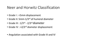
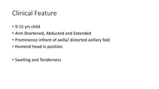
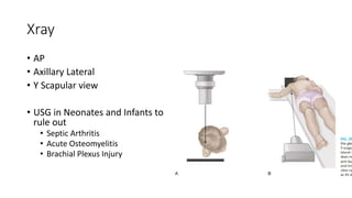
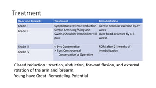
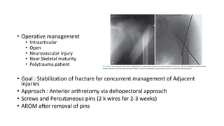
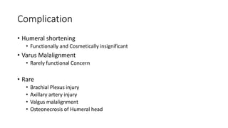
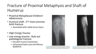
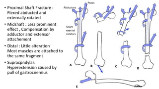
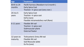
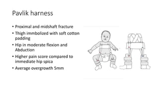
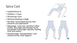
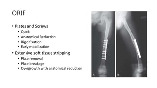
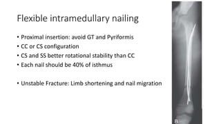
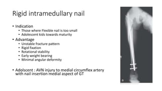
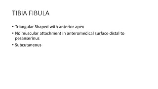
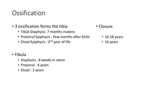
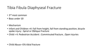
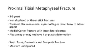
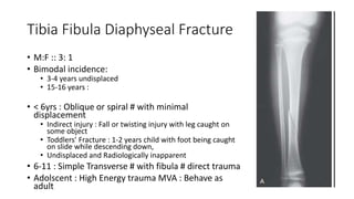
Pediatric Long Bone Fracture
(Humerus, Femur, Tibia)
Presenter Dr Kaushal Raj Kafle
Moderator Asst Prof Dr Sushil Paudel
Humerus : Anatomy
• Three Secondary Ossification Centre
• Humeral head, Greater Tuberosity and
Lesser tuberosity
• Coalesce to single centre by 7 years
of age
• Closes by 14-22 yrs (earlier in girls)
Injuries of Proximal Humeral Physis
• 80% of humeral growth comes from proximal
physis
• 3% of all physeal injuries
• Common in adolescence
• Direct Force: Direct Blow on the postero
lateral aspect of Shoulder
• Indirect Force : Through humeral shaft as in
FOOSH
• Birth Trauma
• Child Abuse
Salter and Harris Classification
• Type I SH : Infant and Small Children
• Type II SH : Older children and
Adolescent
• SH Type III/IV and Glenohumeral
Dislocation : rare due to the universal
ROM at shoulder
Neer and Horwitz Classification
• Grade I : <5mm displacement
• Grade II: 5mm-1/3rd of humeral diameter
• Grade III : 1/3rd - 2/3rd diameter
• Grade IV : >2/3rd diameter displacement
• Angulation associated with Grade III and IV
Clinical Feature
• 9-15 yrs child
• Arm Shortened, Abducted and Extended
• Prominence infront of axilla/ distorted axillary fold
• Humeral head in position
• Swelling and Tenderness
Xray
• AP
• Axillary Lateral
• Y Scapular view
• USG in Neonates and Infants to
rule out
• Septic Arthritis
• Acute Osteomyelitis
• Brachial Plexus Injury
Treatment
Neer and Horwitz Treatment Rehabilitation
Grade I Symptomatic without reduction
Simple Arm sling/ Sling and
Swath./Shoulder immobilizer till
pain
Gentle pendular exercise by 2nd
week
Over head activities by 4-6
weeks
Grade II
Grade III < 6yrs Conservative
> 6 yrs Controversial
Conservative Vs Operative
ROM after 2-3 weeks of
immbolization
Grade IV
Closed reduction : traction, abduction, forward flexion, and external
rotation of the arm and forearm.
Young have Great Remodeling Potential
• Operative management
• Intraarticular
• Open
• Neurovascular injury
• Near Skeletal maturity
• Polytrauma patient
• Goal : Stabilization of fracture for concurrent management of Adjacent
injuries
• Approach : Anterior arthrotomy via deltopectoral approach
• Screws and Percutaneous pins (2 k wires for 2-3 weeks)
• AROM after removal of pins
Complication
• Humeral shortening
• Functionally and Cosmetically insignificant
• Varus Malalignment
• Rarely functional Concern
• Rare
• Brachial Plexus injury
• Axillary artery injury
• Valgus malalignment
• Osteonecrosis of Humeral head
Fracture of Proximal Metaphysis and Shaft of
Humerus
• Proximal Metaphyseal Children>
Adolescence
• Humeral shaft : 2nd most common
birth fracture
• Associated with radial nerve injury
• High Energy Trauma
• Low energy trauma : Rule out
pathological Fracture
• Forceful muscle Contraction
• Unicameral bone cyst and Fibrous
dysplaisia
Diagnosis
• Deformity, Swelling and Pain
• Radial Nerve Injury : Anaesthesia over the dorsum of the hand (1st
and 2nd metacarpal )
• Loss of motor strength of wrist and fingers, thumb extensor and
forearm supinator
•
Treatment
• Infants and newborn
• 1-3 weeks immbolization
• Bandage arm to thorax with modified velpeau bandage or Sling and swathe
• Proper skin care
• Control in alignment : Not necessary
• Palpable callus in 6-8 weeks
• Assess Moro Reflex
• Follow up assessment for Brachial plexus injury
• Proximal Humeral metaphyseal
fracture
• Symptomatic management
• Sling is enough
• Shaft : Closed management
• Coaptation split
• 2-3 weeks : Sling / Hanging
Cast / Fracture Brace
• Circumduction and Pendular
exercises
Operative management
• Open fractures
• Polytrauma
• EX FIX if extensive soft tissue injury
• Intramedullary elastic nails in
polytrauma patients for easy nursing
care
Complications
• Open and vascular injuries
• Radial nerve injury
• Non union less common than adult
Femoral Shaft Fracture
• 1.6 % of all bony Injuries
• Boys : Girls 2.6: 1
• Bimodal Seasonal variation : March and August
• Bimodal Distribution
• Toddler : Trivial Fall
• Adolscent : High Energy Trauma
Diagnosis
• Clear Mechanism of Injury
• Deformity, swelling and localized pain
• Multiple Trauma/ Head Injury / Non ambulating and Severely disabled
child /Patient lacking Sensation (paraplegia /meningomyelocele)
• Careful evaluation needed
• Swelling and Redness may mimic Infection
Associated injuries
• Waddell’s Traid: Femoral Fracture with Abdominal or Thoracic Injury
with Head Injury
• High Velocity Automobile Injury
• Isolated femoral Fracture: Hypotension is rare
• Hematocrit < 30% rarely occurs without Multisystem Injury
• Declining Hematocrit SHOULD NOT be contributed to Closed Femoral
Fracture Until other sources of blood loss have been eliminated
Mechanism of Injury
• Before Ambulating age
• 80% femoral Fracture : ABUSE
• Risk Factors of Abuse
• History Suspicious for Abuse
• Physical or radiological evidence of previous Abuse
• Age under 18 months
• Transverse Fracture are Better Predictor of Non Accidental Trauma
• Older Children: Bone Strong to tolerate Forceful blows
• High Energy trauma : MVA 90%
• Stress Fracture
• Adolescent
• High Intensity Sports: Football, Basketball or running
• Femoral Shaft or Neck Stress Fracture
• Child with thigh pain with Unrecognised stress fracture can have displaced
SOF #
• Extreme Overuse in Non Traditional Sports
• Pathological Fracture : Rare
• Osteogenesis Imperfecta
• Generalised Osteopenia : Cerebral Palsy,
Myelomeningocele
• Neoplasms : Nonossifying Fibroma,
Aneursymal Bone Cyst, Unicameral Cyst,
Eosinophilic Granulom, Osteosarcome
• Subtle Xray Signs : Mixed Lytic Blastic
Lesion disrupting trabecular pattern ,
break in cortex and periosteal reaction
Imaging
• Xray
• Including the Hip and Knee joint
• AP and Lateral View
• MRI/Bone scan : Small Buckle Fracture or Stress Fracture
• Always Look for Communition or undisplaced Butterfly Fragments,
Second fracture, Joint dislocation or pathological features
Classification
• Transverse/ Oblique/ Spiral
• Communitted /Non Communitted
• Open/Closed
• Most Common : Closed Simple non communitted >50%
• Proximal Shaft Fracture :
Flexed abducted and
externally rotated
• Midshaft : Less prominent
effect , Compensation by
adductor and extensor
attachement
• Distal : Little alteration
Most muscles are attached to
the same fragment
• Supracpndylar:
Hyperextension caused by
pull of gastrocnemius
Short
exernal
rotators
Psoas
Abductors
Treatment
• Primary : Age and Fracture Pattern
• Secondary: Childs Weight,Assosciated injury, mechanism of Injury,
Availabilty of equipment, Surgeons experience
• Tertiary: Economic Concern, Post Treatment Care,
Age Treatment
Birth to 24
months
Pavlik harness (Newborn to 6 month )
Early Spica Cast
Traction → spica cast (very rare )
24 mo-5
years
Early Spica Cast
Traction → spica cast
ExFix (rare)
Flexible Intramedullary nail (Rare)
6-11 years Flexible IM nail
Traction → spica cast
Submuscular plane
External fixator
12 years to
maturity
Trohcanteric Entry IM rod
Flexible IM nail
Sub Muscular plate
Ex Fix (rare)
Pavlik harness
• Proximal and midshaft fracture
• Thigh immbolized with soft cotton
padding
• Hip in moderate flexion and
Abduction
• Higher pain score compared to
immediate hip spica
• Average overgrowth 5mm
Spica Cast
• Isolated femur #
• Children < 5 year
• Shortening <2cm
• Minimal Swelling in thigh
• No other assosciated injuries that
prevents cast application
• Advantage : Low cost, excellent safety
profile, Very high rate of good result,
acceptable limb length equality, healing
time and motion
• Disadvantage: Transportation, Cast
intolerance by child, hygiene
Traction and Spica cast
• Rockwood and Wilkins’ fractures in children
Age Varus/val
gus
Angulatio
n
Anterior
posterior
Angulatio
n
Shortening
(mm)
Birth to
24
months
30 30 15
24 mo-5
years
15 20 20
6-11
years
10 15 15
12 years
to
maturity
5 10 10
Rehabilitation
• Spica for 4-8 weeks
• Infants : 4-6 weeks, toodler (8 weeks )
• After cast removal ask parents to allow weight bearing once the child
is comfortable and once the joint stiffness resolves
• Physiotherapy : Not recommended. Aggressive ROM apprehends
child and delays recovery
• Follow up : for 1 year to analyse gait, joint ROM and limb length
Exfix
• Indication
• Open Fracture with poor soft tissue coverage
• Multiple trauma
• Vascular injury requiring immediate revascularization
• Failed Non Surgical management
• Unstable fracture pattern
• Temporarily used to be converted to internal
fixation
• Advantage : easy to apply , angular rotation
correctable in follow ups
• Pin tract infection, Increased incidence of non
union, delayed union and angular deformity
• 5mm overgrowth, Refracture
ORIF
• Plates and Screws
• Quick
• Anatomical Reduction
• Rigid fixation
• Early mobilization
• Extensive soft tissue stripping
• Plate removal
• Plate breakage
• Overgrowth with anatomical reduction
Flexible intramedullary nailing
• Proximal insertion: avoid GT and Pyriformis
• CC or CS configuration
• CS and SS better rotational stability than CC
• Each nail should be 40% of isthmus
• Unstable Fracture: Limb shortening and nail migration
Rigid intramedullary nail
• Indication
• Those where Flexible nail is too small
• Adolescent kids towards maturity
• Advantage
• Unstable fracture pattern
• Rigid fixation
• Rotational stability
• Early weight bearing
• Minimal angular deformity
• Adolscent : AVN injury to medial circumflex artery
with nail insertion medial aspect of GT
Complications
• Limb length Discrepancy
• Angular Deformity
• Rotational deformity
• Delayed Union
• Non Union
• Muscle Weakness
• Infection
• Neurovascular injury
• Compartment Syndrome
TIBIA FIBULA
• Triangular Shaped with anterior apex
• No muscular attachment in anteromedical surface distal to
pesanserinus
• Subcutaneous
Ossification
• 3 ossification forms the tibia
• Tibial Diaphysis: 7 months inutero
• Proximal Epiphysis : Few months after birth
• Distal Epiphysis : 2nd year of life
• Fibula
• Diaphysis : 8 weeks in utero
• Proximal : 4 years
• Distal : 2 years
• Closure
• 16-18 years
• 16 years
Tibia Fibula Diaphyseal Fracture
• 3rd most common
• Boys under 10
• Mechanism
• Infant and Children <4: Fall from height, fall from standing position, bicycle
spoke injury : Spiral or Oblique Fracture
• Child > 4: Pedestrian Accident : Comminuted fracture , Open injuries
• Child Abuse <5% tibial fracture
Proximal Tibial Metaphyseal Fracture
• 3-6 years
• Non displaced or Green stick fractures
• Torsional Stress on medial aspect of leg or direct blow to lateral
aspect
• Medial Cortex fracture with intact lateral cortex
• Fibula may or may not have # or plastic deformation
• Xray : Torus, Greenstick and Complete Fracture
• Most are undisplaced
Treatment
• Conservative
• Reduction under conscious sedation in valgus deformity
• Break the angled Greenstick fracture by bending towards angulation and
slightly overcorrect it
• Long leg cast with three point fixation
• Non weight bearing
• Operative
• Rare with failed closed reduction
• Medial incision over the fracture site , reduction under vision
• Suture the torn periosteum and apply cast
• CRPP with Crossed K wires and Cast
Complication
Valgus deformity 90%
Develops at 6 months and progresses upto 2 year (12-18 mo)
Max deformity : 18O , Resolution within 3 year
Residual 6O with knee slighlty medial to mechanical axis
Counselling
Treat only if residual deformity is
15-20O →well-timed medial proximal tibial epiphysiodesis
> 20O 3 years after the injury →Varus Osteotomy
• Bone overgrowth
• 1cm (max 1.7 cm)
• Overall outcome good but some have knee pain
• Physeal arrest with crush injuries
• Fairly benign early radiographs
Tibia Fibula Diaphyseal Fracture
• M:F :: 3: 1
• Bimodal incidence:
• 3-4 years undisplaced
• 15-16 years :
• < 6yrs : Oblique or spiral # with minimal
displacement
• Indirect injury : Fall or twisting injury with leg caught on
some object
• Toddlers’ Fracture : 1-2 years child with foot being caught
on slide while descending down,
• Undisplaced and Radiologically inapparent
• 6-11 : Simple Transverse # with fibula # direct trauma
• Adolscent : High Energy trauma MVA : Behave as
adult
Treatment : Conservative
• Conservative
• Toddler Fracture : Non displaced Oblique/Spiral Tibia # with intact
fibula
• Better visualized in Oblique views
• Long Leg Cast x 3-4 weeks
• Full weight bearing after cast out
• Follow up Xray unnecessary
• Suspicious case : Long Leg Cast application and 2 week follow up to check for
callus
• Displaced Tibial Fracture With an Intact Fibula
in an Older Child
• Tend to angle into varus due to anterolateral muscle
pulling distal fragment
• Fibula stablises the lateral aspect
• Fluoroscopy Guided reduction and molded to
maintain 3 point bending forces
• Acceptable : 5-10 degree in all plane
• Non Weight bearing for 4-6 weeks
• Change to short leg walking cast
• Recheck weekly for 2-3 week for
remanipulation for malalignment
• Cast wedging if varus angulation > 5O
• Displaced Tibial Fracture With a Fibular Fracture in an Older Child.
• Unstable fracture : Difficult to reduce
• Acceptable
• >50% apposition in
• <5-8o angulation in coronal and sagittal plane
• Residual varus in coronal plane and posterior angulation
• 3 point mold to reduce varus deformity and ankle at 15-20c plantar
flexion to prevent posterior angulation
• Cast extended to above knee with knee in 30-45c Flexion
• Univalve the case, admit, monitor for compartment syndrome
• Close monitoring for 2-3 weeks
• Convert to fracture boot or below knee cast with callus formation
Operative Management
• Rarely Indicated
• Major Indications
• Excessive Fracture instability
• Loss of reduction
• Significant Comminution and Shortening
• Displaced Fracture in skeletally mature child
Options
• Flexible intramedullary nail
• Fluoroscopy guided entry sparing physis
• Lateral and Medial entry with Double C construct
• No rotational Control : Long Leg cast
• Increased incidence of Compartment syndrome
• Functionally excellent outcome with union by 8-
20 weeks
• Closed Reduction Percutaneous pin fixation
• Young patient with rapid healing
• External Fixation
• Highly unstable Closed Tibial #
• 4-6 weeks followed by short leg walking
cast for 3-4 week
• Open Reduction with Internal Fixation
• Routinely not done due to excessive soft
tissue stripping
• Early mobilization, less angular deformilty
compared to Flexible nails
• Locking Rigid Intramedullary nail
• Child towards Skeletal maturity
Open Tibial Diaphyseal Fracture
• Rare in children <5%
• High energy trauma : Automobile hitting pedestrian, cyclist
• Non union and Infection
• 8-10yrs Boys
• Left leg > Right leg
• 58% have associated injuries
• 7% Mortality associated with Trauma
• Children > 11 behave as adult
Management
• Same as for adult
• Debridement, antibiotics and Splinting
• Wound management
• Soft tissue management :
• VAC
• STSG/ FTSG/Flap
• Fracture Stabilization
• Grade I/ Grade II : Long leg cast with Window
• Grossly Contaminated : Ex Fix
• Flexible nails
Indication for amputation
• Unreconstructable Vascular Injury
• Severe muscle injury with extensive bone loss
• Associated nerve injury with no sensation in foot
• Mangled Extremity Severity Scoring >7
Summary
• Proximal physeal and SOH Fractures in pediatrics can be managed
conservatively irrespective of alignment and reduction as it has great
remodeling potential
• The younger the age more deformity is acceptable in femur fracture
• Treatment Modalities in pediatric femur fracture depends on the age
and fracture pattern
• Proximal tibia fracture will develop valgus deformity irrespective of
treatment so counselling is must
• Soft tissue status in the shaft of tibia factor determines the outcome
in tibia fracture
References
• Tachdjian’s Pediatric Orthopaedics 6e
• Rockwood and Wilkins’ fractures in children
Thank you
Next presentation on Pediatric Fracture around ankle by Dr suman
Maharjan

Pediatric Long Bone Fractures.pptx

  • 1.
    Pediatric Long BoneFracture (Humerus, Femur, Tibia) Presenter Dr Kaushal Raj Kafle Moderator Asst Prof Dr Sushil Paudel
  • 2.
    Humerus : Anatomy •Three Secondary Ossification Centre • Humeral head, Greater Tuberosity and Lesser tuberosity • Coalesce to single centre by 7 years of age • Closes by 14-22 yrs (earlier in girls)
  • 3.
    Injuries of ProximalHumeral Physis • 80% of humeral growth comes from proximal physis • 3% of all physeal injuries • Common in adolescence • Direct Force: Direct Blow on the postero lateral aspect of Shoulder • Indirect Force : Through humeral shaft as in FOOSH • Birth Trauma • Child Abuse
  • 4.
    Salter and HarrisClassification • Type I SH : Infant and Small Children • Type II SH : Older children and Adolescent • SH Type III/IV and Glenohumeral Dislocation : rare due to the universal ROM at shoulder
  • 5.
    Neer and HorwitzClassification • Grade I : <5mm displacement • Grade II: 5mm-1/3rd of humeral diameter • Grade III : 1/3rd - 2/3rd diameter • Grade IV : >2/3rd diameter displacement • Angulation associated with Grade III and IV
  • 6.
    Clinical Feature • 9-15yrs child • Arm Shortened, Abducted and Extended • Prominence infront of axilla/ distorted axillary fold • Humeral head in position • Swelling and Tenderness
  • 7.
    Xray • AP • AxillaryLateral • Y Scapular view • USG in Neonates and Infants to rule out • Septic Arthritis • Acute Osteomyelitis • Brachial Plexus Injury
  • 8.
    Treatment Neer and HorwitzTreatment Rehabilitation Grade I Symptomatic without reduction Simple Arm sling/ Sling and Swath./Shoulder immobilizer till pain Gentle pendular exercise by 2nd week Over head activities by 4-6 weeks Grade II Grade III < 6yrs Conservative > 6 yrs Controversial Conservative Vs Operative ROM after 2-3 weeks of immbolization Grade IV Closed reduction : traction, abduction, forward flexion, and external rotation of the arm and forearm. Young have Great Remodeling Potential
  • 9.
    • Operative management •Intraarticular • Open • Neurovascular injury • Near Skeletal maturity • Polytrauma patient • Goal : Stabilization of fracture for concurrent management of Adjacent injuries • Approach : Anterior arthrotomy via deltopectoral approach • Screws and Percutaneous pins (2 k wires for 2-3 weeks) • AROM after removal of pins
  • 10.
    Complication • Humeral shortening •Functionally and Cosmetically insignificant • Varus Malalignment • Rarely functional Concern • Rare • Brachial Plexus injury • Axillary artery injury • Valgus malalignment • Osteonecrosis of Humeral head
  • 11.
    Fracture of ProximalMetaphysis and Shaft of Humerus • Proximal Metaphyseal Children> Adolescence • Humeral shaft : 2nd most common birth fracture • Associated with radial nerve injury • High Energy Trauma • Low energy trauma : Rule out pathological Fracture • Forceful muscle Contraction • Unicameral bone cyst and Fibrous dysplaisia
  • 12.
    Diagnosis • Deformity, Swellingand Pain • Radial Nerve Injury : Anaesthesia over the dorsum of the hand (1st and 2nd metacarpal ) • Loss of motor strength of wrist and fingers, thumb extensor and forearm supinator •
  • 13.
    Treatment • Infants andnewborn • 1-3 weeks immbolization • Bandage arm to thorax with modified velpeau bandage or Sling and swathe • Proper skin care • Control in alignment : Not necessary • Palpable callus in 6-8 weeks • Assess Moro Reflex • Follow up assessment for Brachial plexus injury
  • 14.
    • Proximal Humeralmetaphyseal fracture • Symptomatic management • Sling is enough • Shaft : Closed management • Coaptation split • 2-3 weeks : Sling / Hanging Cast / Fracture Brace • Circumduction and Pendular exercises
  • 15.
    Operative management • Openfractures • Polytrauma • EX FIX if extensive soft tissue injury • Intramedullary elastic nails in polytrauma patients for easy nursing care
  • 16.
    Complications • Open andvascular injuries • Radial nerve injury • Non union less common than adult
  • 17.
    Femoral Shaft Fracture •1.6 % of all bony Injuries • Boys : Girls 2.6: 1 • Bimodal Seasonal variation : March and August • Bimodal Distribution • Toddler : Trivial Fall • Adolscent : High Energy Trauma
  • 18.
    Diagnosis • Clear Mechanismof Injury • Deformity, swelling and localized pain • Multiple Trauma/ Head Injury / Non ambulating and Severely disabled child /Patient lacking Sensation (paraplegia /meningomyelocele) • Careful evaluation needed • Swelling and Redness may mimic Infection
  • 19.
    Associated injuries • Waddell’sTraid: Femoral Fracture with Abdominal or Thoracic Injury with Head Injury • High Velocity Automobile Injury • Isolated femoral Fracture: Hypotension is rare • Hematocrit < 30% rarely occurs without Multisystem Injury • Declining Hematocrit SHOULD NOT be contributed to Closed Femoral Fracture Until other sources of blood loss have been eliminated
  • 20.
    Mechanism of Injury •Before Ambulating age • 80% femoral Fracture : ABUSE • Risk Factors of Abuse • History Suspicious for Abuse • Physical or radiological evidence of previous Abuse • Age under 18 months • Transverse Fracture are Better Predictor of Non Accidental Trauma
  • 21.
    • Older Children:Bone Strong to tolerate Forceful blows • High Energy trauma : MVA 90% • Stress Fracture • Adolescent • High Intensity Sports: Football, Basketball or running • Femoral Shaft or Neck Stress Fracture • Child with thigh pain with Unrecognised stress fracture can have displaced SOF # • Extreme Overuse in Non Traditional Sports
  • 22.
    • Pathological Fracture: Rare • Osteogenesis Imperfecta • Generalised Osteopenia : Cerebral Palsy, Myelomeningocele • Neoplasms : Nonossifying Fibroma, Aneursymal Bone Cyst, Unicameral Cyst, Eosinophilic Granulom, Osteosarcome • Subtle Xray Signs : Mixed Lytic Blastic Lesion disrupting trabecular pattern , break in cortex and periosteal reaction
  • 23.
    Imaging • Xray • Includingthe Hip and Knee joint • AP and Lateral View • MRI/Bone scan : Small Buckle Fracture or Stress Fracture • Always Look for Communition or undisplaced Butterfly Fragments, Second fracture, Joint dislocation or pathological features
  • 24.
    Classification • Transverse/ Oblique/Spiral • Communitted /Non Communitted • Open/Closed • Most Common : Closed Simple non communitted >50%
  • 25.
    • Proximal ShaftFracture : Flexed abducted and externally rotated • Midshaft : Less prominent effect , Compensation by adductor and extensor attachement • Distal : Little alteration Most muscles are attached to the same fragment • Supracpndylar: Hyperextension caused by pull of gastrocnemius Short exernal rotators Psoas Abductors
  • 26.
    Treatment • Primary :Age and Fracture Pattern • Secondary: Childs Weight,Assosciated injury, mechanism of Injury, Availabilty of equipment, Surgeons experience • Tertiary: Economic Concern, Post Treatment Care,
  • 27.
    Age Treatment Birth to24 months Pavlik harness (Newborn to 6 month ) Early Spica Cast Traction → spica cast (very rare ) 24 mo-5 years Early Spica Cast Traction → spica cast ExFix (rare) Flexible Intramedullary nail (Rare) 6-11 years Flexible IM nail Traction → spica cast Submuscular plane External fixator 12 years to maturity Trohcanteric Entry IM rod Flexible IM nail Sub Muscular plate Ex Fix (rare)
  • 28.
    Pavlik harness • Proximaland midshaft fracture • Thigh immbolized with soft cotton padding • Hip in moderate flexion and Abduction • Higher pain score compared to immediate hip spica • Average overgrowth 5mm
  • 29.
    Spica Cast • Isolatedfemur # • Children < 5 year • Shortening <2cm • Minimal Swelling in thigh • No other assosciated injuries that prevents cast application • Advantage : Low cost, excellent safety profile, Very high rate of good result, acceptable limb length equality, healing time and motion • Disadvantage: Transportation, Cast intolerance by child, hygiene
  • 30.
    Traction and Spicacast • Rockwood and Wilkins’ fractures in children
  • 31.
    Age Varus/val gus Angulatio n Anterior posterior Angulatio n Shortening (mm) Birth to 24 months 3030 15 24 mo-5 years 15 20 20 6-11 years 10 15 15 12 years to maturity 5 10 10
  • 32.
    Rehabilitation • Spica for4-8 weeks • Infants : 4-6 weeks, toodler (8 weeks ) • After cast removal ask parents to allow weight bearing once the child is comfortable and once the joint stiffness resolves • Physiotherapy : Not recommended. Aggressive ROM apprehends child and delays recovery • Follow up : for 1 year to analyse gait, joint ROM and limb length
  • 33.
    Exfix • Indication • OpenFracture with poor soft tissue coverage • Multiple trauma • Vascular injury requiring immediate revascularization • Failed Non Surgical management • Unstable fracture pattern • Temporarily used to be converted to internal fixation • Advantage : easy to apply , angular rotation correctable in follow ups • Pin tract infection, Increased incidence of non union, delayed union and angular deformity • 5mm overgrowth, Refracture
  • 34.
    ORIF • Plates andScrews • Quick • Anatomical Reduction • Rigid fixation • Early mobilization • Extensive soft tissue stripping • Plate removal • Plate breakage • Overgrowth with anatomical reduction
  • 35.
    Flexible intramedullary nailing •Proximal insertion: avoid GT and Pyriformis • CC or CS configuration • CS and SS better rotational stability than CC • Each nail should be 40% of isthmus • Unstable Fracture: Limb shortening and nail migration
  • 36.
    Rigid intramedullary nail •Indication • Those where Flexible nail is too small • Adolescent kids towards maturity • Advantage • Unstable fracture pattern • Rigid fixation • Rotational stability • Early weight bearing • Minimal angular deformity • Adolscent : AVN injury to medial circumflex artery with nail insertion medial aspect of GT
  • 37.
    Complications • Limb lengthDiscrepancy • Angular Deformity • Rotational deformity • Delayed Union • Non Union • Muscle Weakness • Infection • Neurovascular injury • Compartment Syndrome
  • 38.
    TIBIA FIBULA • TriangularShaped with anterior apex • No muscular attachment in anteromedical surface distal to pesanserinus • Subcutaneous
  • 39.
    Ossification • 3 ossificationforms the tibia • Tibial Diaphysis: 7 months inutero • Proximal Epiphysis : Few months after birth • Distal Epiphysis : 2nd year of life • Fibula • Diaphysis : 8 weeks in utero • Proximal : 4 years • Distal : 2 years • Closure • 16-18 years • 16 years
  • 40.
    Tibia Fibula DiaphysealFracture • 3rd most common • Boys under 10 • Mechanism • Infant and Children <4: Fall from height, fall from standing position, bicycle spoke injury : Spiral or Oblique Fracture • Child > 4: Pedestrian Accident : Comminuted fracture , Open injuries • Child Abuse <5% tibial fracture
  • 41.
    Proximal Tibial MetaphysealFracture • 3-6 years • Non displaced or Green stick fractures • Torsional Stress on medial aspect of leg or direct blow to lateral aspect • Medial Cortex fracture with intact lateral cortex • Fibula may or may not have # or plastic deformation • Xray : Torus, Greenstick and Complete Fracture • Most are undisplaced
  • 42.
    Treatment • Conservative • Reductionunder conscious sedation in valgus deformity • Break the angled Greenstick fracture by bending towards angulation and slightly overcorrect it • Long leg cast with three point fixation • Non weight bearing • Operative • Rare with failed closed reduction • Medial incision over the fracture site , reduction under vision • Suture the torn periosteum and apply cast • CRPP with Crossed K wires and Cast
  • 43.
    Complication Valgus deformity 90% Developsat 6 months and progresses upto 2 year (12-18 mo) Max deformity : 18O , Resolution within 3 year Residual 6O with knee slighlty medial to mechanical axis Counselling Treat only if residual deformity is 15-20O →well-timed medial proximal tibial epiphysiodesis > 20O 3 years after the injury →Varus Osteotomy
  • 45.
    • Bone overgrowth •1cm (max 1.7 cm) • Overall outcome good but some have knee pain • Physeal arrest with crush injuries • Fairly benign early radiographs
  • 46.
    Tibia Fibula DiaphysealFracture • M:F :: 3: 1 • Bimodal incidence: • 3-4 years undisplaced • 15-16 years : • < 6yrs : Oblique or spiral # with minimal displacement • Indirect injury : Fall or twisting injury with leg caught on some object • Toddlers’ Fracture : 1-2 years child with foot being caught on slide while descending down, • Undisplaced and Radiologically inapparent • 6-11 : Simple Transverse # with fibula # direct trauma • Adolscent : High Energy trauma MVA : Behave as adult
  • 47.
    Treatment : Conservative •Conservative • Toddler Fracture : Non displaced Oblique/Spiral Tibia # with intact fibula • Better visualized in Oblique views • Long Leg Cast x 3-4 weeks • Full weight bearing after cast out • Follow up Xray unnecessary • Suspicious case : Long Leg Cast application and 2 week follow up to check for callus
  • 48.
    • Displaced TibialFracture With an Intact Fibula in an Older Child • Tend to angle into varus due to anterolateral muscle pulling distal fragment • Fibula stablises the lateral aspect • Fluoroscopy Guided reduction and molded to maintain 3 point bending forces • Acceptable : 5-10 degree in all plane • Non Weight bearing for 4-6 weeks • Change to short leg walking cast • Recheck weekly for 2-3 week for remanipulation for malalignment • Cast wedging if varus angulation > 5O
  • 49.
    • Displaced TibialFracture With a Fibular Fracture in an Older Child. • Unstable fracture : Difficult to reduce • Acceptable • >50% apposition in • <5-8o angulation in coronal and sagittal plane • Residual varus in coronal plane and posterior angulation • 3 point mold to reduce varus deformity and ankle at 15-20c plantar flexion to prevent posterior angulation • Cast extended to above knee with knee in 30-45c Flexion • Univalve the case, admit, monitor for compartment syndrome • Close monitoring for 2-3 weeks • Convert to fracture boot or below knee cast with callus formation
  • 51.
    Operative Management • RarelyIndicated • Major Indications • Excessive Fracture instability • Loss of reduction • Significant Comminution and Shortening • Displaced Fracture in skeletally mature child
  • 52.
    Options • Flexible intramedullarynail • Fluoroscopy guided entry sparing physis • Lateral and Medial entry with Double C construct • No rotational Control : Long Leg cast • Increased incidence of Compartment syndrome • Functionally excellent outcome with union by 8- 20 weeks • Closed Reduction Percutaneous pin fixation • Young patient with rapid healing
  • 53.
    • External Fixation •Highly unstable Closed Tibial # • 4-6 weeks followed by short leg walking cast for 3-4 week • Open Reduction with Internal Fixation • Routinely not done due to excessive soft tissue stripping • Early mobilization, less angular deformilty compared to Flexible nails • Locking Rigid Intramedullary nail • Child towards Skeletal maturity
  • 54.
    Open Tibial DiaphysealFracture • Rare in children <5% • High energy trauma : Automobile hitting pedestrian, cyclist • Non union and Infection • 8-10yrs Boys • Left leg > Right leg • 58% have associated injuries • 7% Mortality associated with Trauma • Children > 11 behave as adult
  • 55.
    Management • Same asfor adult • Debridement, antibiotics and Splinting • Wound management • Soft tissue management : • VAC • STSG/ FTSG/Flap • Fracture Stabilization • Grade I/ Grade II : Long leg cast with Window • Grossly Contaminated : Ex Fix • Flexible nails
  • 56.
    Indication for amputation •Unreconstructable Vascular Injury • Severe muscle injury with extensive bone loss • Associated nerve injury with no sensation in foot • Mangled Extremity Severity Scoring >7
  • 57.
    Summary • Proximal physealand SOH Fractures in pediatrics can be managed conservatively irrespective of alignment and reduction as it has great remodeling potential • The younger the age more deformity is acceptable in femur fracture • Treatment Modalities in pediatric femur fracture depends on the age and fracture pattern • Proximal tibia fracture will develop valgus deformity irrespective of treatment so counselling is must • Soft tissue status in the shaft of tibia factor determines the outcome in tibia fracture
  • 58.
    References • Tachdjian’s PediatricOrthopaedics 6e • Rockwood and Wilkins’ fractures in children
  • 59.
    Thank you Next presentationon Pediatric Fracture around ankle by Dr suman Maharjan

Editor's Notes

  • #41 1st Femur 2nd Both bone #