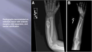
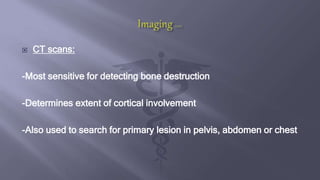
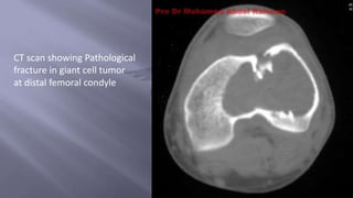
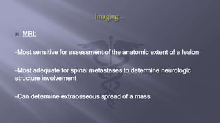
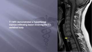
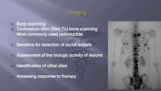
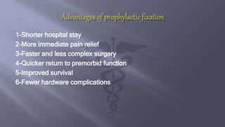
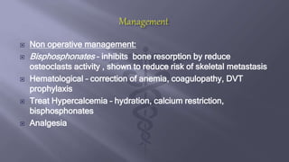
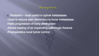
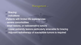
Dr.SAYF ALDEEN HUSSAM
ORTHOPEDIC TRAINEE
BAGHDAD MEDICAL CITY
 Pathological fractures are fractures that occur in abnormal bone.
Although the term can be used in the setting of a generalized
metabolic bone disease, it is usually reserved for fractures
through a focal abnormality. The abnormality may be malignant
or non-malignant in nature.
 Uncorrectable
• Osteogenesis imperfecta
• Polyostotic fibrous dysplasia
• Postmenopausal
• Paget disease
• Osteopetrosis
Systemic- non neoplastic:
 Correctable
• Renal osteodystrophy
• Hyperparathyroidism
• Osteomalacia
• Disuse osteoporosis
 Common sites of metastasis
– Spine
– Pelvis
– Ribs
– Skull
– proximal femur
– Proximal humerus
 Neoplastic:
 Common metastatic cancers
– Breast
– Lung
– Prostate
– thyroid
– kidney
Or noncancerous tumors and cysts
 Pain – most common preceding fracture, night pain, constant dull
in nature, aggravated by activity
 Constitutional symptoms – anorexia, night sweats, weight loss,
fatigue
 Previous history of cancer
 Exposur to Carcinogen – smoking, radiation, occupational toxins
 Spontaneous fracture
 Fracture due to Minor trauma
 Pain at site preceeding fracture
 Multiple recent fractures
 Age > 45 yr.s
 Prior history of malignancy
 Unusual # patterns
 Debilitating pain
 Immobility
 Neurologic deficits – spine metastasis
 Anaemia
 Hypercalcemia
 CBC – anemia of chronic disease
 Calcium – elevated
 Alkaline phosphatase – elevated, non specific
 Tumor markers – PSA, CEA, CA125, TFTs
 N-telopeptide + C-telopeptide –are markers of bone collagen
breakdown measured in serum and urine.
-Measure the overall extent of bone involvement
- Assess the response of the bone to bisphosphonate treatment
 plain radiograph: Imaging Findings
-Fracture line extending through a destructive lesion in the bone
Usually transverse in direction
-Surrounding bone may demonstrate
-Endosteal scalloping
-Cortical destruction
-Permeative pattern
-Frequently associated with a soft tissue mass
We should answer the Enneking's Four Questions for Bony Lesions
1. Where is the lesion located? Epiphysis , metaphysis , diaphysis
2. What is the lesion doing to the bone? transitional zone
3. What is the bone doing to the lesion? periosteal reaction
4. Dose anything suggest histology ? Calcification , ossification
Fracture through a large
metaphyseal Non
Ossifying Fibroma
Radiographs demonstrated an
osteolytic lesion with sclerotic
margins, mild expansion, and
central calcification
 CT scans:
-Most sensitive for detecting bone destruction
-Determines extent of cortical involvement
-Also used to search for primary lesion in pelvis, abdomen or chest
CT scan showing Pathological
fracture in giant cell tumor
at distal femoral condyle
 MRI:
-Most sensitive for assessment of the anatomic extent of a lesion
-Most adequate for spinal metastases to determine neurologic
structure involvement
-Can determine extraosseous spread of a mass
T1 MRI demonstrated a hypointense
marrow-infiltrating lesion involving the T1
vertebral body
 Bone scanning:
 Technetium-99m (99m Tc) bone scanning
Most commonly used radionuclide
 Sensitive for detection of occult lesions
 Assessment of the biologic activity of lesions
 Identification of other sites
 Assessing response to therapy
 Principles of the Open Incisional Biopsy:
1- use longitudinal incision in the extremities allows for extension of
the incision for definitive management
2-Approach do not expose neurovascular structures
3-release tourniquet prior to wound closure
4-Biopsy perform through the involved compartment of the tumor
5- if using a drain, bring drain out of the skin in line with surgical
incision allows drain site to be removed with definitive surgery
6-Cultures for all biopsy to rule out infections that may mimic tumors
on x rays
 Mirel’s criteria useful to determine which lesions at high risk of
fracture and need Prophylactic stabilization
 A score of 8 or more is an indication for prophylactic stabilisation
1-Shorter hospital stay
2-More immediate pain relief
3-Faster and less complex surgery
4-Quicker return to premorbid function
5-Improved survival
6-Fewer hardware complications
 Non operative management:
 Bisphosphonates – inhibits bone resorption by reduce
osteoclasts activity , shown to reduce risk of skeletal metastasis
 Hematological – correction of anemia, coagulopathy, DVT
prophylaxis
 Treat Hypercalcemia – hydration, calcium restriction,
bisphosphonates
 Analgesia
 Radiation – most useful in spinal metastases
-Used to reduce pain secondary to bone metastases
-Halts progression of bony destruction
-Allows healing of an impending pathologic fracture
-Postoperative local tumor control
 Bracing:
 indications
-Patients with limited life expectancies
- severe comorbidities
- small lesions, or radiosensitive tumors
 Upper extremity lesions particularly amenable for bracing
 Adjuvant radiotherapy of susceptible tumors is required
 Operative:
 Indications of surgery
1-Progression of disease after radiation
2- Neurologic compromise caused by bony impingement
3- Radio resistant tumor within the spinal canal
4- Impending fracture
5-Spinal instability caused by a pathologic fracture
6-Progressive deformity
 Principles of surgery:
 Intramedullary device provides better stability.
 Use Bone cement:
– Increases the strength of fixation
– Should not be used to replace segment of bone
 Goal should be to stabilize as much of the bone as possible.
 Need Durable, weight bearing implants
 Bone graft less useful due to prolonged healing time
Pathological fractures managment

Pathological fractures managment

  • 1.
    Dr.SAYF ALDEEN HUSSAM ORTHOPEDICTRAINEE BAGHDAD MEDICAL CITY
  • 2.
     Pathological fracturesare fractures that occur in abnormal bone. Although the term can be used in the setting of a generalized metabolic bone disease, it is usually reserved for fractures through a focal abnormality. The abnormality may be malignant or non-malignant in nature.
  • 3.
     Uncorrectable • Osteogenesisimperfecta • Polyostotic fibrous dysplasia • Postmenopausal • Paget disease • Osteopetrosis Systemic- non neoplastic:  Correctable • Renal osteodystrophy • Hyperparathyroidism • Osteomalacia • Disuse osteoporosis
  • 4.
     Common sitesof metastasis – Spine – Pelvis – Ribs – Skull – proximal femur – Proximal humerus  Neoplastic:  Common metastatic cancers – Breast – Lung – Prostate – thyroid – kidney Or noncancerous tumors and cysts
  • 5.
     Pain –most common preceding fracture, night pain, constant dull in nature, aggravated by activity  Constitutional symptoms – anorexia, night sweats, weight loss, fatigue  Previous history of cancer  Exposur to Carcinogen – smoking, radiation, occupational toxins
  • 6.
     Spontaneous fracture Fracture due to Minor trauma  Pain at site preceeding fracture  Multiple recent fractures  Age > 45 yr.s  Prior history of malignancy  Unusual # patterns
  • 7.
     Debilitating pain Immobility  Neurologic deficits – spine metastasis  Anaemia  Hypercalcemia
  • 8.
     CBC –anemia of chronic disease  Calcium – elevated  Alkaline phosphatase – elevated, non specific  Tumor markers – PSA, CEA, CA125, TFTs  N-telopeptide + C-telopeptide –are markers of bone collagen breakdown measured in serum and urine. -Measure the overall extent of bone involvement - Assess the response of the bone to bisphosphonate treatment
  • 9.
     plain radiograph:Imaging Findings -Fracture line extending through a destructive lesion in the bone Usually transverse in direction -Surrounding bone may demonstrate -Endosteal scalloping -Cortical destruction -Permeative pattern -Frequently associated with a soft tissue mass
  • 10.
    We should answerthe Enneking's Four Questions for Bony Lesions 1. Where is the lesion located? Epiphysis , metaphysis , diaphysis 2. What is the lesion doing to the bone? transitional zone 3. What is the bone doing to the lesion? periosteal reaction 4. Dose anything suggest histology ? Calcification , ossification
  • 12.
    Fracture through alarge metaphyseal Non Ossifying Fibroma
  • 14.
    Radiographs demonstrated an osteolyticlesion with sclerotic margins, mild expansion, and central calcification
  • 15.
     CT scans: -Mostsensitive for detecting bone destruction -Determines extent of cortical involvement -Also used to search for primary lesion in pelvis, abdomen or chest
  • 16.
    CT scan showingPathological fracture in giant cell tumor at distal femoral condyle
  • 17.
     MRI: -Most sensitivefor assessment of the anatomic extent of a lesion -Most adequate for spinal metastases to determine neurologic structure involvement -Can determine extraosseous spread of a mass
  • 18.
    T1 MRI demonstrateda hypointense marrow-infiltrating lesion involving the T1 vertebral body
  • 19.
     Bone scanning: Technetium-99m (99m Tc) bone scanning Most commonly used radionuclide  Sensitive for detection of occult lesions  Assessment of the biologic activity of lesions  Identification of other sites  Assessing response to therapy
  • 20.
     Principles ofthe Open Incisional Biopsy: 1- use longitudinal incision in the extremities allows for extension of the incision for definitive management 2-Approach do not expose neurovascular structures 3-release tourniquet prior to wound closure 4-Biopsy perform through the involved compartment of the tumor 5- if using a drain, bring drain out of the skin in line with surgical incision allows drain site to be removed with definitive surgery 6-Cultures for all biopsy to rule out infections that may mimic tumors on x rays
  • 21.
     Mirel’s criteriauseful to determine which lesions at high risk of fracture and need Prophylactic stabilization  A score of 8 or more is an indication for prophylactic stabilisation
  • 22.
    1-Shorter hospital stay 2-Moreimmediate pain relief 3-Faster and less complex surgery 4-Quicker return to premorbid function 5-Improved survival 6-Fewer hardware complications
  • 23.
     Non operativemanagement:  Bisphosphonates – inhibits bone resorption by reduce osteoclasts activity , shown to reduce risk of skeletal metastasis  Hematological – correction of anemia, coagulopathy, DVT prophylaxis  Treat Hypercalcemia – hydration, calcium restriction, bisphosphonates  Analgesia
  • 24.
     Radiation –most useful in spinal metastases -Used to reduce pain secondary to bone metastases -Halts progression of bony destruction -Allows healing of an impending pathologic fracture -Postoperative local tumor control
  • 25.
     Bracing:  indications -Patientswith limited life expectancies - severe comorbidities - small lesions, or radiosensitive tumors  Upper extremity lesions particularly amenable for bracing  Adjuvant radiotherapy of susceptible tumors is required
  • 26.
     Operative:  Indicationsof surgery 1-Progression of disease after radiation 2- Neurologic compromise caused by bony impingement 3- Radio resistant tumor within the spinal canal 4- Impending fracture 5-Spinal instability caused by a pathologic fracture 6-Progressive deformity
  • 27.
     Principles ofsurgery:  Intramedullary device provides better stability.  Use Bone cement: – Increases the strength of fixation – Should not be used to replace segment of bone  Goal should be to stabilize as much of the bone as possible.  Need Durable, weight bearing implants  Bone graft less useful due to prolonged healing time