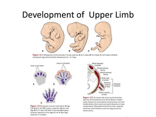
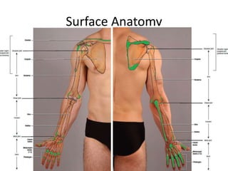
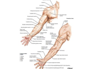
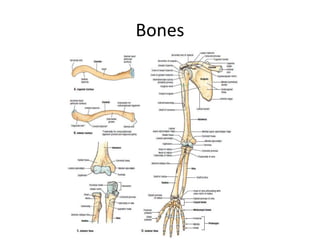
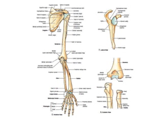
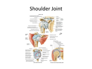
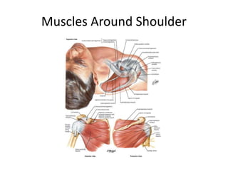
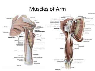
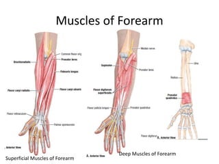
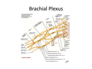
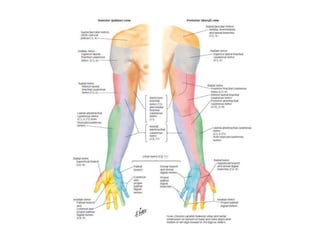
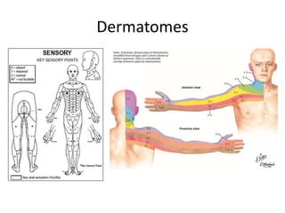
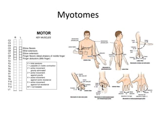
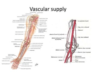
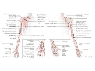
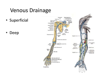
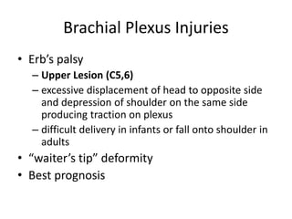
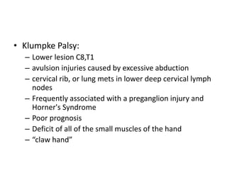
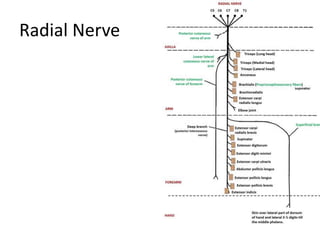
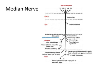
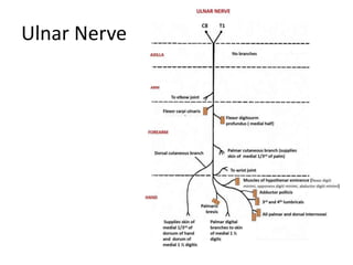
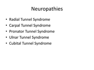
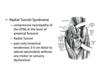
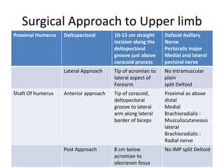
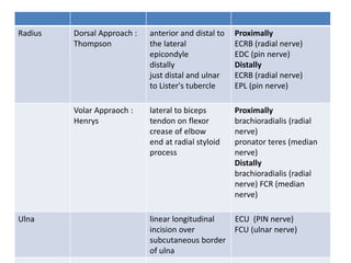
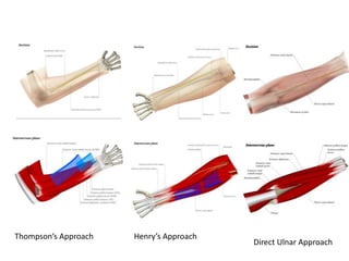
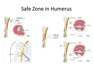
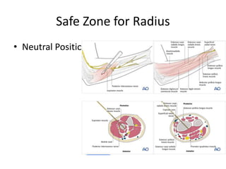
Applied Anatomy of Upper Limb
Presenter Dr Kaushal Raj Kafle
Moderator Dr Nischal Ghimire
Overview
• Development of Upper Limb
• Surface Anatomy
• Bones and Joints of Upper limb
• Muscles and Compartments
• Neurovascular Structures of Upperlimb
• Surgical anatomy of Upper limb
Development of Upper Limb
• Bone Age
• Limb Defect
– Meromelia / Amelia
– Micromelia
– Phocomelia
– Brachydactly
– Cleft Hand
– Congenital Absence of Radius
– Amniotic Bands
Surface Anatomy
Bones
Shoulder Joint
Shoulder Joint
• Dislocation
– Ball and Socket Joint with Wide ROM
– Unstable Joint
– Inferior dislocation attributed to unprotected in
Inferior aspect
– Anteriorly : Subscapularis , Superiorly :
Supraspinatous, Posteriorly : Teres Minor and
infraspinatous
Shoulder Arthrocentesis
Shoulder arthroscopy
• Posterior portal
• Anterior portal
• Lateral portal
Elbow
Elbow Arthrocentesis
• Elbow flexed at 90O
• Locate the capitulum and
head of radius
• Palpable depression with
elbow flexed and forearm
semipronated
Pulled Elbow
• Annular Ligament
– Funnel shaped in adult
– Vertical in Children
• Students Elbow :
– Subcutaneous olecranon bursitis
• Tennis Elbow
– Lateral Epicondylitis
• Golfers Elbow
– Medial epicondyliltis
Wrist
Muscles and Compartments
Muscles Around Shoulder
Muscles of Arm
Muscles of Forearm
Superficial Muscles of Forearm
Deep Muscles of Forearm
Compartment Syndrome
• Pressure inside the closed osteofascial
compartment increases to such an extent that
there is a compromise of microcirculation,
leading to tissue damage
• Traumatic and Atraumatic
Brachial Plexus
Dermatomes
Myotomes
Vascular supply
Venous Drainage
• Superficial
• Deep
Brachial Plexus Injuries
• Erb’s palsy
– Upper Lesion (C5,6)
– excessive displacement of head to opposite side
and depression of shoulder on the same side
producing traction on plexus
– difficult delivery in infants or fall onto shoulder in
adults
• “waiter’s tip” deformity
• Best prognosis
• Klumpke Palsy:
– Lower lesion C8,T1
– avulsion injuries caused by excessive abduction
– cervical rib, or lung mets in lower deep cervical lymph
nodes
– Frequently associated with a preganglion injury and
Horner's Syndrome
– Poor prognosis
– Deficit of all of the small muscles of the hand
– “claw hand”
Radial Nerve
Median Nerve
Ulnar Nerve
Neuropathies
• Radial Tunnel Syndrome
• Carpal Tunnel Syndrome
• Pronator Tunnel Syndrome
• Ulnar Tunnel Syndrome
• Cubital Tunnel Syndrome
• Radial Tunnel Syndrome
– compressive neuropathy of
the (PIN) at the level of
proximal forearm
– Radial Tunnel
– pain only (maximal
tenderness 3-5 cm distal to
lateral epicondyle) without
any motor or sensory
dysfunction
Surgical Approach to Upper limb
Proximal Humerus Deltopectoral 10-15 cm straight
incision along the
deltopectoral
groove just above
coracoid process
Deltoid Axillary
Nerve
Pectoralis major
Medial and lateral
pectoral nerve
Lateral Approach Tip of acromian to
lateral aspect of
Forearm
No intramuscular
plain
split Deltoid
Shaft Of humerus Anterior approach Tip of coracoid,
deltopectoral
groove to lateral
arm along lateral
border of biceps
Proximal as above
distal
Medial
Brachioradialis :
Musculocutaneous
lateral
Brachioradialis :
Radial nerve
Post Approach 8 cm below
acromian to
olecranon fossa
No IMP split Deltoid
Radius Dorsal Approach :
Thompson
anterior and distal to
the lateral
epicondyle
distally
just distal and ulnar
to Lister's tubercle
Proximally
ECRB (radial nerve)
EDC (pin nerve)
Distally
ECRB (radial nerve)
EPL (pin nerve)
Volar Appraoch :
Henrys
lateral to biceps
tendon on flexor
crease of elbow
end at radial styloid
process
Proximally
brachioradialis (radial
nerve)
pronator teres (median
nerve)
Distally
brachioradialis (radial
nerve) FCR (median
nerve)
Ulna linear longitudinal
incision over
subcutaneous border
of ulna
ECU (PIN nerve)
FCU (ulnar nerve)
• THomsp
Thompson’s Approach Henry’s Approach
Direct Ulnar Approach
Safe Zone in Humerus
Safe Zone for Radius
• Neutral Position
Ulna
• Pronation
• Between the extensor carpi ulnaris and the
flexor carpi ulnaris
Summary
• Upper limb is the non weight bearing limb
• Stability is sacrified to mobility
• Knowlegde of structure without the
understanding of function is almost useless
clinically because the aim of the treatment is
to preserve or restore the function
References
• Atlas of Human Anatomy, Franklin H Netter, 7e
• Grant’s Atlas of Anatomy, 13e
• Lasts Anatomy 12e
• Langman’s Embryology, 12e
• Moore and Dalley’s Clinically Oriented
anatomy
• Campbell’s Operative Orthopedics, 14e
• Internet
• Thank You
• Next Presentation

Applied anatomy of Upper limb

  • 1.
    Applied Anatomy ofUpper Limb Presenter Dr Kaushal Raj Kafle Moderator Dr Nischal Ghimire
  • 2.
    Overview • Development ofUpper Limb • Surface Anatomy • Bones and Joints of Upper limb • Muscles and Compartments • Neurovascular Structures of Upperlimb • Surgical anatomy of Upper limb
  • 3.
  • 4.
    • Bone Age •Limb Defect – Meromelia / Amelia – Micromelia – Phocomelia – Brachydactly – Cleft Hand – Congenital Absence of Radius – Amniotic Bands
  • 5.
  • 7.
  • 9.
  • 10.
    Shoulder Joint • Dislocation –Ball and Socket Joint with Wide ROM – Unstable Joint – Inferior dislocation attributed to unprotected in Inferior aspect – Anteriorly : Subscapularis , Superiorly : Supraspinatous, Posteriorly : Teres Minor and infraspinatous
  • 11.
  • 12.
    Shoulder arthroscopy • Posteriorportal • Anterior portal • Lateral portal
  • 13.
  • 14.
    Elbow Arthrocentesis • Elbowflexed at 90O • Locate the capitulum and head of radius • Palpable depression with elbow flexed and forearm semipronated
  • 15.
    Pulled Elbow • AnnularLigament – Funnel shaped in adult – Vertical in Children
  • 16.
    • Students Elbow: – Subcutaneous olecranon bursitis • Tennis Elbow – Lateral Epicondylitis • Golfers Elbow – Medial epicondyliltis
  • 18.
  • 19.
  • 20.
  • 21.
  • 22.
    Muscles of Forearm SuperficialMuscles of Forearm Deep Muscles of Forearm
  • 23.
    Compartment Syndrome • Pressureinside the closed osteofascial compartment increases to such an extent that there is a compromise of microcirculation, leading to tissue damage • Traumatic and Atraumatic
  • 24.
  • 26.
  • 27.
  • 28.
  • 31.
  • 32.
    Brachial Plexus Injuries •Erb’s palsy – Upper Lesion (C5,6) – excessive displacement of head to opposite side and depression of shoulder on the same side producing traction on plexus – difficult delivery in infants or fall onto shoulder in adults • “waiter’s tip” deformity • Best prognosis
  • 33.
    • Klumpke Palsy: –Lower lesion C8,T1 – avulsion injuries caused by excessive abduction – cervical rib, or lung mets in lower deep cervical lymph nodes – Frequently associated with a preganglion injury and Horner's Syndrome – Poor prognosis – Deficit of all of the small muscles of the hand – “claw hand”
  • 34.
  • 35.
  • 36.
  • 37.
    Neuropathies • Radial TunnelSyndrome • Carpal Tunnel Syndrome • Pronator Tunnel Syndrome • Ulnar Tunnel Syndrome • Cubital Tunnel Syndrome
  • 38.
    • Radial TunnelSyndrome – compressive neuropathy of the (PIN) at the level of proximal forearm – Radial Tunnel – pain only (maximal tenderness 3-5 cm distal to lateral epicondyle) without any motor or sensory dysfunction
  • 39.
    Surgical Approach toUpper limb Proximal Humerus Deltopectoral 10-15 cm straight incision along the deltopectoral groove just above coracoid process Deltoid Axillary Nerve Pectoralis major Medial and lateral pectoral nerve Lateral Approach Tip of acromian to lateral aspect of Forearm No intramuscular plain split Deltoid Shaft Of humerus Anterior approach Tip of coracoid, deltopectoral groove to lateral arm along lateral border of biceps Proximal as above distal Medial Brachioradialis : Musculocutaneous lateral Brachioradialis : Radial nerve Post Approach 8 cm below acromian to olecranon fossa No IMP split Deltoid
  • 40.
    Radius Dorsal Approach: Thompson anterior and distal to the lateral epicondyle distally just distal and ulnar to Lister's tubercle Proximally ECRB (radial nerve) EDC (pin nerve) Distally ECRB (radial nerve) EPL (pin nerve) Volar Appraoch : Henrys lateral to biceps tendon on flexor crease of elbow end at radial styloid process Proximally brachioradialis (radial nerve) pronator teres (median nerve) Distally brachioradialis (radial nerve) FCR (median nerve) Ulna linear longitudinal incision over subcutaneous border of ulna ECU (PIN nerve) FCU (ulnar nerve)
  • 41.
    • THomsp Thompson’s ApproachHenry’s Approach Direct Ulnar Approach
  • 42.
    Safe Zone inHumerus
  • 43.
    Safe Zone forRadius • Neutral Position
  • 44.
    Ulna • Pronation • Betweenthe extensor carpi ulnaris and the flexor carpi ulnaris
  • 45.
    Summary • Upper limbis the non weight bearing limb • Stability is sacrified to mobility • Knowlegde of structure without the understanding of function is almost useless clinically because the aim of the treatment is to preserve or restore the function
  • 46.
    References • Atlas ofHuman Anatomy, Franklin H Netter, 7e • Grant’s Atlas of Anatomy, 13e • Lasts Anatomy 12e • Langman’s Embryology, 12e • Moore and Dalley’s Clinically Oriented anatomy • Campbell’s Operative Orthopedics, 14e • Internet
  • 47.
  • 48.

Editor's Notes

  • #24 Fractures of the forearm - including both diaphyseal forearm fractures and fractures of the distal radius[3] (most common) Crush injuries Penetrating trauma Non-traumatic Reperfusion injury[4] Angioplasty or angiography Intravenous line extravasations Injection of illicit drugs Coagulopathies or bleeding disorders Hematoma in patients treated with anticoagulants Constrictive dressings or casts Burns Insect bites
  • #35 C5 deficiency        -axillary nerve deficiency (weakness in deltoid, teres minor)       -suprascapular nerve deficiency (weakness in supraspinatus, infraspinatus)       -musculocutaneous nerve deficiency (weakness to biceps) C6 deficiency        -radial nerve deficiency (weakness in brachioradialis, supinator)
  • #36 claw hand”     -wrist held in extreme extension because of the unopposed wrist extensors     -hyperextension of MCP due to loss of hand intrinsics      -flexion of IP joints due to loss of hand intrinsics
  • #37 Posterior wall axilla courses on the posterior wall of the axilla (on subscapularis, latissimus dorsi, teres major) 3 Branches in axilla posterior cutaneous nerve of the arm branch to long head of triceps branch to medial head of triceps Triangular interval it then runs through the triangular interval with profunda brachii artery in posterior compartment between long head of triceps and humerus  Spiral groove   next it courses through the spiral groove between lateral and medial heads of triceps bottom line = Safe zone posteriorly of 10 cm distal to the lateral acromion and 10 cm proximal to lateral epicondyle branches in spiral groove inferior lateral cutaneous nerve of the arm posterior cutaneous nerve of the forearm branch to lateral head of triceps branch to medial head of triceps and anconeus Lateral intermuscular septa next it passes through the lateral intermuscular septa never less than 7.5 cm above the distal articular surface.  runs between brachialis and brachioradialis (anterior to lateral epicondyle) gives branches to supply: lateral brachialis, brachioradialis, ECRL, ECRB Terminal branches level of radiohumeral joint line, divides into terminal branches  superficial sensory branch  Deep branch/PIN   ECRB branch
  • #38 Anterior compartment of arm anterior compartment (anteromedial to humerus) runs with brachial artery (lateral in upper arm / medial at elbow) no branches in the arm Forearm enters the forearm between the pronator teres and biceps tendon travels between flexor digitorum superficialis (FDS) and flexor digitorum profundus (FDP) then emerges between the FDS and flexor pollicis longus (FPL) Hand the nerve then enters the hand via the carpal tunnel, along with the tendons of the FDS, FDP and FPL Terminal branches anterior interosseous branch (AIN) innervates the deep volar compartment of forearm except the ulnar half of the FDP palmar cutaneous branch  supplies sensory innervation to lateral palm recurrent branch (to thenar compartment) digital cutaneous branches supply the radial 3 1/2 digits (palmar) can also supply the index, long, and ring fingers dorsally
  • #41 Radial Tunnel 5 cm in length from the level of the radiocapitellar joint, extending distally past the proximal edge of the supinator boundaries lateral brachioradialis ECRL ECRB medial biceps tendon brachialis floor capsule of the radiocapitellar joint
  • #42 Carpal tunnel is narrowest at the level of the hook of the hamate Compressive Neuropathy at the level of wrist Predisposing factor : female, obese, pregnant, RA, Hypothyroidism, smoking, alcoholism, repeated motion Pathologic (inflamed) synovium - most common cause of idiopathic CTS Paresthesia in thumb, index, middle finger and radial half of ring finger Thenar Atrophy, positive Phanel test, tinel test and Dunkan Test
  • #43 5 potential sites of entrapment include supracondylar process   residual osseous structure on distal humerus present in 1% of population ligament of Struthers   travels from tip of supracondylar process to medial epicondyle not to be confused with arcade of Struthers which is a site of ulnar compression neuropathy in cubital tunnel syndrome bicipital aponeurosis (a.k.a. lacertus fibrosus)   between ulnar and humeral heads of pronator teres   FDS aponeurotic arch   Physical exam provocative tests are specific for different sites of entrapment positive Tinel sign in the proximal anterior forearm but no Tinel sign at wrist nor provocative symptoms with wrist flexion as would be seen in CTS resisted elbow flexion with forearm supination (compression at bicipital aponeurosis) resisted forearm pronation with elbow extended (compression at two heads of pronator teres) resisted contraction of FDS to middle finger (compression at FDS fibrous arch)
  • #45 ites of compression (proximal to distal) medial intermuscular septum most proximal site, 8cm proximal to medial epicondyle Arcade of Struthers  medial epicondyle (osteophytes) cubital tunnel retinaculum (Osborne's ligament) anconeus epitrochlearis muscle replaces Osborne's ligament in 11% of population, causing static compression  aponeurosis of the two heads of the FCU (arcuate ligament) often continuous with Osbourne's ligament deep flexor/pronator aponeurosis  most distal site, 4 cm distal to medial epicondyle elbow flexion reduce cubital tunnel volume because   FCU aponeurosis tenses Osborne's ligament becomes taught MCL bulges into cubital tunnel the internal anatomy of the ulnar nerve explains the predominance of hand symptoms in cubital tunnel syndrome  fibers to FCU and FDP are central and hand intrinsic fibers are peripheral
  • #50 The axillary nerve runs dorsolaterally around the humeral metaphysis, about 5-7 cm below the acromion. Pins or screws are inserted from a (antero-) lateral direction through the deltoid muscle. Avoid damage to the axillary nerve (as shown in the next illustration) and the long biceps tendon. The tips of the pins should just perforate the far cortex. If too deep, the tips can injure the medial neurovascular bundle. Classically, pins are inserted from a posterior (alternatively from a posterolateral or posteromedial) direction through the triceps muscle, to stay within the safe zone and to avoid damage to the radial nerve. Avoid penetration of the olecranon fossa. The tips of the pins or screws should just perforate the far cortex. Placement too deep can damage the median nerve and/or brachial artery. The safe zone in the distal third is usually not accessible in polytraumatized patients, who are supine. In addition, a posteriorly inserted pin may irritate the triceps tendon and may be uncomfortable to position the arm in bed. Therefore, consider placing the pin laterally within the dangerous zone of the radial nerve. To reduce radial nerve injuries, use incisions which are large enough to ensure palpation and/or direct visualization of the radial nerve (no stab incisions).
  • #51 Beware of the superficial radial nerve and outcropping muscles or extensor tendons. This illustration shows the distal third of the forearm. Any pins need to be inserted under direct vision using retractors down to the bone to avoid the superficial branch of the radial nerve. A skin incision is made and then progressively deepened by spreading a forceps and advancing the retractors layer by layer until the bone is reached. Except for the radial nerve, all previously mentioned structures run in the flexor compartment anteriorly. Note: the cephalic vein runs parallel to the superficial radial nerve in the distal part of the forearm. Pitfall: The more proximal the surgical field, the greater the risk of damage to important structures! Usually pin insertion is via the posterolateral aspect of the radius. Avoid anterior pin insertion.