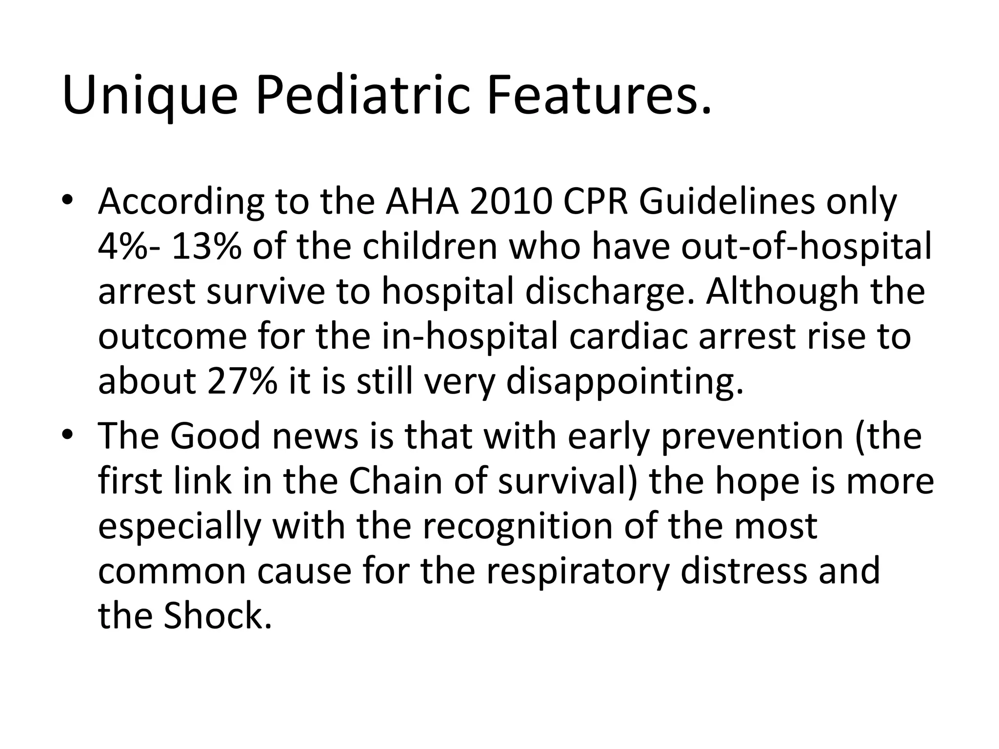
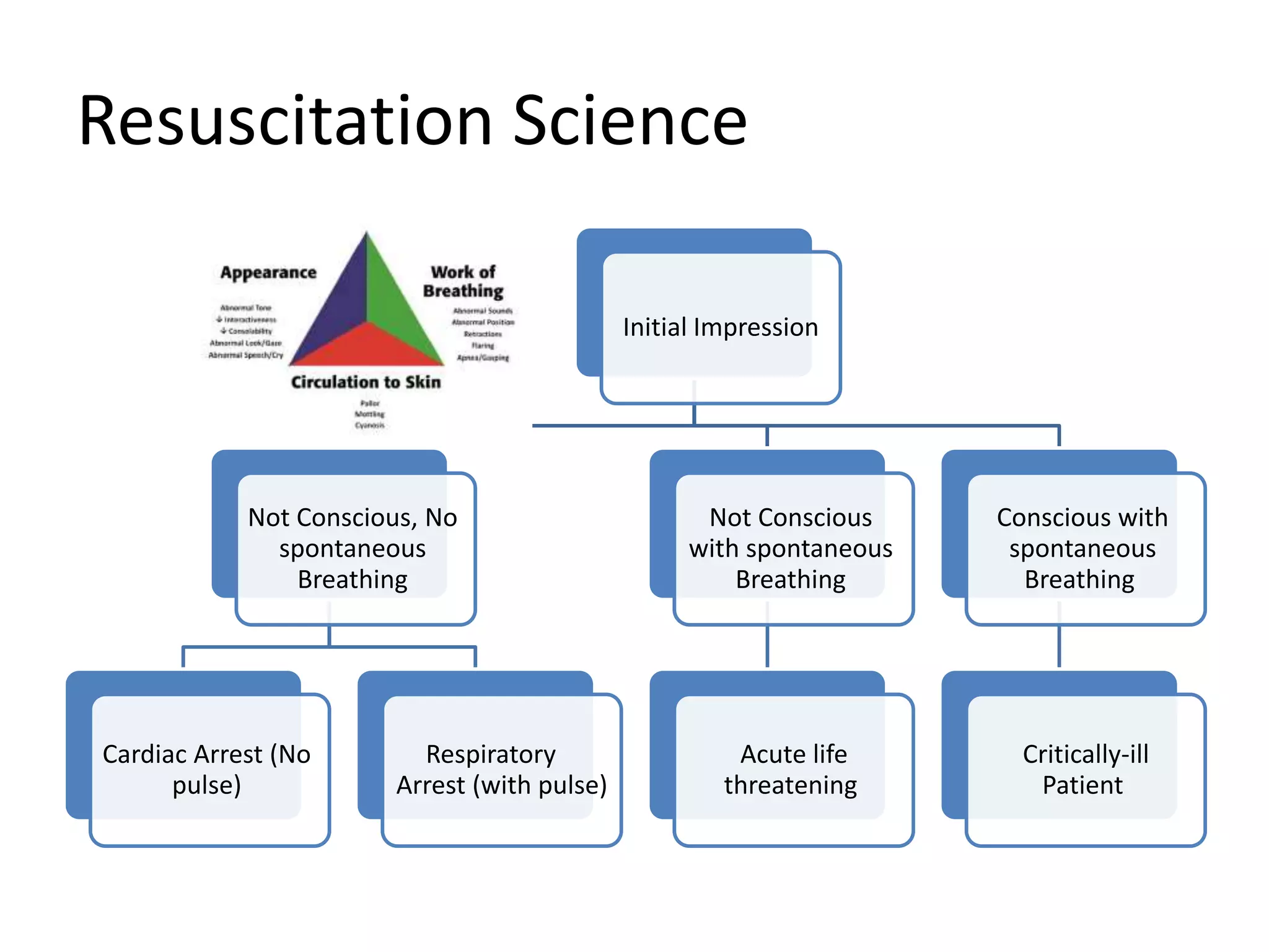
Dr. May Amr
Muhammad
AHA Senior Instructor
(BLS, FA, ACLS, PALS,
PEARS programs).
Resuscitation Science
• Resuscitation is the process of correcting
physiological disorders in an acutely unwell
patient (either arrested or an acute life
threatening condition).
• Although basic life support skills are essential
for all healthcare providers, the action taken
prior to the arrest is essential for either arrest
prevention or for better outcomes.
1.CHILD.
2.INFANT.
3
Who is ……….?
Unique Pediatric Features.
• According to the AHA 2010 CPR Guidelines only
4%- 13% of the children who have out-of-hospital
arrest survive to hospital discharge. Although the
outcome for the in-hospital cardiac arrest rise to
about 27% it is still very disappointing.
• The Good news is that with early prevention (the
first link in the Chain of survival) the hope is more
especially with the recognition of the most
common cause for the respiratory distress and
the Shock.
Resuscitation Science
Initial Impression
Not Conscious, No
spontaneous
Breathing
Cardiac Arrest (No
pulse)
Respiratory
Arrest (with pulse)
Not Conscious
with spontaneous
Breathing
Acute life
threatening
Conscious with
spontaneous
Breathing
Critically-ill
Patient
Pediatric Pre-arrest conditions
R
e
s
p
i
r
a
t
o
r
y
Upper Airway Obstruction
Lower Airway Obstruction
Lung Tissue Disease
Disorder Control of Breathing
Pediatric Pre-arrest conditions
S
h
o
c
k
Hypovolemic
Obstructive
Distributive
Cardiogenic
Pediatric Pre-arrest conditions
C
a
r
d
i
a
c
Tachycardia
Bradycardia
Acute Life threatening Conditions
• In the all age groups especially the pediatric
age group the medical conditions that affects
the conscious level with subsequent affection
of the airway are categorized as acute life
threatening conditions.
• This affection could rise from either central
disorder of breathing or severe bradycardia.
Pediatric Chain of Survival
Systematic Approach
• Open
• Clear
• Maintainable
AirwayA
• Respiratory Rate and Pattern.
• Respiratory Effort
• Chest Movement and Expansion.
• Abnormal Lung and Airway Sounds.
• Oxygen Saturation by Pulse Oximeter (≥94%)
BreathingB
• Heart Rate
• Pulse (Central and Peripheral)
• Capillary Refill Time
• Skin Color and Temperature.
• Blood Pressure.
CirculationC
• AVPU
• Pupils Size and Reaction to light.
• Random Blood Sugar.
DisabilityD
• Skin Appearance
• Temperature.
ExposureE
Systematic Approach
• Why this approach?
Following the Systematic gives both the healthcare provider an
evidence based system and the patient the maximum
opportunity for the success.
For the patient it guarantee the beginning with the most acute
life threatening condition that kills first.
WHAT KILLS FIRST,
TREATED FIRST.
Airway
• Airway Anatomy Differences:
Airway
• Opening the Airway:
Airway
• Opening the Airway:
Airway
• Clearing the Airway:
Airway
• Maintaining Airway Patency:
Airway
• Maintaining Airway Patency (Adjuvants):
Airway
• Maintaining Airway Patency (Adjuvants):
Airway
• Maintaining Airway Patency (Advanced):
1- Supraglotic Devices:
Airway
• Maintaining Airway Patency (Advanced):
1- Infraglotic Devices:
Breathing
• Oxygen Delivery Systems:
1- Low-Flow systems:
Breathing
• Oxygen Delivery Systems:
2- High-Flow systems:
Breathing
• Assisted Ventilations (Bag-Valve Mask Device):
Breathing
• Assisted Ventilations (Bag-Valve Mask Device):
Breathing
• Monitoring:
Breathing
• N.B: Waveform Capnography:
Pediatric Pre-arrest conditions
R
e
s
p
i
r
a
t
o
r
y
Upper Airway Obstruction
Lower Airway Obstruction
Lung Tissue Disease
Disorder Control of Breathing
Respiratory Problems:
• Upper Airway Obstruction:
1. Stridor, Barking cough, Snoring.
• Lower Airway Obstruction:
1-Wheezes, Prolonged Expiration.
• Lung Tissue Disease:
1- Grunting, crackles.
• Disorder Control of Breathing
1- Irregular respiratory pattern, inadequate
respiration.
Respiratory Problems:
• Upper Airway Obstruction:
Management:
• Lower Airway Obstruction:
Management:
• Lung Tissue Disease:
Management:
• Disorder Control of Breathing
Management:
Circulation
• Heart Rate:
Circulation
• Heart Rate:
Circulation
• Heart Rhythm
Circulation
• Circulation Access
1- Intra-venous Cannulation:
Circulation
• Circulation Access:
2-Intra-ossous Cannulation
Pediatric Pre-arrest conditions
S
h
o
c
k
Hypovolemic
Obstructive
Distributive
Cardiogenic
Circulatory Problems:
• Hypovolemic
1-weak peripheral pulse, delayed capillary refill.
• Distributive
1- (May be) Strong peripheral pulse, brisk
capillary refill.
Pediatric Pre-arrest conditions
C
a
r
d
i
a
c
Tachycardia
Bradycardia
Cardiac Problems (Bradycardia)
Disability
The child is awake giving appropriate
response.(according to age).
Alert
The child response only when called by name or
on loudness.
Voice
The child response only to painful stimuli as
rubbing the chest bone with finger knuckles.
Pain
The child doesn’t respond to any stimulus.
Unrespons
ive
Exposure
Possible ProblemAppearanceTypes of Purple Skin
Low platelet count.Tiny dotsPetichiea
Severe infection or
septic shock.
Larger SpotsPurpura
Pediatric Chain of Survival
BLS Algorithm
C •Chest Compressions
A •Airway
B •Breathing
44
BLS Guidelines (Since 2010)
• 2- Emphasis on High Quality CPR:
I. START within 10 sec. of arrest recognition.
II. PUSH HARD: Compression rate AT LEAST 100
-120 min.
III.PUSH FAST: Compression depth AT LEAST 5
and not more than 6 cm in children and 4 cm
in infants or 1/3 of the AP chest diameter).
IV. ALLOW COMPLETE CHEST RECOIL.
V. Minimize interruptions.
VI. Give effective breaths.
VII.Avoid excessive ventilations.
VIII.Early use of the feedback devices.
45
BLS in Guidelines (since2010)
3- The routine use of cricoid pressure is
no more recommended.
4- No Look, Listen, and Feel .
5- De- emphasis on the pulse check (from
5-10 sec only and if any doubt start chest
compressions).
6-Use of AED for infants:
Manual is preferred than Automated.
If not available use the pediatric dose
attenuator.
If not available use the Adult AED.
7- Team Approach to CPR.
46
BLS in Guidelines (since 2010)
1.Check Response
Child PEDIATRIC
47
48
BLS
2.Activate EMS and ask for AED.
Child Infant
49
BLS
3. Scan for Breathing and Check PULSE simultaneously.
Hand & Body Position. Don’t Forget.
I. START within 10 sec. of
arrest recognition.
II. PUSH HARD: Compression
rate of 100 -120/ min.
III. PUSH FAST: Compression
depth of 5-6 cm in adults, (5
-6 cm in children and 4-5 cm
in infants or 1/3 of the AP
chest diameter).
IV. ALLOW COMPLETE CHEST
RECOIL.
V. Minimize interruptions.
50
BLS
4. Start Chest Compressions (Child).
CHILD INFANT
51
BLS
4. Start Chest Compressions (CLILD & INFANT).
I. Open Airway (Head Tilt – Chin Lift / Jaw
Thrust).
II. Give Breath (Mouth to Barrier/ Mouth to
mouth).
III. Use of airway Adjuvant.
IV. Use of Advanced Airways.
52
BLS
5. Give Breath.
Power Button.
53
BLS
6. Use AED.
ADULT PEDIARTIC
54
BLS
6. Use AED (Lead Position).
Questions
Take Home
Pediatric Bls for nurses

Pediatric Bls for nurses

Editor's Notes

  • #45 It was……. Only delay breathing for app. 18 sec or less in adult (in child and infant less than 9 sec.)
  • #51 For Adults.
  • #55 Special situations: 1-Hairy Chest. 2-