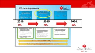
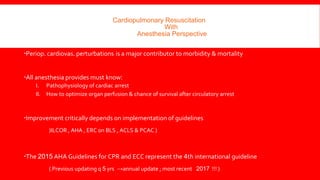
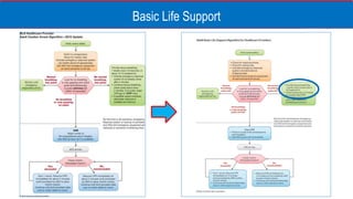
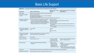
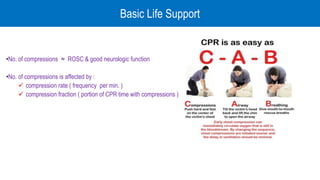
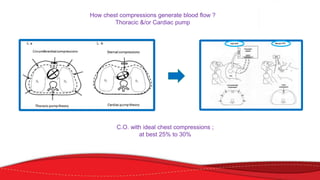
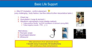
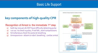
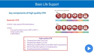
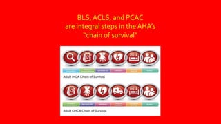
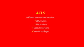
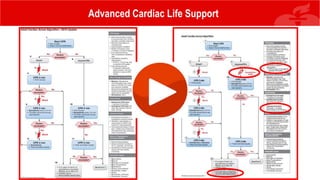
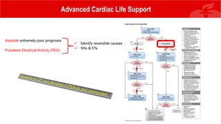
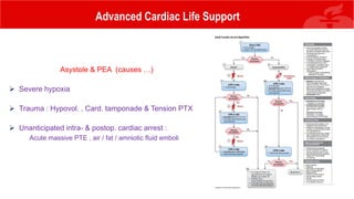
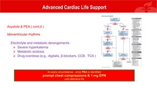
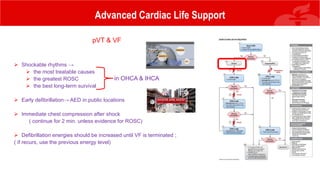
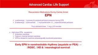
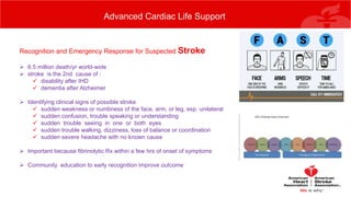
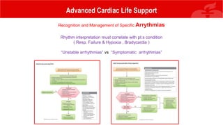
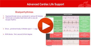
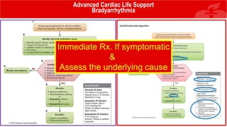
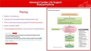
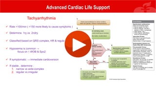
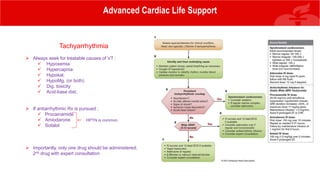
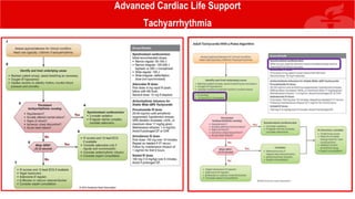
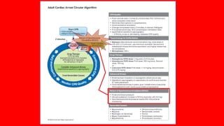
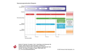
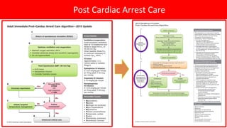
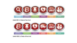
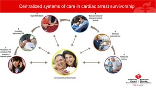
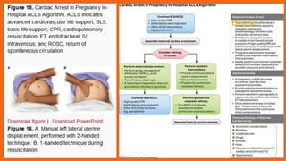
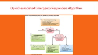
Cardiopulmonary Resuscitation
with
Anesthesia perspective
Shiraz University of Medical Sciences – Shiraz , Iran
April 2021
Mansoor Masjedi MD
Associate prof. of anesthesia , Critical care consultant
2021
2010 2015 2020
31% 46% 62%
5
Abbreviations used in this presentation
 ILCOR: International Liaison Committee on Resuscitation
 AHA : American Heart Association
 ERC : European Resuscitation Council
 FCTC : Fars Cardiopulmonary Resuscitation Training Center
 OHCA : Out of Hospital Cardiac Arrest
 IHCAtserrA caidraC latipsoH nI :
 BLS :Basic Life Support
 ACLS :Advanced Cardiovascular Life Support
 PALS : Pediatric Advanced Life Support
 NRP : Neonatal Resuscitation Program
 HCW : Health Care Worker
 AED :Automated External Defibrillator
 ROSC : Return Of Spontaneous Circulation
Abbreviation
s
Cardiopulmonary Resuscitation
With
Anesthesia Perspective
Periop. cardiovas. perturbations is a major contributor to morbidity & mortality
All anesthesia provides must know:
I. Pathophysiology of cardiac arrest
II. How to optimize organ perfusion & chance of survival after circulatory arrest
Improvement critically depends on implementation of guidelines
(ILCOR , AHA , ERC on BLS , ACLS & PCAC )
The 2015 AHA Guidelines for CPR and ECC represent the 4th international guideline
( Previous updating q 5 yrs →annual update ; most recent 2017 !!! )
Cardiopulmonary Resuscitation
With
Anesthesia Perspective
 >
500,000 deaths/yr in USA
 70
% of OHCAs occur at home, 50% unwitnessed
Significant advances in resuscitation science, but still:
 OHCA ≈
10.4%
) DH litnu SME yb RPC (
IHCA ≈
22.3% to 25.5% HD
Sudden cardiac arrest (SCA)
A complex and dynamic process ;
Forward blood flow continues until pressure equilibrium reaches
1. Aorta & Rt. atrium ( systemic )
2. Pulmonary artery & lt. atrium ( pulmonary )
As pressure gradient diminishes,
lt heart filling is decreased, Rt heart filling is increased
& venous capacitance vessels become increasingly distended
Arterial & venous equilibration (≈ 5 min. after no-flow cardiac arrest)
coronary & cerebral blood flows stop
Sudden cardiac arrest (SCA)
The goal of CPR is to :
1. Maintain O2 & blood supply to vital organs
2. Restore spontaneous circulation
3. Minimize post-resuscitation organ injury
4. Improve survival & neurologic outcome
Basic Life Support
Basic Life Support
Basic Life Support
Basic Life Support
•No. of compressions ≈ ROSC & good neurologic function
•No. of compressions is affected by :
 compression rate ( frequency per min. )
 compression fraction ( portion of CPR time with compressions )
How chest compressions generate blood flow ?
Thoracic &/or Cardiac pump
C.O. with ideal chest compressions ;
at best 25% to 30%
Basic Life Support
Automated External Defibrillators and Manual Defibrillation
VF & PVT are the most common cardiac arrhythmias encountered during witnessed
cardiac arrest
CPR prolongs tissue viability and the duration of VF by providing oxygen and energy
substrate, but cannot convert the arrhythmia to an organized rhythm in most
circumstances
Basic Life Support
1) Uninterrupted chest compressions while ;
 Attach defib. Pads
 Rhythm analysis is not occurring
2) Resume Chest compressions immediately after shock
3) Rhythm analysis after 2 min. of compressions & breathings
4) Defibrillation only for VF & rapid VT
Basic Life Support
Single versus Stacked Defibrillation
 2-min. chest compressions after each shock vs immediate successive shocks
for persistent VF
The rationale : when VF is terminated → a brief period of asystole or PEA ( no perfusion ) →
necessitating chest compressions to provide organ perfusion & circulation of drugs
 Stacked defibrillation , only during ;
1. cardiac surgery
2. cardiac cath. Lab.
( where invasive monitoring & defibrillation pads are in place already )
Basic Life Support
Determination of Efficiency of Cardiopulmonary Resuscitation
 Optimal assessment without interrupting chest compressions during CPR & ROSC;
 ETCO2 (quantitative waveform with CMV )
 Invasive arterial BP monitoring
 Other monitorings
 coronary perfusion pressure
 arterial relaxation pressure
 central venous oxygen saturation
 2010 AHA Guideline ;
PETCO2 >10 pre-intubation & > 20 mmHg post-intubation predicted ROSC
No trials showed titrating efforts during CPR , improved survival or neurologic outcome
Basic Life Support
Airway Management and Ventilation
 In early minutes after card. arrest, oxygen delivery is limited more by blood flow than AO2
 Chest compressions are the priority except asphyxiation, drowning or suffocation
 HCPs must determine; BMV , ETT or SGA ( Inadequate evidence to show a difference in survival)
 Use maximum FiO2 during CPR
2015 AHA/Guidelines
Either a BVM or an advanced airway may be used in both IHCA & OHCA
Basic Life Support
 After ET Intubation , confirm placement
(very challenging ; body habitus, low-flow status & other resuscitative tasks )
1. Chest rise
2. Auscultation ( lungs & stomach )
3. Waveform capnography (most reliable method)
 False-positive results : air/CO2 insufflation of stomach during BMV
 False-negative results : PTE , ↓C.O.
Alternatives ;
4. Fiberoptic scope
5. Esophageal detector
6. Ultrasound
After placement of an advanced airway
1 breath every 6 seconds (10 breaths/min)
with continuous chest compressions
Basic Life Support
key components of high-quality CPR
Recognition of Arrest is the immediate 1st step
1) both lay rescuers & HCP have difficulty detecting pulse
2) <10 sec. to check a pulse, if not felt , start compressions
3) Simultaneous check for pulse & breathing
4) Unresponsive + absent or abnl. breathing ₌ cardiac arrest
Basic Life Support
key components of high-quality CPR
Bystander CPR
OHCA : high-quality CPR & defibrillation
IHCA :
recognition before arrest ( MET or RRT ) +
high-quality CPR +
defibrillation
High-quality CPR
1. Compression rate ( 100-120)
2. Compression depth ( 5-6 cm)
3. Allowing complete chest recoil after each compression
4. Minimizing interruptions in compressions
5. Avoiding excessive ventilation
Advanced Cardiac
Life Support
BLS, ACLS, and PCAC
are integral steps in theAHA’s
“chain of survival”
ACLS
Different interventions based on
ECG rhythm
Medications
Special situations
New technologies
Advanced Cardiac Life Support
Asystole extremely poor prognosis
Pulseless Electrical Activity (PEA)
 Identify reversible causes
 5Hs & 5Ts
Advanced Cardiac Life Support
Asystole & PEA (causes …)
 Severe hypoxia
 Trauma : Hypovol. , Card. tamponade & Tension PTX
 Unanticipated intra- & postop. cardiac arrest :
Acute massive PTE , air / fat / amniotic fluid emboli
Advanced Cardiac Life Support
Asystole & PEA ( cont,d )
Idioventricular rhythms
Electrolyte and metabolic derangements :
 Severe hyperkalemia
 Metabolic acidosis
 Drug overdose (e.g., digitalis, β-blockers, CCB , TCA )
In every circumstance , once PEA is identified
prompt chest compressions & 1 mg EPN
until definitive Rx
Advanced Cardiac Life Support
pVT & VF
 Shockable rhythms →
 the most treatable causes
 the greatest ROSC
 the best long-term survival
 Early defibrillation→ AED in public locations
 Immediate chest compression after shock
( continue for 2 min. unless evidence for ROSC)
 Defibrillation energies should be increased until VF is terminated ;
( if recurs, use the previous energy level)
in OHCA & IHCA
Advanced Cardiac Life Support
Resuscitation Medications During Cardiac Arrest
EPN
 α-adrenergic : ↑coronary & cerebral perfusion pressure during CPR
 β-adrenergic : controversial ; ↑ myocardial work & ↓ subendocardial perfusion
Thus standard-dose ; 1 mg q 3-5 minutes
 High-dose EPN , exceptions :
 β-blocker overdose
 Calcium channel blocker overdose
 EPN is titrated to real-time physiologically monitored parameters
Early EPN in nonshockable rhythms (asystole or PEA) →
↑ROSC, ↑HD & ↑neurological survival
Advanced Cardiac Life Support
Antiarrthymia Medications
 During shock-refractory VF/pVT →
restoration & maintenance of ROSC
 Amiodarone in VF/pVT if unresponsive to CPR, defib. & vasopressor
( lidocaine as an alternative )
 Routine use of Mg for VF/pVT is not recommended
 Routine use of NaHCO3 in cardiac arrest is not recommended
Advanced Cardiac Life Support
 CPR in Mechanical Circulatory Support
MCS : ↑ed use in End-stage heart failure ( LV , RV , Both & TAH* )
*TAH : Total Artificial Heart
 Hemodynami-cally stable PEA ( pseudo-PEA)
 Difficult :
Lack of a pulse → NIBP , SpO2 ???!!!
 Assess tissue perfusion ;
skin color , capillary refill & mental status
Advanced Cardiac Life Support
 CPR in Mechanical Circulatory Support
 Total Artificial Heart
 No ECG
 Chest compressions : ineffective
 Antiarrhythmic drugs & D/C shock : futile
 Vasopressors used in ACLS are contraindicated
(↑ afterload → complete hemodynamic collapse & worsen TAH function)
The only therapeutic option : restore mechanical function of the device
 One liter IV N/S
 Assist ventilation
 Transport to hospital ASAP
Advanced Cardiac Life Support
CPR Using a Mechanical Cardiopulmonary Resuscitation Device
 Manual chest compressions limitations :
 30% of C.O. at best
 Fatigue
 Varying skill levels & training
 Pauses during defibrillation & switch of rescuers
 During transport
 Initial studies with MCD
 improved organ perfusion pressures
 enhanced cerebral blood flow
 higher end-tidal CO2
 A recent large multicenter randomized controlled trial showed no survival advantage ?!
(long pauses : device application time 36.0 seconds)
AHA Guideline 2015
manual chest compressions : standard of care
but
mechanical CPR devices , alternate in specific settings
(e.g., limited rescuers , prolonged CPR, during hypothermic cardiac arrest, during preparation for ECPR)
FEEL free to do echo in CPR - M.Masjedi MD
American Heart Association Scientific Sessions 2017
Role of POCUS in CA
IHCA/OHCA
FEEL free to do echo in CPR - M.Masjedi MD
Are we doing CPR right ?
AHA ACLS 2015 Recommendation - Updated
 Myocardial contractility
 Treatable causes of cardiac
arrest
 Hypovolemia
 Tension Pneumothorax
 Pulmonary embolism
 Pericardial tamponade
 Monitoring
 Prognosis
If a qualified sonographer is present
& Ultrasound does not interfere with the standard protocol
Then
It may be cosidered as an adjunct
Advanced Cardiac Life Support
Card./Resp. arrest & Opioid overdose
 AHA Guideline for opioid addicts:
If unresponsive & no Nl breathing but a pulse → BLS + IM or intranasal naloxone
 Ideal dose of naloxone is unknown ; 0.04 to 0.4 mg IV or IM
 Goal of Rx ;
 Provide patent airway & ventilation
 Prevent resp. & card. arrest without provoking severe opioid withdrawal
Advanced Cardiac Life Support
Recognition and Emergency Response for Suspected Stroke
 6.5 million death/yr world-wide
 stroke is the 2nd cause of :
 disability after IHD
 dementia after Alzheimer
 Identifying clinical signs of possible stroke
 sudden weakness or numbness of the face, arm, or leg, esp. unilateral
 sudden confusion, trouble speaking or understanding
 sudden trouble seeing in one or both eyes
 sudden trouble walking, dizziness, loss of balance or coordination
 sudden severe headache with no known cause
 Important because fibrinolytic Rx within a few hrs of onset of symptoms
 Community education to early recognition improve outcome
Advanced Cardiac Life Support
Recognition and Management of Specific Arrythmias
Rhythm interpretation must correlate with pt.s condition
( Resp. Failure & Hypoxia , Bradycardia )
“Unstable arrhythmias” vs “Symptomatic arrhythmias”
Advanced Cardiac Life Support
Bradyarrhythmias
1. Supraventricular (sinus, junctional or various AV blocks)
2. Ventricular (complete heart block with a very slow idiovent.
escape rhythm)
 Sinus , junctional brady. & Mobitz type I ≈ ↑ vagal tone
 AVN blocks : first, second & third degree
Advanced Cardiac Life Support
Bradyarrhythmia
Immediate Rx. If symptomatic
&
Assess the underlying cause
Pacing
 Sedation in all awake pts
 If pacing is not successful consider acidemia & elect. abnl.
 TCP is a temporary measure be prepared for transvenous pacing
 Expert consultation ASAP
 Transesophageal atrial pacing : Intraop supravent. bradyarrhythmias
Advanced Cardiac Life Support
Bradyarrhythmia
Advanced Cardiac Life Support
Tachyarrhythmia
 Rate >100/min ( >150 more likely to cause symptoms )
 Determine: 1ry vs 2ndry
 Classified based on QRS complex, HR & regularity
 Hypoxemia is common →
focus on ↑ WOB & Spo2
 If symptomatic → immediate cardioversion
 If stable , determine :
1. narrow vs wide-complex
2. regular vs irregular
Advanced Cardiac Life Support
Tachyarrhythmia
 Always seek for treatable causes of VT :
 Hypoxemia
 Hypercapnia
 Hypokal.
 HypoMg. (or both)
 Dig. toxicity
 Acid-base dist.
 If antiarrhythmic Rx is pursued ;
 Procainamide
 Amiodarone
 Sotalol
 Importantly, only one drug should be administered;
2nd drug with expert consultation
HPTN is common
Advanced Cardiac Life Support
Tachyarrhythmia
Post Cardiac Arrest Care
 Hypoxemia , ischemia & reperfusion → MODS
 PCAC :
 Dx. & Rx. of Cause of cardiac arrest
 Assessment & mitigation of ischemia-reperfusion injury
 Severity vary widely among organ systems & pts
 Care must be tailored
Ashish R. Panchal. Circulation. Part 3: Adult Basic and Advanced Life
Support: 2020 American Heart Association Guidelines for
Cardiopulmonary Resuscitation and Emergency Cardiovascular Care,
Volume: 142, Issue: 16_suppl_2, Pages: S366-S468, DOI:
(10.1161/CIR.0000000000000916) © 2020 American Heart Association, Inc.
Post Cardiac Arrest Care
Recovery expectations
and
survivorship plans
Centralized systems of care in cardiac arrest survivorship
Roadmap to recovery in cardiac arrest survivorship
Early bird catches the worm Early morning walk to the hospital
Cardiac Arrest
in
special situations
Opioid-associated Emergency Responders Algorithm
23/08/1442

CPR with anesthesia perspective 2021

  • 1.
    Cardiopulmonary Resuscitation with Anesthesia perspective ShirazUniversity of Medical Sciences – Shiraz , Iran April 2021 Mansoor Masjedi MD Associate prof. of anesthesia , Critical care consultant
  • 2.
  • 4.
  • 5.
    5 Abbreviations used inthis presentation  ILCOR: International Liaison Committee on Resuscitation  AHA : American Heart Association  ERC : European Resuscitation Council  FCTC : Fars Cardiopulmonary Resuscitation Training Center  OHCA : Out of Hospital Cardiac Arrest  IHCAtserrA caidraC latipsoH nI :  BLS :Basic Life Support  ACLS :Advanced Cardiovascular Life Support  PALS : Pediatric Advanced Life Support  NRP : Neonatal Resuscitation Program  HCW : Health Care Worker  AED :Automated External Defibrillator  ROSC : Return Of Spontaneous Circulation Abbreviation s
  • 6.
    Cardiopulmonary Resuscitation With Anesthesia Perspective Periop.cardiovas. perturbations is a major contributor to morbidity & mortality All anesthesia provides must know: I. Pathophysiology of cardiac arrest II. How to optimize organ perfusion & chance of survival after circulatory arrest Improvement critically depends on implementation of guidelines (ILCOR , AHA , ERC on BLS , ACLS & PCAC ) The 2015 AHA Guidelines for CPR and ECC represent the 4th international guideline ( Previous updating q 5 yrs →annual update ; most recent 2017 !!! )
  • 7.
    Cardiopulmonary Resuscitation With Anesthesia Perspective > 500,000 deaths/yr in USA  70 % of OHCAs occur at home, 50% unwitnessed Significant advances in resuscitation science, but still:  OHCA ≈ 10.4% ) DH litnu SME yb RPC ( IHCA ≈ 22.3% to 25.5% HD
  • 8.
    Sudden cardiac arrest(SCA) A complex and dynamic process ; Forward blood flow continues until pressure equilibrium reaches 1. Aorta & Rt. atrium ( systemic ) 2. Pulmonary artery & lt. atrium ( pulmonary ) As pressure gradient diminishes, lt heart filling is decreased, Rt heart filling is increased & venous capacitance vessels become increasingly distended Arterial & venous equilibration (≈ 5 min. after no-flow cardiac arrest) coronary & cerebral blood flows stop
  • 9.
    Sudden cardiac arrest(SCA) The goal of CPR is to : 1. Maintain O2 & blood supply to vital organs 2. Restore spontaneous circulation 3. Minimize post-resuscitation organ injury 4. Improve survival & neurologic outcome
  • 10.
  • 11.
  • 12.
  • 13.
    Basic Life Support •No.of compressions ≈ ROSC & good neurologic function •No. of compressions is affected by :  compression rate ( frequency per min. )  compression fraction ( portion of CPR time with compressions )
  • 14.
    How chest compressionsgenerate blood flow ? Thoracic &/or Cardiac pump C.O. with ideal chest compressions ; at best 25% to 30%
  • 15.
    Basic Life Support AutomatedExternal Defibrillators and Manual Defibrillation VF & PVT are the most common cardiac arrhythmias encountered during witnessed cardiac arrest CPR prolongs tissue viability and the duration of VF by providing oxygen and energy substrate, but cannot convert the arrhythmia to an organized rhythm in most circumstances
  • 16.
    Basic Life Support 1)Uninterrupted chest compressions while ;  Attach defib. Pads  Rhythm analysis is not occurring 2) Resume Chest compressions immediately after shock 3) Rhythm analysis after 2 min. of compressions & breathings 4) Defibrillation only for VF & rapid VT
  • 17.
    Basic Life Support Singleversus Stacked Defibrillation  2-min. chest compressions after each shock vs immediate successive shocks for persistent VF The rationale : when VF is terminated → a brief period of asystole or PEA ( no perfusion ) → necessitating chest compressions to provide organ perfusion & circulation of drugs  Stacked defibrillation , only during ; 1. cardiac surgery 2. cardiac cath. Lab. ( where invasive monitoring & defibrillation pads are in place already )
  • 18.
    Basic Life Support Determinationof Efficiency of Cardiopulmonary Resuscitation  Optimal assessment without interrupting chest compressions during CPR & ROSC;  ETCO2 (quantitative waveform with CMV )  Invasive arterial BP monitoring  Other monitorings  coronary perfusion pressure  arterial relaxation pressure  central venous oxygen saturation  2010 AHA Guideline ; PETCO2 >10 pre-intubation & > 20 mmHg post-intubation predicted ROSC No trials showed titrating efforts during CPR , improved survival or neurologic outcome
  • 19.
    Basic Life Support AirwayManagement and Ventilation  In early minutes after card. arrest, oxygen delivery is limited more by blood flow than AO2  Chest compressions are the priority except asphyxiation, drowning or suffocation  HCPs must determine; BMV , ETT or SGA ( Inadequate evidence to show a difference in survival)  Use maximum FiO2 during CPR 2015 AHA/Guidelines Either a BVM or an advanced airway may be used in both IHCA & OHCA
  • 20.
    Basic Life Support After ET Intubation , confirm placement (very challenging ; body habitus, low-flow status & other resuscitative tasks ) 1. Chest rise 2. Auscultation ( lungs & stomach ) 3. Waveform capnography (most reliable method)  False-positive results : air/CO2 insufflation of stomach during BMV  False-negative results : PTE , ↓C.O. Alternatives ; 4. Fiberoptic scope 5. Esophageal detector 6. Ultrasound After placement of an advanced airway 1 breath every 6 seconds (10 breaths/min) with continuous chest compressions
  • 21.
    Basic Life Support keycomponents of high-quality CPR Recognition of Arrest is the immediate 1st step 1) both lay rescuers & HCP have difficulty detecting pulse 2) <10 sec. to check a pulse, if not felt , start compressions 3) Simultaneous check for pulse & breathing 4) Unresponsive + absent or abnl. breathing ₌ cardiac arrest
  • 22.
    Basic Life Support keycomponents of high-quality CPR Bystander CPR OHCA : high-quality CPR & defibrillation IHCA : recognition before arrest ( MET or RRT ) + high-quality CPR + defibrillation High-quality CPR 1. Compression rate ( 100-120) 2. Compression depth ( 5-6 cm) 3. Allowing complete chest recoil after each compression 4. Minimizing interruptions in compressions 5. Avoiding excessive ventilation
  • 23.
  • 24.
    BLS, ACLS, andPCAC are integral steps in theAHA’s “chain of survival”
  • 25.
    ACLS Different interventions basedon ECG rhythm Medications Special situations New technologies
  • 26.
  • 27.
    Advanced Cardiac LifeSupport Asystole extremely poor prognosis Pulseless Electrical Activity (PEA)  Identify reversible causes  5Hs & 5Ts
  • 28.
    Advanced Cardiac LifeSupport Asystole & PEA (causes …)  Severe hypoxia  Trauma : Hypovol. , Card. tamponade & Tension PTX  Unanticipated intra- & postop. cardiac arrest : Acute massive PTE , air / fat / amniotic fluid emboli
  • 29.
    Advanced Cardiac LifeSupport Asystole & PEA ( cont,d ) Idioventricular rhythms Electrolyte and metabolic derangements :  Severe hyperkalemia  Metabolic acidosis  Drug overdose (e.g., digitalis, β-blockers, CCB , TCA ) In every circumstance , once PEA is identified prompt chest compressions & 1 mg EPN until definitive Rx
  • 30.
    Advanced Cardiac LifeSupport pVT & VF  Shockable rhythms →  the most treatable causes  the greatest ROSC  the best long-term survival  Early defibrillation→ AED in public locations  Immediate chest compression after shock ( continue for 2 min. unless evidence for ROSC)  Defibrillation energies should be increased until VF is terminated ; ( if recurs, use the previous energy level) in OHCA & IHCA
  • 31.
    Advanced Cardiac LifeSupport Resuscitation Medications During Cardiac Arrest EPN  α-adrenergic : ↑coronary & cerebral perfusion pressure during CPR  β-adrenergic : controversial ; ↑ myocardial work & ↓ subendocardial perfusion Thus standard-dose ; 1 mg q 3-5 minutes  High-dose EPN , exceptions :  β-blocker overdose  Calcium channel blocker overdose  EPN is titrated to real-time physiologically monitored parameters Early EPN in nonshockable rhythms (asystole or PEA) → ↑ROSC, ↑HD & ↑neurological survival
  • 32.
    Advanced Cardiac LifeSupport Antiarrthymia Medications  During shock-refractory VF/pVT → restoration & maintenance of ROSC  Amiodarone in VF/pVT if unresponsive to CPR, defib. & vasopressor ( lidocaine as an alternative )  Routine use of Mg for VF/pVT is not recommended  Routine use of NaHCO3 in cardiac arrest is not recommended
  • 34.
    Advanced Cardiac LifeSupport  CPR in Mechanical Circulatory Support MCS : ↑ed use in End-stage heart failure ( LV , RV , Both & TAH* ) *TAH : Total Artificial Heart  Hemodynami-cally stable PEA ( pseudo-PEA)  Difficult : Lack of a pulse → NIBP , SpO2 ???!!!  Assess tissue perfusion ; skin color , capillary refill & mental status
  • 35.
    Advanced Cardiac LifeSupport  CPR in Mechanical Circulatory Support  Total Artificial Heart  No ECG  Chest compressions : ineffective  Antiarrhythmic drugs & D/C shock : futile  Vasopressors used in ACLS are contraindicated (↑ afterload → complete hemodynamic collapse & worsen TAH function) The only therapeutic option : restore mechanical function of the device  One liter IV N/S  Assist ventilation  Transport to hospital ASAP
  • 36.
    Advanced Cardiac LifeSupport CPR Using a Mechanical Cardiopulmonary Resuscitation Device  Manual chest compressions limitations :  30% of C.O. at best  Fatigue  Varying skill levels & training  Pauses during defibrillation & switch of rescuers  During transport  Initial studies with MCD  improved organ perfusion pressures  enhanced cerebral blood flow  higher end-tidal CO2  A recent large multicenter randomized controlled trial showed no survival advantage ?! (long pauses : device application time 36.0 seconds) AHA Guideline 2015 manual chest compressions : standard of care but mechanical CPR devices , alternate in specific settings (e.g., limited rescuers , prolonged CPR, during hypothermic cardiac arrest, during preparation for ECPR)
  • 37.
    FEEL free todo echo in CPR - M.Masjedi MD American Heart Association Scientific Sessions 2017 Role of POCUS in CA IHCA/OHCA
  • 38.
    FEEL free todo echo in CPR - M.Masjedi MD Are we doing CPR right ?
  • 39.
    AHA ACLS 2015Recommendation - Updated  Myocardial contractility  Treatable causes of cardiac arrest  Hypovolemia  Tension Pneumothorax  Pulmonary embolism  Pericardial tamponade  Monitoring  Prognosis If a qualified sonographer is present & Ultrasound does not interfere with the standard protocol Then It may be cosidered as an adjunct
  • 40.
    Advanced Cardiac LifeSupport Card./Resp. arrest & Opioid overdose  AHA Guideline for opioid addicts: If unresponsive & no Nl breathing but a pulse → BLS + IM or intranasal naloxone  Ideal dose of naloxone is unknown ; 0.04 to 0.4 mg IV or IM  Goal of Rx ;  Provide patent airway & ventilation  Prevent resp. & card. arrest without provoking severe opioid withdrawal
  • 41.
    Advanced Cardiac LifeSupport Recognition and Emergency Response for Suspected Stroke  6.5 million death/yr world-wide  stroke is the 2nd cause of :  disability after IHD  dementia after Alzheimer  Identifying clinical signs of possible stroke  sudden weakness or numbness of the face, arm, or leg, esp. unilateral  sudden confusion, trouble speaking or understanding  sudden trouble seeing in one or both eyes  sudden trouble walking, dizziness, loss of balance or coordination  sudden severe headache with no known cause  Important because fibrinolytic Rx within a few hrs of onset of symptoms  Community education to early recognition improve outcome
  • 42.
    Advanced Cardiac LifeSupport Recognition and Management of Specific Arrythmias Rhythm interpretation must correlate with pt.s condition ( Resp. Failure & Hypoxia , Bradycardia ) “Unstable arrhythmias” vs “Symptomatic arrhythmias”
  • 43.
    Advanced Cardiac LifeSupport Bradyarrhythmias 1. Supraventricular (sinus, junctional or various AV blocks) 2. Ventricular (complete heart block with a very slow idiovent. escape rhythm)  Sinus , junctional brady. & Mobitz type I ≈ ↑ vagal tone  AVN blocks : first, second & third degree
  • 44.
    Advanced Cardiac LifeSupport Bradyarrhythmia Immediate Rx. If symptomatic & Assess the underlying cause
  • 45.
    Pacing  Sedation inall awake pts  If pacing is not successful consider acidemia & elect. abnl.  TCP is a temporary measure be prepared for transvenous pacing  Expert consultation ASAP  Transesophageal atrial pacing : Intraop supravent. bradyarrhythmias Advanced Cardiac Life Support Bradyarrhythmia
  • 46.
    Advanced Cardiac LifeSupport Tachyarrhythmia  Rate >100/min ( >150 more likely to cause symptoms )  Determine: 1ry vs 2ndry  Classified based on QRS complex, HR & regularity  Hypoxemia is common → focus on ↑ WOB & Spo2  If symptomatic → immediate cardioversion  If stable , determine : 1. narrow vs wide-complex 2. regular vs irregular
  • 47.
    Advanced Cardiac LifeSupport Tachyarrhythmia  Always seek for treatable causes of VT :  Hypoxemia  Hypercapnia  Hypokal.  HypoMg. (or both)  Dig. toxicity  Acid-base dist.  If antiarrhythmic Rx is pursued ;  Procainamide  Amiodarone  Sotalol  Importantly, only one drug should be administered; 2nd drug with expert consultation HPTN is common
  • 48.
    Advanced Cardiac LifeSupport Tachyarrhythmia
  • 49.
  • 53.
    Post Cardiac ArrestCare  Hypoxemia , ischemia & reperfusion → MODS  PCAC :  Dx. & Rx. of Cause of cardiac arrest  Assessment & mitigation of ischemia-reperfusion injury  Severity vary widely among organ systems & pts  Care must be tailored
  • 54.
    Ashish R. Panchal.Circulation. Part 3: Adult Basic and Advanced Life Support: 2020 American Heart Association Guidelines for Cardiopulmonary Resuscitation and Emergency Cardiovascular Care, Volume: 142, Issue: 16_suppl_2, Pages: S366-S468, DOI: (10.1161/CIR.0000000000000916) © 2020 American Heart Association, Inc.
  • 55.
  • 56.
  • 58.
    Centralized systems ofcare in cardiac arrest survivorship
  • 59.
    Roadmap to recoveryin cardiac arrest survivorship
  • 60.
    Early bird catchesthe worm Early morning walk to the hospital
  • 61.
  • 63.
  • 64.