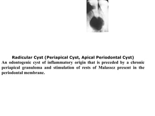
This lecture, which oriented to the level of mind of undergraduate students, discuss the topic of pulpectomy, its indications, contraindications, and procedural steps.
Visit us on Facebook:
https://www.facebook.com/iraqi.Dental.Academy
This lecture, which oriented to the level of mind of undergraduate students, discuss the topic of pulpectomy, its indications, contraindications, and procedural steps.
Visit us on Facebook:
https://www.facebook.com/iraqi.Dental.Academy
Definition: The ideal tooth extraction is painless removal of the whole tooth or tooth root with minimal trauma to the investing tissues so that the wound heals uneventfully and no post operative prosthetic problem is created.
All the mistakes are rectified.Complete and precise knowledge about EXODONTIA .I would like to again focus on compatibility of this ppt;some pictures differ from original one.Animations and Transitions added are not visible .Good for beginners to understand and remember.Images give you better way to grasp.Enjoy and have fun watching this ppt.
Exodontia /certified fixed orthodontic courses by Indian dental academy Indian dental academy
The Indian Dental Academy is the Leader in continuing dental education , training dentists in all aspects of dentistry and offering a wide range of dental certified courses in different formats.
Deep Bite| Braces Treatment| Certification Courses in Fixed Orthodontics in D...Dr. Rajat Sachdeva
Deep Bite
Excessive Overlaping of upper front teeth over the lower front teeth is deep bite.
Orthodontic Treatment through braces, Invisalign, Damon's Braces, Traditional braces, Orthognathic Surgeries.
Restorative and periodontal therapy, Habit Breaking appliances.
All the procedure performing by experienced one.
Dr. Sachdeva's Dental Institute, where you will learn to perform the procedures impeccably.
To Learn More, Call us:-+919818894041,01142464041
Follow Our Link:-
Google link:
https://business.google.com/dashboard/l/04970356233769420071
Facebook link for Dental Courses:
https://www.facebook.com/dentalcoursesdelhi/
Facebook link for Dental Treatments:
https://www.facebook.com/sachdevadental/
You tube Link:
https://www.youtube.com/user/drrajatsachdeva
Linkedin link:
https://www.linkedin.com/in/drrajatsachdeva/
Slideshare:
https://www.slideshare.net/drrajatsachdeva
Twitter Page :
https://twitter.com/drrajatsachdeva
Instagram page :
https://www.instagram.com/surgicalmasterrajat/
Practo Profile :
https://www.practo.com/delhi/doctor/dr-rajat-sachdeva-dentist
Blogger Profile :
http://drrajatsachdeva.blogspot.com/
Facial Aesthetics Facebook Page :
https://www.facebook.com/facialaesthetics.delhi
Facial Aesthetics you tube channel :
http://www.youtube.com/channel/UCheM4wF9nWGXJYOmScvsQNw
The Indian Dental Academy is the Leader in continuing dental education , training dentists in all aspects of dentistry and
offering a wide range of dental certified courses in different formats.for more details please visit
www.indiandentalacademy.com
Lung Cancer: Artificial Intelligence, Synergetics, Complex System Analysis, S...Oleg Kshivets
RESULTS: Overall life span (LS) was 2252.1±1742.5 days and cumulative 5-year survival (5YS) reached 73.2%, 10 years – 64.8%, 20 years – 42.5%. 513 LCP lived more than 5 years (LS=3124.6±1525.6 days), 148 LCP – more than 10 years (LS=5054.4±1504.1 days).199 LCP died because of LC (LS=562.7±374.5 days). 5YS of LCP after bi/lobectomies was significantly superior in comparison with LCP after pneumonectomies (78.1% vs.63.7%, P=0.00001 by log-rank test). AT significantly improved 5YS (66.3% vs. 34.8%) (P=0.00000 by log-rank test) only for LCP with N1-2. Cox modeling displayed that 5YS of LCP significantly depended on: phase transition (PT) early-invasive LC in terms of synergetics, PT N0—N12, cell ratio factors (ratio between cancer cells- CC and blood cells subpopulations), G1-3, histology, glucose, AT, blood cell circuit, prothrombin index, heparin tolerance, recalcification time (P=0.000-0.038). Neural networks, genetic algorithm selection and bootstrap simulation revealed relationships between 5YS and PT early-invasive LC (rank=1), PT N0—N12 (rank=2), thrombocytes/CC (3), erythrocytes/CC (4), eosinophils/CC (5), healthy cells/CC (6), lymphocytes/CC (7), segmented neutrophils/CC (8), stick neutrophils/CC (9), monocytes/CC (10); leucocytes/CC (11). Correct prediction of 5YS was 100% by neural networks computing (area under ROC curve=1.0; error=0.0).
CONCLUSIONS: 5YS of LCP after radical procedures significantly depended on: 1) PT early-invasive cancer; 2) PT N0--N12; 3) cell ratio factors; 4) blood cell circuit; 5) biochemical factors; 6) hemostasis system; 7) AT; 8) LC characteristics; 9) LC cell dynamics; 10) surgery type: lobectomy/pneumonectomy; 11) anthropometric data. Optimal diagnosis and treatment strategies for LC are: 1) screening and early detection of LC; 2) availability of experienced thoracic surgeons because of complexity of radical procedures; 3) aggressive en block surgery and adequate lymph node dissection for completeness; 4) precise prediction; 5) adjuvant chemoimmunoradiotherapy for LCP with unfavorable prognosis.
The prostate is an exocrine gland of the male mammalian reproductive system
It is a walnut-sized gland that forms part of the male reproductive system and is located in front of the rectum and just below the urinary bladder
Function is to store and secrete a clear, slightly alkaline fluid that constitutes 10-30% of the volume of the seminal fluid that along with the spermatozoa, constitutes semen
A healthy human prostate measures (4cm-vertical, by 3cm-horizontal, 2cm ant-post ).
It surrounds the urethra just below the urinary bladder. It has anterior, median, posterior and two lateral lobes
It’s work is regulated by androgens which are responsible for male sex characteristics
Generalised disease of the prostate due to hormonal derangement which leads to non malignant enlargement of the gland (increase in the number of epithelial cells and stromal tissue)to cause compression of the urethra leading to symptoms (LUTS
Explore natural remedies for syphilis treatment in Singapore. Discover alternative therapies, herbal remedies, and lifestyle changes that may complement conventional treatments. Learn about holistic approaches to managing syphilis symptoms and supporting overall health.
NVBDCP.pptx Nation vector borne disease control programSapna Thakur
NVBDCP was launched in 2003-2004 . Vector-Borne Disease: Disease that results from an infection transmitted to humans and other animals by blood-feeding arthropods, such as mosquitoes, ticks, and fleas. Examples of vector-borne diseases include Dengue fever, West Nile Virus, Lyme disease, and malaria.
Prix Galien International 2024 Forum ProgramLevi Shapiro
June 20, 2024, Prix Galien International and Jerusalem Ethics Forum in ROME. Detailed agenda including panels:
- ADVANCES IN CARDIOLOGY: A NEW PARADIGM IS COMING
- WOMEN’S HEALTH: FERTILITY PRESERVATION
- WHAT’S NEW IN THE TREATMENT OF INFECTIOUS,
ONCOLOGICAL AND INFLAMMATORY SKIN DISEASES?
- ARTIFICIAL INTELLIGENCE AND ETHICS
- GENE THERAPY
- BEYOND BORDERS: GLOBAL INITIATIVES FOR DEMOCRATIZING LIFE SCIENCE TECHNOLOGIES AND PROMOTING ACCESS TO HEALTHCARE
- ETHICAL CHALLENGES IN LIFE SCIENCES
- Prix Galien International Awards Ceremony
Title: Sense of Taste
Presenter: Dr. Faiza, Assistant Professor of Physiology
Qualifications:
MBBS (Best Graduate, AIMC Lahore)
FCPS Physiology
ICMT, CHPE, DHPE (STMU)
MPH (GC University, Faisalabad)
MBA (Virtual University of Pakistan)
Learning Objectives:
Describe the structure and function of taste buds.
Describe the relationship between the taste threshold and taste index of common substances.
Explain the chemical basis and signal transduction of taste perception for each type of primary taste sensation.
Recognize different abnormalities of taste perception and their causes.
Key Topics:
Significance of Taste Sensation:
Differentiation between pleasant and harmful food
Influence on behavior
Selection of food based on metabolic needs
Receptors of Taste:
Taste buds on the tongue
Influence of sense of smell, texture of food, and pain stimulation (e.g., by pepper)
Primary and Secondary Taste Sensations:
Primary taste sensations: Sweet, Sour, Salty, Bitter, Umami
Chemical basis and signal transduction mechanisms for each taste
Taste Threshold and Index:
Taste threshold values for Sweet (sucrose), Salty (NaCl), Sour (HCl), and Bitter (Quinine)
Taste index relationship: Inversely proportional to taste threshold
Taste Blindness:
Inability to taste certain substances, particularly thiourea compounds
Example: Phenylthiocarbamide
Structure and Function of Taste Buds:
Composition: Epithelial cells, Sustentacular/Supporting cells, Taste cells, Basal cells
Features: Taste pores, Taste hairs/microvilli, and Taste nerve fibers
Location of Taste Buds:
Found in papillae of the tongue (Fungiform, Circumvallate, Foliate)
Also present on the palate, tonsillar pillars, epiglottis, and proximal esophagus
Mechanism of Taste Stimulation:
Interaction of taste substances with receptors on microvilli
Signal transduction pathways for Umami, Sweet, Bitter, Sour, and Salty tastes
Taste Sensitivity and Adaptation:
Decrease in sensitivity with age
Rapid adaptation of taste sensation
Role of Saliva in Taste:
Dissolution of tastants to reach receptors
Washing away the stimulus
Taste Preferences and Aversions:
Mechanisms behind taste preference and aversion
Influence of receptors and neural pathways
Impact of Sensory Nerve Damage:
Degeneration of taste buds if the sensory nerve fiber is cut
Abnormalities of Taste Detection:
Conditions: Ageusia, Hypogeusia, Dysgeusia (parageusia)
Causes: Nerve damage, neurological disorders, infections, poor oral hygiene, adverse drug effects, deficiencies, aging, tobacco use, altered neurotransmitter levels
Neurotransmitters and Taste Threshold:
Effects of serotonin (5-HT) and norepinephrine (NE) on taste sensitivity
Supertasters:
25% of the population with heightened sensitivity to taste, especially bitterness
Increased number of fungiform papillae
Recomendações da OMS sobre cuidados maternos e neonatais para uma experiência pós-natal positiva.
Em consonância com os ODS – Objetivos do Desenvolvimento Sustentável e a Estratégia Global para a Saúde das Mulheres, Crianças e Adolescentes, e aplicando uma abordagem baseada nos direitos humanos, os esforços de cuidados pós-natais devem expandir-se para além da cobertura e da simples sobrevivência, de modo a incluir cuidados de qualidade.
Estas diretrizes visam melhorar a qualidade dos cuidados pós-natais essenciais e de rotina prestados às mulheres e aos recém-nascidos, com o objetivo final de melhorar a saúde e o bem-estar materno e neonatal.
Uma “experiência pós-natal positiva” é um resultado importante para todas as mulheres que dão à luz e para os seus recém-nascidos, estabelecendo as bases para a melhoria da saúde e do bem-estar a curto e longo prazo. Uma experiência pós-natal positiva é definida como aquela em que as mulheres, pessoas que gestam, os recém-nascidos, os casais, os pais, os cuidadores e as famílias recebem informação consistente, garantia e apoio de profissionais de saúde motivados; e onde um sistema de saúde flexível e com recursos reconheça as necessidades das mulheres e dos bebês e respeite o seu contexto cultural.
Estas diretrizes consolidadas apresentam algumas recomendações novas e já bem fundamentadas sobre cuidados pós-natais de rotina para mulheres e neonatos que recebem cuidados no pós-parto em unidades de saúde ou na comunidade, independentemente dos recursos disponíveis.
É fornecido um conjunto abrangente de recomendações para cuidados durante o período puerperal, com ênfase nos cuidados essenciais que todas as mulheres e recém-nascidos devem receber, e com a devida atenção à qualidade dos cuidados; isto é, a entrega e a experiência do cuidado recebido. Estas diretrizes atualizam e ampliam as recomendações da OMS de 2014 sobre cuidados pós-natais da mãe e do recém-nascido e complementam as atuais diretrizes da OMS sobre a gestão de complicações pós-natais.
O estabelecimento da amamentação e o manejo das principais intercorrências é contemplada.
Recomendamos muito.
Vamos discutir essas recomendações no nosso curso de pós-graduação em Aleitamento no Instituto Ciclos.
Esta publicação só está disponível em inglês até o momento.
Prof. Marcus Renato de Carvalho
www.agostodourado.com
Knee anatomy and clinical tests 2024.pdfvimalpl1234
This includes all relevant anatomy and clinical tests compiled from standard textbooks, Campbell,netter etc..It is comprehensive and best suited for orthopaedicians and orthopaedic residents.
- Video recording of this lecture in English language: https://youtu.be/lK81BzxMqdo
- Video recording of this lecture in Arabic language: https://youtu.be/Ve4P0COk9OI
- Link to download the book free: https://nephrotube.blogspot.com/p/nephrotube-nephrology-books.html
- Link to NephroTube website: www.NephroTube.com
- Link to NephroTube social media accounts: https://nephrotube.blogspot.com/p/join-nephrotube-on-social-media.html
Ethanol (CH3CH2OH), or beverage alcohol, is a two-carbon alcohol
that is rapidly distributed in the body and brain. Ethanol alters many
neurochemical systems and has rewarding and addictive properties. It
is the oldest recreational drug and likely contributes to more morbidity,
mortality, and public health costs than all illicit drugs combined. The
5th edition of the Diagnostic and Statistical Manual of Mental Disorders
(DSM-5) integrates alcohol abuse and alcohol dependence into a single
disorder called alcohol use disorder (AUD), with mild, moderate,
and severe subclassifications (American Psychiatric Association, 2013).
In the DSM-5, all types of substance abuse and dependence have been
combined into a single substance use disorder (SUD) on a continuum
from mild to severe. A diagnosis of AUD requires that at least two of
the 11 DSM-5 behaviors be present within a 12-month period (mild
AUD: 2–3 criteria; moderate AUD: 4–5 criteria; severe AUD: 6–11 criteria).
The four main behavioral effects of AUD are impaired control over
drinking, negative social consequences, risky use, and altered physiological
effects (tolerance, withdrawal). This chapter presents an overview
of the prevalence and harmful consequences of AUD in the U.S.,
the systemic nature of the disease, neurocircuitry and stages of AUD,
comorbidities, fetal alcohol spectrum disorders, genetic risk factors, and
pharmacotherapies for AUD.
ARTIFICIAL INTELLIGENCE IN HEALTHCARE.pdfAnujkumaranit
Artificial intelligence (AI) refers to the simulation of human intelligence processes by machines, especially computer systems. It encompasses tasks such as learning, reasoning, problem-solving, perception, and language understanding. AI technologies are revolutionizing various fields, from healthcare to finance, by enabling machines to perform tasks that typically require human intelligence.
New Directions in Targeted Therapeutic Approaches for Older Adults With Mantl...i3 Health
i3 Health is pleased to make the speaker slides from this activity available for use as a non-accredited self-study or teaching resource.
This slide deck presented by Dr. Kami Maddocks, Professor-Clinical in the Division of Hematology and
Associate Division Director for Ambulatory Operations
The Ohio State University Comprehensive Cancer Center, will provide insight into new directions in targeted therapeutic approaches for older adults with mantle cell lymphoma.
STATEMENT OF NEED
Mantle cell lymphoma (MCL) is a rare, aggressive B-cell non-Hodgkin lymphoma (NHL) accounting for 5% to 7% of all lymphomas. Its prognosis ranges from indolent disease that does not require treatment for years to very aggressive disease, which is associated with poor survival (Silkenstedt et al, 2021). Typically, MCL is diagnosed at advanced stage and in older patients who cannot tolerate intensive therapy (NCCN, 2022). Although recent advances have slightly increased remission rates, recurrence and relapse remain very common, leading to a median overall survival between 3 and 6 years (LLS, 2021). Though there are several effective options, progress is still needed towards establishing an accepted frontline approach for MCL (Castellino et al, 2022). Treatment selection and management of MCL are complicated by the heterogeneity of prognosis, advanced age and comorbidities of patients, and lack of an established standard approach for treatment, making it vital that clinicians be familiar with the latest research and advances in this area. In this activity chaired by Michael Wang, MD, Professor in the Department of Lymphoma & Myeloma at MD Anderson Cancer Center, expert faculty will discuss prognostic factors informing treatment, the promising results of recent trials in new therapeutic approaches, and the implications of treatment resistance in therapeutic selection for MCL.
Target Audience
Hematology/oncology fellows, attending faculty, and other health care professionals involved in the treatment of patients with mantle cell lymphoma (MCL).
Learning Objectives
1.) Identify clinical and biological prognostic factors that can guide treatment decision making for older adults with MCL
2.) Evaluate emerging data on targeted therapeutic approaches for treatment-naive and relapsed/refractory MCL and their applicability to older adults
3.) Assess mechanisms of resistance to targeted therapies for MCL and their implications for treatment selection
New Drug Discovery and Development .....NEHA GUPTA
The "New Drug Discovery and Development" process involves the identification, design, testing, and manufacturing of novel pharmaceutical compounds with the aim of introducing new and improved treatments for various medical conditions. This comprehensive endeavor encompasses various stages, including target identification, preclinical studies, clinical trials, regulatory approval, and post-market surveillance. It involves multidisciplinary collaboration among scientists, researchers, clinicians, regulatory experts, and pharmaceutical companies to bring innovative therapies to market and address unmet medical needs.
1. 1)Odontogenic infections & its complications
2)Extraction of erupted and unerupted teeth
3)Management of fractured primary tooth roots
4)Management of unerupte; impacted& supernumery
teeth
5)Management of oral lesions in the newborn as
Epstein's pearls, dental lamina cysts, Bohn's
nodules, and congenital epulis (Neumann's tumor)
6) Surgical opening of eruption cyst
7) Surgical excision of mucocele.
8) Oral structural anomalies as maxillary frenum,
mandibular labial frenum, mandibular lingual
frenum. Frenuloplasty and frenectomy (for
ankyloglossia)
2. • It is a localized, purulent form of apical
periodontitis.
• A fluctuant buccal or palatal swelling, with
or without a draining fistula.
• Regional adenopathy is usually present.
• If pus is draining, pain usually is not
severe.
1. Antibiotics are not necessary unless
concurrent cellulitis is present.
2. Acute incision and drainage of a fluctuant
area.
3.Definitive therapy is root canal treatment
or extraction.
3. •Cellulitis follow apical periodontitis
•Diffuse, tense, painful swelling of the
affected tissues occurs.
•Regional lymph-adenopathy, fever.
•The infection can spread into the major
facial spaces of the head and neck, with
the attendant risk of airway compromise.
4. • Maxillary infection spread to the
periorbital area, increasing the risk of
serious complications that include loss of
vision, cavernous sinus thrombosis, and
central nervous system involvement
5. • It is inflammation of the soft tissues
surrounding the crown of a partially erupted
tooth (wisdom tooth).
• Bacterial plaque and food debris accumulate
beneath the flap of gum covering the
partially erupted tooth.
• Inflammatory edema, as trauma from the
opposing tooth, leads to swelling of the flap,
pain, tenderness, and a bad taste caused by
pus oozing from beneath the flap.
• Regional lymph-adenopathy is common, and
cellulites and trismus can occur.
6. EXTRACTION
• It is painless removal of the whole tooth or
tooth root with minimal trauma to the investing
tissue, So that wounds heals uneventfully
without post operative problems.
• Avoid injury to soft tissues as tongue, lips,
gingiva & cheeks.
• Avoid injury to underlying developing
permanent & other hard tissues as bone&
adjacent or opposing teeth.
• Use radiograph to determine:-
• Size & shape of roots.
• Amount & directions of root resorption.
• Position & stage of development of
underlying permanent tooth.
• Any pathology.
7. Differences between Primary and
Permanent Teeth
• Size primary teeth are smaller in every
dimensions than permanent.
• Shape- crown of primary teeth are more
bulbous. The furcation of primary molar
root is positioned more cervically than
permanent.
• Physiology- root of primary teeth resorb
naturally where as in the permanent
resorption is normally a sign of pathology.
• Support- the bone of alveolus is much
more elastic in the younger patient.
8. Modification to Extraction
technique in children
• Type of forceps :- the beaks & handles are
smaller, & to accommodate more bulbous
crown the beaks are more curved in forceps
designed for removal of primary teeth.
• The wide splaying of primary molars roots
means that more expansion of the socket is
required.
• Due to relatively cervical position of the
bifurcation in primary molars it is injudicious to
use forceps with deeply plunging beaks.
• Avoid blind investigation of primary socket.
• Because of physiological resorption it is often
preferable to leave small fragments in situ if
root fractures.
9. Indication for extraction of deciduous
teeth
• Badly carious can not be restored.
• Over retained primary teeth preventing
eruption of permanent successor.
• Infection of periapical area can not be treated
without extraction.
• For orthodontic purpose.
• Supernumerary teeth if not needed in dental
arch.
• In traumatic injury to teeth if vertical fracture
occur.
• Ankylosed primary teeth that have permanent
successor and fails to exfoliate normally.
• Impacted teeth.
• Ectopically positioned can not be brought
into function.
10. Contraindications for extractions of
teeth in children
• Child having bleeding disorder.
• Acute infections like stomatitis& acute Vincent’s
infections.
• Herpetic stomatitis.
• Acute pericementitis.
• Acute dentoalveolar abscess.
• Acute cellulitis.
• Malignancy.
• Teeth getting irradiation.
• Acute or chronic heart disease, Congenital heart
disease & Kidney disease.
11. Pre - Operative Preparation of The
Parent & Child
• PARENT-
1. Parental Consent before the procedure.
2. Instruct the parent not to discuss with the
child what the dentist will do.
• CHILD-
1. Armamentarium should be kept behind the
chair.
2. Never hold needle in front of child always
hidden by fingers.
3. Before giving the LA, explain to the child
(sensation of pinching or an ant biting felt).
4. Child realizes the difference between
pressure & pain.
12.
13.
14.
15.
16.
17. Extraction Technique
• Patient Position
The child should be seated in a dental chair
reclined 30° to the vertical for extraction
under LA & under GA- supine position.
18. Operator Position
When removing upper teeth under LA the operator
stand in front of the patient with straight back & the
patient mouth at a level just below the operator
shoulder.
A RIGHT handed operator removes lower left teeth from
similar position in front of the patient except that the
patient mouth is at a height just below the operator
elbow. When removing the teeth from the lower right ,
the E RIGHT handed operator stand behind the patient
with the chair as low as possible to allow good vision.
Extraction Technique
19. • The non-working hand
1. It retract soft tissues to allow visibility &
access.
2. It protects the tissues if the instruments
slips.
3. It provides resistance to extraction
forces on the mandible to prevent
dislocation.
4. It provides feel to the operator during the
extraction & gives information about
resistance to removal.
20. Upper primary & permanent anteriors
When teeth are in normal position:
forceps used– for primary teeth– upper primary
anterior
OR upper primary root forceps.
for permanent teeth– upper straight forceps
force applied– applying the forceps beaks to the root
then Using clockwise & anticlockwise rotating about
long axis.
21. • Labially placed upper lateral incisor & canine
have vary little buccal support & are easily
removed, either by using straight forceps
Applied mesially & distally & using a slight
rotatory movement or By the use of elevator.
Most commonly used elevator are WARWICK
JAMES & COUPLANDS elevator.
• Palatally positioned lateral incisors & canine
are usually not accessible with forceps & thus
elevator are used.
22. Upper primary molars
• These teeth display
the most widely
splayed roots so
considerable
• Expansion of socket
is required.
• Forceps used– upper
primary molar forceps
• Force applied– initially
palatally to expand the
socket then continous
• Buccaly directed
force.
23. Upper premolars
• Forcep used – upper premolar forceps
• Removed by the buccal expansion
Upper permanent molars
• Forceps– left & right upper molar forceps
• Removed by expanding the socket in the
buccal direction
Lower primary anterior
• Forceps– lower primary anterior or root
forceps
• Extracted same as upper anterior.
24. • A gauze sponge is partially unfolded &
placed over the tongue and gently
covering the oro-pharynx as a protective
screen against swallowing or aspirating
an extracted tooth.
25. Lower permanent anterior
• Root of lower incisors are thin mesiodistally &
rotation is likely
• To cause root fracture so the most effective method
of removal
• Is to apply lower root forceps & expand the socket
labially.
• Permanent lower canine may be delivered by
rotatory movement or
• By buccal expansion.
26. Lower primary molars
• Forceps– lower primary molar
forcep.
• two pointed beaks which engage the
bifurcation.
• Buccolingual expansion of socket
27. Lower premolars
Forcep– lower premolar forceps
Removed by rotatory movement around the long axis
of root
Lower permanent molars
Two designs of forceps used –1.lower molar forceps
-2.forcep of cowhorn design
Lower molar forcep have two pointed beaks which are
applied in the Region of bifurcation buccally &
lingually.
Applied the forceps & move the tooth in buccal
direction to expand the buccal cortical plate.
When buccal expansion is not sufficient to deliver the
tooth then the forceps should be moved to expand the
28. • The No. 301 straight elevate‘ is inserted into the
cleft and rotated slightly splitting the tooth in half.
One-half is removed with the No. 151 forceps.
• A No. 330 FG bur is placed adjacent to the buccal
groove to initiate buccal to lingual sectioning.
• The cleft extends from the buccal groove through
the lingual groove, dividing the crown in half
bucca-lingually. Its depth is to the floor of the
pulp chamber extending buccally and lingually
into the free gingival space.
• The remaining half of tooth is removed.
29. ROOT AS THE RESIDUUM OF A
SEVERE CARIOUS PROCESS
• Root remnants of a seven destructive
carious process. The radiograph indicates
the roots supported in inflammatory tissue.
• Straight elevator is used to elevate the
distal root remnant of a right mandibular
second primary molar.
30. Management of impacted teeth
• Impacted teeth (including
supernumeraries) are treated in children
for several reasons
1. Symptomatic (eg. pain)
2. Radiographic sign of pathology
(eg.dentigerous cyst formation)
3. Part of an orthodontics treatment plan
31. • Impacted central incisors either
“simple” the tip of the impacted tooth
is near the adjacent cemento-enamel
junctions or “complex” where the
impacted tooth is positioned high in
the vestibule.
Central incisors usually are impacted
labially.
32. Extraction Of Impacted Teeth
Flap design
Flap should –
1. be mucoperiosteol.
2. Be cut 90 degree to bone.
3. Have a good blood supply.
4. Avoid damage to imp. Structures
5. Allow atraumatic reflection.
6. Provide adequate access and visibility.
7. Permit reapposition of the wound margins
over sound bone.
33. • If the incisal edge of the maxillary central
incisor is positioned coronal (incisal) to the
mucogingival junction,
• an apically positioned flap,
• flap/closed eruption technique, or
• a gingivectomy procedure.
• If the patient has insufficient attached
gingiva, an apically positioned flap is chosen.
• If sufficient gingiva is present, a gingivectomy
uncovering may be appropriate.
• If the incisal edge is positioned apical to the
CEJ, a flap/closed eruption technique should
be employed.
34. Flap for buccaly placed teeth
2 designs–
Ist Design-
Gingival margin as the horizontal component and a
vertical relief incision into the depth of the buccal
sulcus
IInd Design –
Semilunar incision, at least 5 mm of attached
gingiva should be maintained at the narrowest
point to ensure a good blood supply to marginal
gingiva.
35. Flap for palatally/lingually placed teeth –
Palatally positioned teeth are best removed
via an incision that follows the palatal
gingival margin. Such an incision maintain
the integrity of greater palatine nerve &
vessels.
In the lower jaw adequate access to the
lingual side is obtained by raising the
lingual gingiva & reflected mucosa via an
incision run around the lingual gingival
margin
36. 1. Maxillary Canine-Labial Impaction
2. Maxillary Canine- Intra-Alveolar Impaction
3. Maxillary Canine- Simple Palatal Impaction
4. Maxillary Canine – Complex Palatal Impaction is
oriented horizontally .
• A full-thickness palatal flap is reflected from the molar
through the midline.
• Bone is removed from the crown of the impacted tooth,
being very careful not to damage the roots of the
central lateral incisor, especially around the apices of
these teeth. The area is isolated to achieve a dry field
for bracketing.
5. Mandibular Canine
6. Mandibular Second Premolar
37. Flap/Closed Eruption Technique
• A crestal incision is made and buccal and/or
lingual flaps are reflected.
• Appropriate bone removal is accomplished,
• A bracket or chain is attached to the impacted
tooth.
• The flaps are returned to their original location
for complete closure.
• The chain passes under the flap, exits at the mid-
crestal incision area, and is attached to the arch
wire.
This technique is best used with high labially
impacted teeth and teeth that are impacted in
the mid-alveolar area.
38. Bone Removal
this may be carried out using a hand piece
and bur or by the use of chisels.
Tooth Removal
once sufficient bone has been removed to
allow identification of the tooth to be
extracted & exposure of the greatest
diameter of its crown, the tooth should be
elevated.
Suturing
Palatally /lingually placed
teeth
39. POST OPERATIVE COMPLICATIONS
• DRY SOCKET
• ASPIRATION OR SWALLOWING OF
TOOTH
• POSTOPERATIVE BLEEDING
• PAIN
• SWELLING
• INFECTION
40. POST OPERATIVE INSTRUCTION
• FOR CHILD-
1. The child should not be dismissed until blood
clot formed
2. Hold a small cotton roll between his teeth for ½
an hour.
3. Not to bite his lip.
4. Do not disturb the area where tooth was
removed.
5. Do not rinse mouth for 24 hors after extraction.
• FOR PARENT-
1. Reinforce the child for instructions that already
given to the child.
2. Light meal with no hard food.
3. Analgesics is prescribed if the extraction was
traumatic & antibiotic coverage is done if the
area was infected.
41. Structural Anomalies
Maxillary Frenum
A high or prominent maxillary frenum in children,
associated with a diastema.
Recent trends significantly fewer frenectomies. Treatment is
necessary when the attachment exerts a traumatic force on
the gingiva or it causes a diastema remain after eruption of
the perm canines.
• Treatment delayed until the permanent incisors and cuspids
have erupted and had an opportunity to close naturally.
• If a frenum is present and the papilla blanches when the
upper lip is pulled, removal.
• Frenectomy performed after orthodontic treatment is
completed and the diastema is closed.
42. Labial frenuloplasty
• Local anesthesia.
• Curved hemostat placed close to the lip.
• Frenulum is cut along the hemostat.
• Lateral incisions for removal of fibrous
frenulum.
43. A high frenum present on the labial aspect of the
mandibular ridge. This is most in the central incisor
area and frequently occurs in individuals where the
vestibule is shallow.
The mandibular anterior frenum, as it is known,
occasionally inserts into the free or marginal gingival
tissue. Movements of the lower lip cause the frenum to
pull on the fibers inserting into the free marginal
tissue, can lead to food and plaque accumulation.
Early treatment is indicated to prevent subsequent
inflammation, recession, pocket formation, and
possible loss of the alveolar bone and/or tooth.
44. Mandibular Lingual Frenum/Ankyloglossia
Ankyloglossia is a developmental anomaly of the
tongue characterized by a short, thick lingual frenum
resulting in limitation of tongue movement. 2types.
Total ankyloglossia is rare& the tongue is completely
fused to the floor of the mouth.
Partial ankyloglossia is variable
A short lingual frenum can inhibit tongue movement and
create deglutition problems. Frenectomy for functional
problems should be considered on an individual basis. If
evaluation shows that function will be improved by
surgery, treatment should be considered.
45. Ankyloglossia also can lead to problems with
breastfeeding, speech, malocclusion, and potential
periodontal problems.
When indicated, frenuloplasty to facilitate
breastfeeding. Improving tongue mobility and speech.
Therapy by a qualified speech therapist have been
completed.
Ankyloglossia also has been associated with Class
III malocclusion. The abnormal tongue position may
affect skeletal development.
46. Lingual frenuloplasty
• Lingual frenulum restricting the
movements of the tongue.
• Local anesthesia.
• Curved hemostat is placed close to the
tongue.
• Frenulum is cut.
47.
48. It is important to observe the eruption of
these permanent teeth. Occasionally they
will erupt out of position if the primary
tooth is retained. To prevent the mal-
positioning of the permanent tooth it
might be necessary to remove the
retained primary tooth.
Retained primary tooth
49.
50. Supernumerary teeth and hyperdontia are an excess in tooth
number. Supernumerary teeth are thought to be related to
disturbances in the initiation and proliferation stages of dental
development. Or syndrome associated (cleidocranial dysplasia)
or familial.
Supernumerary teeth can occur in either the primary or
permanent dentition. In 33% of the cases, a supernumerary
tooth in the primary dentition is followed by the supernumerary
tooth complement in the permanent dentition. The permanent
dentition being affected 5 times more frequently than the
primary dentition and males being affected twice as frequently
as females.
51. 90% of single tooth supernumerary are in the
maxillary arch.
The anterior midline of the maxilla as a mesiodens.
A mesiodens suspected if there is an asymmetric
eruption pattern of the maxillary incisors, delayed
eruption of the maxillary incisors with or without any
over-retained primary incisors, or ectopic eruption of a
maxillary incisor.
The diagnosis confirmed with radiographs
including occlusal, periapical, or panoramic films. 2
periapical radiographs or the tube shift technique
(buccal object rule or Clark's rule).
52. Complications of supernumerary teeth can include:_
delayed and/or lack of eruption of permanent tooth,
crowding, Displacement
resorption of adjacent teeth,
dentigerous cyst formation,
pericoronal space ossification, and
crown resorption.
A mesiodens that is conical in shape and is not inverted has a
better chance for eruption than a mesiodens that is tubercular
in shape and is inverted.
53. Surgical management depend on the size, shape,
and number of supernumeraries and the patient's
dental development.
The treatment objective for a non-erupting primary
tooth mesiodens differs in that the removal of these
teeth usually is not recommended as the surgical
intervention may disrupt or damage the underlying
developing permanent teeth.
Erupted primary tooth mesiodens typically are left
to shed normally upon the eruption of the permanent
dentition
54. • Extraction of an unerupted primary or permanent
tooth mesiodens is recommended during the mixed
dentition to allow the normal eruptive force of the
permanent incisor to bring itself into the oral
cavity. Waiting until the adjacent incisors have at
least two thirds root development will present less
risk to the developing teeth but still allow
spontaneous eruption of the incisors.
• In 75% of the cases, extraction of the mesiodens
during the mixed dentition results in spontaneous
eruption and alignment of the adjacent teeth.
• If the adjacent teeth do not erupt within 6 to 12
months, surgical exposure and orthodontic
treatment may be necessary to aid their eruption.
55. Oral pathologies occurring in newborn children include Epstein's
pearls, dental lamina cysts, Bohn's nodules, and congenital epulis.
Epstein's pearls are common and found in about 75 to 80% of
newborns. They occur in the median palatal raphe area, as a result of
trapped epithelial remnants along the line of fusion of the palatal
halves.
Dental lamina cysts, found on the crests of the dental ridges,
most commonly are seen bilaterally in the region of the first primary
molars. They result from remnants of the dental lamina.
Bohn's nodules are remnants of salivary gland epithelium and
usually are found on the buccal and lingual aspects of the ridge,
away from the midline.
Epstein's pearls, Bohn's nodules, and dental lamina cysts typically
present as asymptomatic 1 to 3 mm nodules or papules. They are
smooth, whitish in appearance, and filled with keratin. No treatment
is required as these cysts usually disappear during the first 3 months
of life.
56. Natal teeth have been defined as those teeth present
at birth, and neonatal teeth are those that erupt during
the first 30 days of life.
The occurrence of natal and neonatal teeth is rare
The teeth most often affected are the mandibular
primary incisors. Although many theories exist as to why
the teeth occur, The superficial position of the tooth germ
associated with a hereditary factor seems to be the most
accepted possibility.
If the tooth is not excessively mobile or causing
feeding problems, it should be preserved and maintained
in healthy condition if at all possible. Close monitoring is
indicated to ensure that the tooth remains stable.
57. This baby was seen because of early exfoliation of teeth.
A biopsy of the gingiva adjacent to the mandibular
incisors rendered the diagnosis of Langerhans's cell
histiocytosis. This child developed the acute
disseminated variety.
58. Riga-Fede caused by the natal or neonatal tooth
rubbing the ventral surface of the tongue during
feeding and causing ulceration. Failure to diagnose and
properly treat this lesion result in dehydration &
inadequate nutrient intake for the infant.
Treatment is smoothing rough incisal edges or
placing resin over the edge of the tooth. or, extraction.
when deciding to extract a natal or neonatal tooth is
the potential for hemorrhage. Unless the child is at
least 10 days old, consultation with the pediatrician
regarding adequate hemostasis may be indicated prior
to extraction.
59. Congenital epulis of the newborn, also known as granular cell
tumor or Neumann's tumor, is a rare benign tumor seen only in
newborns.
This lesion is typically a protuberant mass arising from the
gingival mucosa. It is most often found on the anterior maxillary
ridge.
Patients typically present with feeding and/or respiratory
problems. Congenital epulis has a marked predilection for females at
8-10:1.
Treatment normally consists of surgical excision. The newborn
usually heals well and no future complications or treatment should
be expected
60. Eruption Cyst (Eruption Hematoma)
The eruption cyst is a soft tissue cyst that results from a
separation of the dental follicle from the crown of an erupting tooth.
Fluid accumulation occurs within this created follicular space.
Eruption cysts most commonly are found in the mandibular
molar region. Color of these lesions can range from normal to blue-
black or brown depending on the amount of blood in the cystic fluid.
The blood is secondary to trauma. If trauma is intense, these blood
filled lesions sometimes are referred to as eruption hematomas.
Because the tooth erupts through the lesion, no treatment is
necessary. If the cyst does not rupture spontaneously or the lesion
becomes infected, the roof of the cyst may be opened surgically.
61. Dentigerous cyst associated with mesiodens
Dentigerous Cyst (Follicular Cyst)
An odontogenic cyst that surrounds the crown f an impacted tooth;
caused by fluid accumulation between the reduced enamel
epithelium and the enamel surface, resulting in a cyst in which the
crown is located within the lumen and root (s) outside
62. Radicular Cyst (Periapical Cyst, Apical Periodontal Cyst)
An odontogenic cyst of inflammatory origin that is preceded by a chronic
periapical granuloma and stimulation of rests of Malassez present in the
periodontal membrane.
63. Calcifying Odontogenic Cyst (Gorlin Cyst, Odntogenic Ghost Cell Tumor)
A rare, well-circumscribed,
solid or cystic lesion derived
from odontogenic epitheliium
that resembles follicular
ameloblastoma but contains
"ghost cells" and spherical
calcifications.
64. The mucocele is a common lesion in children and adolescents
resulting from the rupture of a minor salivary gland excretory duct
with subsequent spillage of mucin into the surrounding connective
tissues that may later be surrounded in a fibrous capsule.
Most mucoceles are well-circumscribed bluish translucent
fluctuant swellings (although deeper and long-standing lesions may
range from normal in color to having a whitish keratinized surface)
that are firm to palpation.
Local mechanical trauma to the minor salivary gland is often the
cause of rupture.
At least 75% of cases are found on the lower lip, usually lateral
to the midline. Mucoceles also can be found on the buccal mucosa,
ventral surface of the tongue, retromolar region, and floor of the
mouth (ranula).
Superficial mucoceles and some mucoceles are short-lived lesions
that burst spontaneously, leaving shallow ulcers that heal within a
few days.
Many lesions, however, require local surgical excision with the
removal of adjacent minor salivary glands to minimize the risk of
recurrence.
65. Accessory accumulation of thyroid tissue that is usually
functional within the body of the posterior tongue.
ٍSurgical excision with possible thyroid replacement
therapy
66. Periapical radiograph of complex composite odontoma
Resorption of the roots
of the maxillary central
and lateral incisor
caused by complex
composite odontome
67. This infant had a neuroectodermal tumor of infancy. The
radiograph shows a large osteolitic lesion which is displacing
teeth germs. Surgical treatment of this neoplasm also
implicated ablation of those germs with permanent loss of the
primary and permanent teeth affected.
68. SOFT AND HARD TISSUE NEOPLASMS. A vast
number of either benign or malignant
neoplasms as well as several tumor like
conditions developing in the soft or hard
tissues in the vicinity of teeth can be
responsible for the partial loss of teeth either
by directly interfering with tooth
development or as a consequence to therapy.
Examples include: neuroectodermal tumor of
infancy, central giant cell granuloma, fibrous
dysplasia, sarcomas, etc
69. LANGERHANS' CELL HISTIOCYTOSIS is possibly a
neoplastic proliferation of Langerhans'
histiocytes which is presently classified as
follows: ACUTE DISSEMINATED (Letterer-Siwe
disease) it exclusively affects infants and it
manifests clinically with hepatosplenomegaly
and diffuse radiolucent bone lesions. The bone
lesions in the jaws produce alveolar bone
resorption with consequent teeth and/or teeth
germs exofoliation. Radiographically this finding
has been described as "floating teeth". There is
also marked gingivitis and soft tissue necrosis.
This type is generally fatal
70. Surgical Procedures for handicapped
Ensure that patients with congenital heart disease
receive antimicrobial prophylaxis before dental
procedures
Consider surgical correction of gingival hyperplasia
(overgrown gums) as a result of phenytoin
Refer for surgical correction of cleft lip and palate at
age 2-3 months
Consider bone grafting to enhance jaw size and
dental arch stability
Control body movement in mentally handicap
71. Electrical burns of the oral cavity are
acquired most commonly through direct
contact with a live wire, plug, socket, or
household appliance. In young children,
this typically occurs when a wall socket
is explored with the mouth or a wire is
chewed upon.
72. 1. Early treatment of electrical burns includes tetanus
prophylaxis and the administration of both systemic
and topical antibiotics to provide coverage against
a broad spectrum of microorganisms.
2. The wounds are left open to the air to allow the
formation of a hardened, dry eschar. Creams,
salves, and dressings are contraindicated.
3. Instead, saline or chlorhexidine is used to cleanse
the wound just prior to application of a topical
antibiotic.
4. Though not universally accepted, removable
appliances often are fabricated to apply lateral
force at the commissure to prevent tissue
contraction and assure normal width and contours.
73. 5. Nutritional maintenance is essential and, while naso-gastric
tubes may serve as a vital aid until normal oral function is
restored, there is no evidence that immediately resuming
normal masticatory function and oral feeding increases the
risk of infection.
6. Intralesional steroid injections along with vitamin E creams
and massage therapy are used in the later stages of burn
management to prevent microstomia and retraction of the
orbicularis oris muscle .
74. Complication expect after surgery:-
1. There may be bleeding from the mouth if teeth have been
removed child may nausea and vomiting may occur .
2. The child may experience discomfort in the mouth, jaw or neck.
3. There may be a slight rise in temperature for 24 hours.
4. The child may be sleep for several hours after returning home.
Rest and quiet activities are important after surgery. The child may
be unsteady for a while due to the medication given. It is
important to closely supervise any activity for the remainder of
the day. Limit the day to indoor activities.