Downloaded 199 times


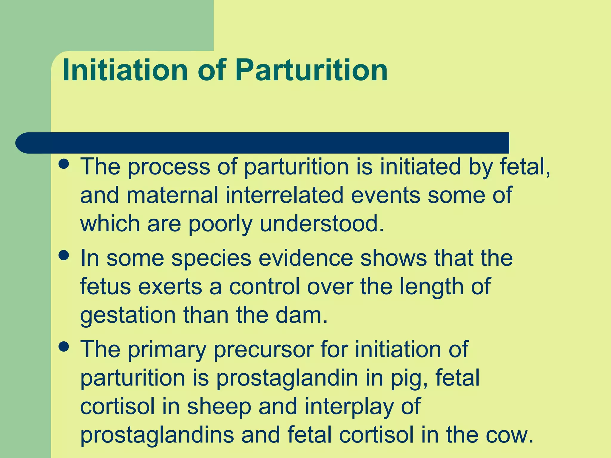


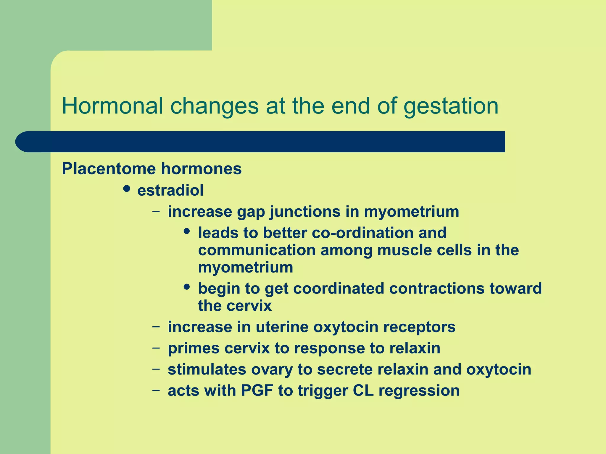


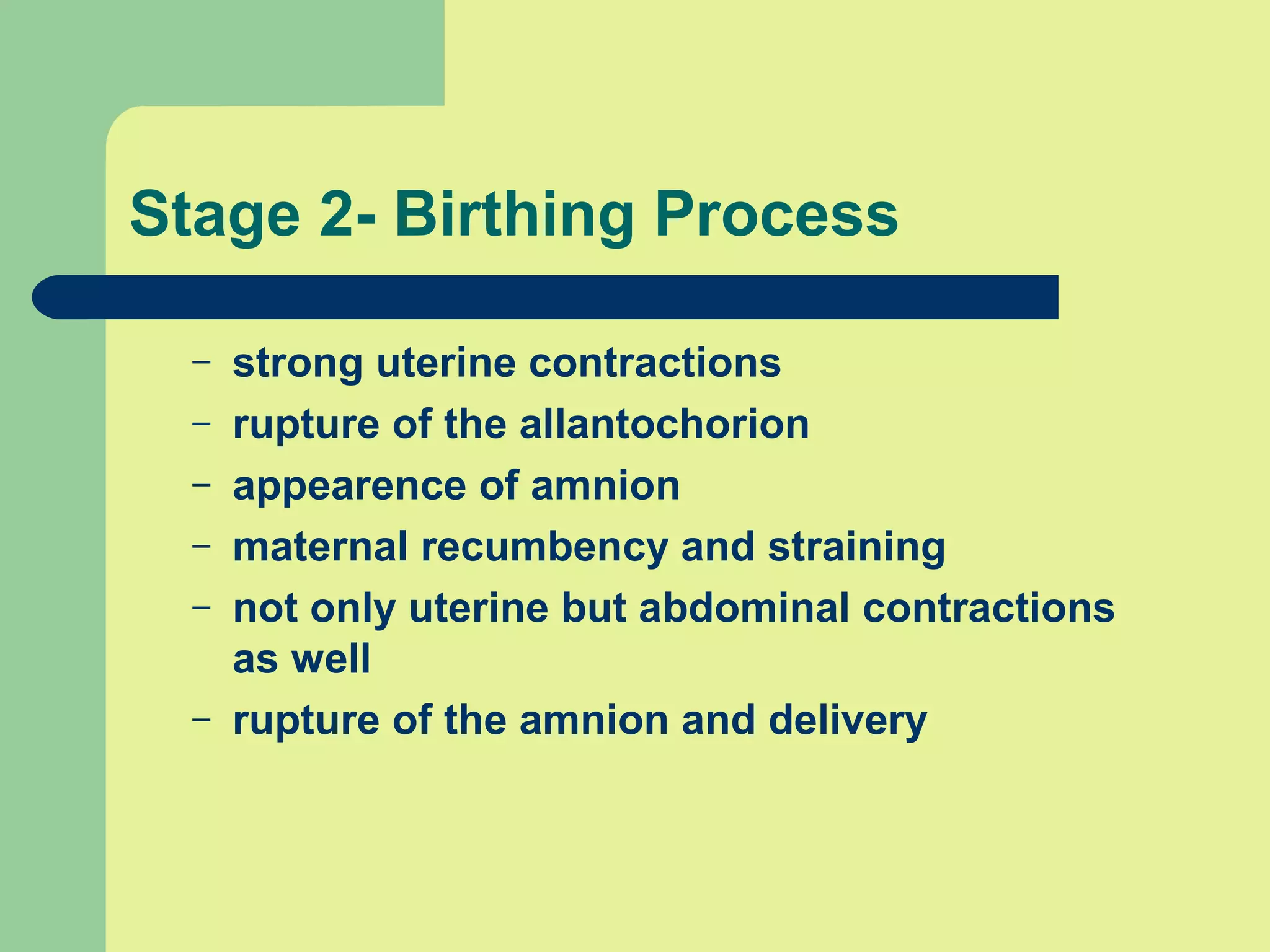


This document discusses diseases, accidents, and dystocia during gestation in livestock. It covers abortion, the average length of gestation for different species, infectious and non-infectious causes of abortion, the stages of parturition (birth), signs of approaching parturition, hormonal changes that initiate parturition, fetal positioning, induction of labor, and causes of dystocia (difficult birth). Dystocia can occur if there are issues with the birth canal size, fetal size/position, or lack of expulsive forces during delivery. Early intervention is important if dystocia is detected.