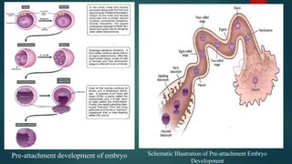
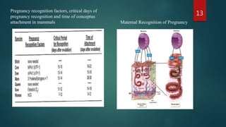
PREGNANCYAND ITS
TERMINATION
PREPARED BY
SUSHIL NEUPANE
M.V.SC (THERIOGENOLOGY)
INSTITUTE OF AGRICULTURE AND ANIMAL SCIENCE
TRIBHUVAN UNIVERSITY
KATHMANDU, NEPAL
1
Pregnancy
 Pregnancy is the term used to describe the period in which a fetus develops inside
uterus.
2
Method of Pregnancy Diagnosis
Method of PD: Rectal Palpation 3
Introduction
A conceptus is defined as the product of conception. It includes:
 The embryo during the early embryonic stage
 The embryo and extraembryonic membranes during the preimplantation stage
 The fetus and placenta during the post-attachment phase.
Four steps must be achieved before the embryo can attach to the uterus. They are:
 Development within the confines of the zona pellucida
 Hatching of the blastocyst from the zona pellucida
 Formation of the extraembryonic membranes
 Maternal recognition of pregnancy
4
Pre-attachment development of embryo Schematic Illustration of Pre-attachment Embryo
Development
5
Hatching of the blastocyst is governed by three forces. They are:
• growth and fluid accumulation within the blastocyst
• production of enzymes by the trophoblastic cells
• contraction of the blastocyst
The extraembryonic membranes of the pre-attachment embryo consist of the:
• yolk sac
• chorion
• amnion
• allantois
6
Implantation of Embryo in Domestic Animals
Schematic Diagram Illustrating the Typical Development of Extraembryonic Membranes in Mammals
7
 Formation of the extraembryonic membranes is an obligatory step in the
acquisition of the embryo's ability to attach to the uterus of the dam.
 The extraembryonic membranes are a set of four anatomically distinct
membranes that originate from the trophoblast, endoderm, mesoderm and
the embryo.
 The trophoblast, along with the primitive endoderm and mesoderm, give
rise to the chorion and the amnion.
 The yolk sac develops from the primitive endoderm.
 The chorion will eventually attach to the uterus, while the amnion will
provide a fluid-filled protective sac for the developing fetus.
8
 When amnion is developing, a small evagination from the posterior
region of the primitive gut begins to form. This sac-like evagination is
referred to as the allantois.
 The allantois is a fluid-filled sac that collects liquid waste from the
embryo. As the embryo grows, the allantois continues to expand and
eventually will make contact with the chorion.
 When the allantois reaches a certain volume, it presses against the
chorion and eventually fuses with it. When fusion takes place the two
membranes are called the allantochorion.
 The allantochorionic membrane is the fetal contribution to the placenta
and will provide the surface for attachments to the endometrium.
9
Maternal Recognition of Pregnancy
 The critical series of events by which the conceptus initially signals its
presence to the dam and enables pregnancy to continue is referred to as
maternal recognition of pregnancy.
 Maternal recognition of pregnancy must occur prior to luteolysis.
 Progesterone must be maintained at sufficiently high levels so that
embryogenesis and attachment of the developing conceptus to the
endometrium can take place.
 The embryo enters the uterus between days 2 and 5 after ovulation.
 If an adequate signal is not delivered in a timely manner, the dam will
experience luteolysis, progesterone concentrations will decline and
pregnancy will be terminated.
10
Maternal Recognition of Pregnancy
 IFN-tau is secreted by the trophoblastic cells of the blastocyst (cow
and ewe).
 IFN tau acts on the endometrial cells of the uterus to inhibit the
production of oxytocin receptors so that oxytocin cannot stimulate
PGF2α synthesis.
 In addition, IFN-tau causes secretion of proteins from the uterine
glands.
 Finally, IFN-tau can leave the uterus via the uterine vein to affect the
ovary and circulating immune cells.
 ln the ewe and the cow, the free-floating blastocyst produces specific
proteins that provide the signal for prevention of luteolysis.
11
Maternal Recognition of Pregnancy
 The specific proteins were once called ovine trophoblastic protein 1 ( oTP-1)
and bovine trophoblastic protein 1 (bTP- I).
 In the pregnant sow, the blastocyst produces estradiol that causes the PGF2α to
be rerouted into the uterine lumen, where it is destroyed, thus preventing
luteolysis.
 The equine conceptus must make extensive contact with the endometrial surface
to initiate and complete maternal recognition of pregnancy.
 In the woman, maternal recognition of pregnancy is provided by a hormone
called human chorionic gonadotropin (hCG).
 Maternal recognition of pregnancy in the dog and the cat probably does not
require a signal from the conceptus.
12
Pregnancy recognition factors, critical days of
pregnancy recognition and time of conceptus
attachment in mammals Maternal Recognition of Pregnancy
13
Termination of Pregnancy
 Embryonic Death
 Abortion
 Mummification
 Maceration
 Dystocia
 Parturition
14
Embryonic Death
 Embryonic death or mortality denotes the death of fertilized ova and embryo up
to the end of implantation.
 It refers to the losses which occur in the period between fertilization and the
completion of the stage of differentiation at approximately day 45.
 In early embryonic death, embryo dies before maternal recognition of pregnancy
(MRP) therefore extension of lifespan of corpus luteum does not commence.
 Early embryonic mortality is a major source of embryonic and economic loss
through repeat breeding and increased cost of artificial insemination.
 EED also leads to extended calving intervals and prolonged dry period resulting
in reduced life time milk production and reduced net calf production.
 Embryonic mortality is classified as Very Early Embryonic Mortality (day 0 to
7), Early Embryonic Mortality (day 7 to 24), Late Embryonic Mortality and
Early Fetal mortality (days 24 to 285).
15
Causes of Embryonic Death
1. Genetic Factors: Expression of lethal genes can cause death of the embryo within
the first 5 days of pregnancy.
2. Chromosome Abnormalities
a. Numerical abnormalities
i. Aneuploidy: In females, X chromosomal aneuploidy occurs like Turner's
syndrome (XO) and Triple X syndrome (XXX).
ii. Polyploidy: Polyploidy arises when there is a failure of the block to polyspermy
or if there is retention of the first or second (or both) polar bodies during
oogenesis.
b. Structural Abnormalities: It depends upon whether genetic material has been lost
(deletions) or just rearranged (insertions, inversions and translocations). The best
characterized chromosomal abnormality in cattle is the 1/29 Robertsonian
translocation, found in various breeds worldwide.
16
Causes of Embryonic Death
3. Endocrine Factors
 A decreased level of progesterone also results in EEM.
 Injections of estrogen at the time of estrum or within several days after ovulation
will affect the transport of the fertilized ova in the oviduct resulting in too rapid
transport or tubal locking of the ova and death of the zygote.
4. Nutritional Factors
a. Effects of Energy and Protein
b. Effects of Toxins
c. Excesses of Protein
d. Macro – Minerals and vitamins
e. Genital Infections
17
Diagnosis And Treatment
Diagnosis
 Examining Embryos
 Determining Progesterone in Blood, Milk and Saliva
 Pregnancy Associated Glycoprotein (PAG) Test
Treatment
 Supplementing Progesterone/Progestogen
 Use of hCG
 PMSG Administration
 GnRH Treatment
18
Abortion
 Abortion is the termination of pregnancy at a stage where the expelled fetus is of
recognizable size ranging from 45 to 260 days of gestation and not viable.
 Abortion is defined as a condition in which fetus is delivered live or dead before reaching
the stage of viability where the delivered fetus is visible by naked eyes.
19
A. Infectious Causes of Abortion
Abortion 20
A. Abortion due to infectious agents
Abortion
B. Physical Causes
 Douching, Infusing or inseminating Pregnant animals
 Trauma
 Rupture of amniotic vesicle by RP
 Removal of CL
 Torsion of Uterus
 Marked Stress
C. Chemical , Drugs and Poisonous plants
 Nitrates, arsenic, pine needles
21
Abortion
D. Hormonal Factors
a. High level of estrogen
b. Glucocorticosteriods deficiency
c. Progesterone deficiency
E. Nutritional Factors
a. Malnutrition
b. Vitamin A deficiency
c. Iodine deficiency
F. Miscellaneous
a. Twinning
b. Allergens and anaphylactic reaction
c. Tumors
22
Mummification
 Sequlae of a dead fetus is putrification due to autolytic changes in body tissue which
involve release of digestive enzymes from digestive tract and liver of the fetus and
putrefaction by microbes.
 There are number of pre- requisite for mummification process which includes:
a. Death of fetus post ossification.
b. Rapid dehydration of uterine environment.
c. Anaerobic environment of uterus.
d. Sterile uterine environment by means of closed cervix and intact endometrium.
 Classified as papyraceous and hematic type.
 In papyraceous, fetal skin appears like parchment paper without any exudate, which is
reported in non-ruminants
 In hematic, fetal skin surrounded by viscous chocolate coloured adhesive material,
reported in cattle and buffaloes.
23
Etiology
Infectious agents
 Leptospirosis
 Bovine Viral Diarrhea (BVD)
 Neospora caninum
Non-infectious agents
 Disturbed hormone level
 Chromosomal abnormality
 Twisting of umbilical cord
 Uterine torsion
 Defective placentation.
24
Clinical Presentation And
Diagnosis
 Majority of cases complains about prolong
gestation period without sign of parturition
 Per rectal palpation as well as
Ultrasonographic examination reveals
compact, firm and immobile mass without
fetal fluid and placentomes.
 Per vaginal examination ends with closed
cervix.
 Animal appears normal but may sometime
show weight loss along with reduced feed
intake and milk yield.
25
Treatment
By injection of PGF2α
Hysterectomy : caesarian
operation using caudal flank
laparotomy
Preventive measures
Regular hormone profiling
Maintenance of hygiene
and sanitation
Prophylaxis
Maceration
 Fetal death, putrification, luteolysis and failure of abortion are known as maceration.
 Characterized by fetal death and incomplete abortion to occur as a result of uterine
inertia and intrauterine infections.
 Incomplete abortion after the 3rd month of gestation is the main reason for a retained
fetal bony mass in the uterus of cows and buffaloes.
 History of intermittent/frequent straining for several days accompanied by fetid reddish
grey vulvar discharge.
 Noticed by the owners when foul smelling pus is discharged by a pregnant animal,
sometime pyrexia and anorexia
 Diagnosed by the history, finding of a piece of bone lodged in the cervix, rectal
palpation.
26
Dystocia
 Dystocia has been defined as “a birth that reduces calf viability, causes
maternal injury and requires assistance”.
 Have a huge economic impact on producers due to calf mortality, increased
veterinary costs, decreased production, reduced fertility, and in extreme cases,
injury to or death of the dam.
 The incidence of dystocia in dairy cattle was reported to range from 2 to 22
percent, while the proportion of assisted calving was higher, ranging from 10
to 50 percent
27
Etiology of dystocia
Maternal causes of dystocia
a. Incomplete dilatation of cervix
b. Narrow pelvis
c. Uterine torsion
d. Uterine inertia
Fetal causes of dystocia
a. Fetal maldisposition
b. Fetal oversize
c. Fetal monstrosities
28
Treatment and prevention
Use of:
• For incomplete dilatation of cervix, Oxytocin 50 IU along with Diethyl Stilbestrol 30mg.
• For smooth muscle relaxation, Valethamate Bromide ( Injection Epidocin) 80mg IM may be
helpful in some cases otherwise caesarian section should be performed.
• Torsion of uterus can be detorted within 36 hours by Schaffer’s Method otherwise caesarian section
is the best treatment of choice.
• Primary Uterine Inertia: Calcium Borogluconate 400ml IV & Oxytocin 20-60 IU Intramuscularly.
• Secondry uterine inertia: Dystocia may be relieved by (a) Force extraction (b) Extraction after
correction (c) Foetotomy (d) Caesarian Section.
• Oxytetracycline (injectable and intrauterine) post handling.
Prevention and Control
General Nutrition
Mineral Supplementation
29
Parturition
 Parturition is defined as “the act or process of giving birth”.
 It is an event that must be timed to match fetal maturation and its
ability to survive outside of the uterus (Wood, 1999).
 At the time of parturition, an increase in estradiol production and a
decline in progesterone is essential for calving to occur (Streyl,
2011) as these hormonal changes combine to depolarize uterine
myometrial cells and thereby increase uterine motility (Wood, 1999).
30
Stage I of parturition
 Stage I is initiated by the fetus and is highly dependent on the activation of the fetal
hypothalamus-pituitary-adrenal (HPA) axis.
 Fetal hypercortisolemia induces intrauterine prostaglandin synthesis via estrogen-
independent and estrogen-dependent pathways (Whittle, 2000).
 The estrogen-independent pathway occurs within the trophoblastic tissue and leads to
elevations in fetal plasma prostaglandin E2 (PGE2) concentrations
 The estrogen-dependent pathway occurs within the maternal endometrium leading to
increases in the production and release of maternal plasma prostaglandin F2α
(PGF2α).
 The first stage of parturition is complete once the fetus enters the cervical canal at
which stage the chorioallantoic sac usually ruptures.
 Stage I of parturition usually lasts approximately 6 h but variation among cattle
occurs and this stage may last longer in heifers.
31
Stage II of parturition
 Oxytocin facilitates myometrial contractility and its secretion increases as the pressure
against the cervix continues to increase due to the Ferguson reflex.
 Each bout of straining consists of 5 to 7 contractions and increases to 8 to 10 contractions
as the process advances.
 The intact amniotic sac appears at the vulva shortly after the rupture of the
chorioallantois.
 A sign of progress in stage II of parturition is a recumbent dam that is straining
intermittently but strongly.
 On average, the second stage of parturition lasts 2 to 4 h in multiparous cows, but it can
be longer in heifers as additional effort is required to dilate the birth canal (Norman and
Youngquist 2007).
 Maximal force is needed to deliver the fetal head through the vulva and in larger calves,
additional abdominal pressure is needed to deliver the shoulders and hindquarters.
32
Stage III of parturition
 Detachment and expulsion of the fetal membranes requires the separation of cotyledon
villi from the caruncle crypts
 Facilitated by vasoconstriction of the villi arteries associated with continued myometrial
contractions.
 The majority of cattle will pass their placenta within 6 h after parturition (Van Werven,
1992)
 The placenta is considered retained if it has not been expelled within 12 h of calving
(Berglund, 1987).
33
34
Stages of labor and related events in farm
animals
35
Stages of labor and related events in farm
animals
36
Average duration of stages of labor in farm
animals (hours)
References
 Beagley, J. C., Whitman, K. J., Baptiste, K. E., & Scherzer, J. (2010). Physiology and treatment of retained fetal membranes in cattle. Journal of
veterinary internal medicine, 24(2), 261-268.
 Berglund, B., & Philipsson, J. (1987). External signs of preparation for calving and course of parturition in Swedish dairy cattle breeds. Animal
Reproduction Science, 15(1-2), 61-79.
 Comline, R., L. Hall, R. Lavelle, P. Nathanielsz and M. Silver (1974). "Parturition in the cow: endocrine changes in animals with chronically implanted
catheters in the foetal and maternal circulations." Journal of Endocrinology 63(3): 451-472.
 Drost, M., & Holm, L. W. (1968). Prolonged gestation in ewes after foetal adrenalectomy. Journal of Endocrinology, 40(3), 293-296.
 Holm, L. W., Parker, H. R., & Galligan, S. J. (1961). Adrenal insufficiency in postmature Holstein calves. American journal of obstetrics and
gynecology, 81(5), 1000-1008.
 Hydbring, E., Madej, A., MacDonald, E., Drugge-Boholm, G., Berglund, B., & Olsson, K. (1999). Hormonal changes during parturition in heifers and
goats are related to the phases and severity of labour. Journal of Endocrinology, 160(1), 75-86.
 Janowski, T., Zduńczyk, S., Małecki-Tepicht, J., Barański, W., & Raś, A. (2002). Mammary secretion of oestrogens in the cow. Domestic Animal
Endocrinology, 23(1-2), 125-137.
 Musah, A. I., Schwabe, C., Willham, R. L., & Anderson, L. L. (1988). Dystocia, pelvic and cervical dilatation in beef heifers after induction of
parturition with relaxin combined with cloprostenol or dexamethasone. Animal Reproduction Science, 16(3-4), 237-248.
 Proudfoot, K. L., Huzzey, J. M., & Von Keyserlingk, M. A. G. (2009). The effect of dystocia on the dry matter intake and behavior of Holstein
cows. Journal of dairy science, 92(10), 4937-4944.
 Senger, P. L. (2003). Placentation, the endocrinology of gestation and parturition. Pathways to pregnancy and parturition, 2, 304-325.
37
References
 Shah, K. D., Nakao, T., & Kubota, H. (2006). Plasma estrone sulphate (E1S) and estradiol-17β (E2β) profiles during
pregnancy and their relationship with the relaxation of sacrosciatic ligament, and prediction of calving time in
Holstein–Friesian cattle. Animal reproduction science, 95(1-2), 38-53.
 Streyl, D., Sauter-Louis, C., Braunert, A., Lange, D., Weber, F., & Zerbe, H. (2011). Establishment of a standard
operating procedure for predicting the time of calving in cattle. Journal of veterinary science, 12(2), 177-185.
 Taverne, M. A. M., Breeveld-Dwarkasing, V. N. A., van Dissel-Emiliani, F. M. F., Bevers, M. M., De Jong, R., &
Van der Weijden, G. C. (2002). Between prepartum luteolysis and onset of expulsion. Domestic animal
endocrinology, 23(1-2), 329-337.
 Van Werven, T., Schukken, Y. H., Lloyd, J., Brand, A., Heeringa, H. T., & Shea, M. (1992). The effects of duration
of retained placenta on reproduction, milk production, postpartum disease and culling rate. Theriogenology, 37(6),
1191-1203.
 Whittle, W., A. Holloway, S. Lye, W. Gibb and J. Challis (2000). "Prostaglandin Production at the Onset of Ovine
Parturition Is Regulated by Both Estrogen-Independent and Estrogen-Dependent Pathways." Endocrinology
141(10): 3783-3791.
 Wood, C. E. (1999). Control of parturition in ruminants. Journal of reproduction and fertility. Supplement, 54,
115-126.
 Youngquist, R. S., & Threlfall, W. R. (2007). Current Therapy in Large Animal Theriogenology, Saunders.
38
39

Pregnancy and its termination

  • 1.
    PREGNANCYAND ITS TERMINATION PREPARED BY SUSHILNEUPANE M.V.SC (THERIOGENOLOGY) INSTITUTE OF AGRICULTURE AND ANIMAL SCIENCE TRIBHUVAN UNIVERSITY KATHMANDU, NEPAL 1
  • 2.
    Pregnancy  Pregnancy isthe term used to describe the period in which a fetus develops inside uterus. 2 Method of Pregnancy Diagnosis
  • 3.
    Method of PD:Rectal Palpation 3
  • 4.
    Introduction A conceptus isdefined as the product of conception. It includes:  The embryo during the early embryonic stage  The embryo and extraembryonic membranes during the preimplantation stage  The fetus and placenta during the post-attachment phase. Four steps must be achieved before the embryo can attach to the uterus. They are:  Development within the confines of the zona pellucida  Hatching of the blastocyst from the zona pellucida  Formation of the extraembryonic membranes  Maternal recognition of pregnancy 4
  • 5.
    Pre-attachment development ofembryo Schematic Illustration of Pre-attachment Embryo Development 5
  • 6.
    Hatching of theblastocyst is governed by three forces. They are: • growth and fluid accumulation within the blastocyst • production of enzymes by the trophoblastic cells • contraction of the blastocyst The extraembryonic membranes of the pre-attachment embryo consist of the: • yolk sac • chorion • amnion • allantois 6 Implantation of Embryo in Domestic Animals
  • 7.
    Schematic Diagram Illustratingthe Typical Development of Extraembryonic Membranes in Mammals 7
  • 8.
     Formation ofthe extraembryonic membranes is an obligatory step in the acquisition of the embryo's ability to attach to the uterus of the dam.  The extraembryonic membranes are a set of four anatomically distinct membranes that originate from the trophoblast, endoderm, mesoderm and the embryo.  The trophoblast, along with the primitive endoderm and mesoderm, give rise to the chorion and the amnion.  The yolk sac develops from the primitive endoderm.  The chorion will eventually attach to the uterus, while the amnion will provide a fluid-filled protective sac for the developing fetus. 8
  • 9.
     When amnionis developing, a small evagination from the posterior region of the primitive gut begins to form. This sac-like evagination is referred to as the allantois.  The allantois is a fluid-filled sac that collects liquid waste from the embryo. As the embryo grows, the allantois continues to expand and eventually will make contact with the chorion.  When the allantois reaches a certain volume, it presses against the chorion and eventually fuses with it. When fusion takes place the two membranes are called the allantochorion.  The allantochorionic membrane is the fetal contribution to the placenta and will provide the surface for attachments to the endometrium. 9
  • 10.
    Maternal Recognition ofPregnancy  The critical series of events by which the conceptus initially signals its presence to the dam and enables pregnancy to continue is referred to as maternal recognition of pregnancy.  Maternal recognition of pregnancy must occur prior to luteolysis.  Progesterone must be maintained at sufficiently high levels so that embryogenesis and attachment of the developing conceptus to the endometrium can take place.  The embryo enters the uterus between days 2 and 5 after ovulation.  If an adequate signal is not delivered in a timely manner, the dam will experience luteolysis, progesterone concentrations will decline and pregnancy will be terminated. 10
  • 11.
    Maternal Recognition ofPregnancy  IFN-tau is secreted by the trophoblastic cells of the blastocyst (cow and ewe).  IFN tau acts on the endometrial cells of the uterus to inhibit the production of oxytocin receptors so that oxytocin cannot stimulate PGF2α synthesis.  In addition, IFN-tau causes secretion of proteins from the uterine glands.  Finally, IFN-tau can leave the uterus via the uterine vein to affect the ovary and circulating immune cells.  ln the ewe and the cow, the free-floating blastocyst produces specific proteins that provide the signal for prevention of luteolysis. 11
  • 12.
    Maternal Recognition ofPregnancy  The specific proteins were once called ovine trophoblastic protein 1 ( oTP-1) and bovine trophoblastic protein 1 (bTP- I).  In the pregnant sow, the blastocyst produces estradiol that causes the PGF2α to be rerouted into the uterine lumen, where it is destroyed, thus preventing luteolysis.  The equine conceptus must make extensive contact with the endometrial surface to initiate and complete maternal recognition of pregnancy.  In the woman, maternal recognition of pregnancy is provided by a hormone called human chorionic gonadotropin (hCG).  Maternal recognition of pregnancy in the dog and the cat probably does not require a signal from the conceptus. 12
  • 13.
    Pregnancy recognition factors,critical days of pregnancy recognition and time of conceptus attachment in mammals Maternal Recognition of Pregnancy 13
  • 14.
    Termination of Pregnancy Embryonic Death  Abortion  Mummification  Maceration  Dystocia  Parturition 14
  • 15.
    Embryonic Death  Embryonicdeath or mortality denotes the death of fertilized ova and embryo up to the end of implantation.  It refers to the losses which occur in the period between fertilization and the completion of the stage of differentiation at approximately day 45.  In early embryonic death, embryo dies before maternal recognition of pregnancy (MRP) therefore extension of lifespan of corpus luteum does not commence.  Early embryonic mortality is a major source of embryonic and economic loss through repeat breeding and increased cost of artificial insemination.  EED also leads to extended calving intervals and prolonged dry period resulting in reduced life time milk production and reduced net calf production.  Embryonic mortality is classified as Very Early Embryonic Mortality (day 0 to 7), Early Embryonic Mortality (day 7 to 24), Late Embryonic Mortality and Early Fetal mortality (days 24 to 285). 15
  • 16.
    Causes of EmbryonicDeath 1. Genetic Factors: Expression of lethal genes can cause death of the embryo within the first 5 days of pregnancy. 2. Chromosome Abnormalities a. Numerical abnormalities i. Aneuploidy: In females, X chromosomal aneuploidy occurs like Turner's syndrome (XO) and Triple X syndrome (XXX). ii. Polyploidy: Polyploidy arises when there is a failure of the block to polyspermy or if there is retention of the first or second (or both) polar bodies during oogenesis. b. Structural Abnormalities: It depends upon whether genetic material has been lost (deletions) or just rearranged (insertions, inversions and translocations). The best characterized chromosomal abnormality in cattle is the 1/29 Robertsonian translocation, found in various breeds worldwide. 16
  • 17.
    Causes of EmbryonicDeath 3. Endocrine Factors  A decreased level of progesterone also results in EEM.  Injections of estrogen at the time of estrum or within several days after ovulation will affect the transport of the fertilized ova in the oviduct resulting in too rapid transport or tubal locking of the ova and death of the zygote. 4. Nutritional Factors a. Effects of Energy and Protein b. Effects of Toxins c. Excesses of Protein d. Macro – Minerals and vitamins e. Genital Infections 17
  • 18.
    Diagnosis And Treatment Diagnosis Examining Embryos  Determining Progesterone in Blood, Milk and Saliva  Pregnancy Associated Glycoprotein (PAG) Test Treatment  Supplementing Progesterone/Progestogen  Use of hCG  PMSG Administration  GnRH Treatment 18
  • 19.
    Abortion  Abortion isthe termination of pregnancy at a stage where the expelled fetus is of recognizable size ranging from 45 to 260 days of gestation and not viable.  Abortion is defined as a condition in which fetus is delivered live or dead before reaching the stage of viability where the delivered fetus is visible by naked eyes. 19 A. Infectious Causes of Abortion
  • 20.
    Abortion 20 A. Abortiondue to infectious agents
  • 21.
    Abortion B. Physical Causes Douching, Infusing or inseminating Pregnant animals  Trauma  Rupture of amniotic vesicle by RP  Removal of CL  Torsion of Uterus  Marked Stress C. Chemical , Drugs and Poisonous plants  Nitrates, arsenic, pine needles 21
  • 22.
    Abortion D. Hormonal Factors a.High level of estrogen b. Glucocorticosteriods deficiency c. Progesterone deficiency E. Nutritional Factors a. Malnutrition b. Vitamin A deficiency c. Iodine deficiency F. Miscellaneous a. Twinning b. Allergens and anaphylactic reaction c. Tumors 22
  • 23.
    Mummification  Sequlae ofa dead fetus is putrification due to autolytic changes in body tissue which involve release of digestive enzymes from digestive tract and liver of the fetus and putrefaction by microbes.  There are number of pre- requisite for mummification process which includes: a. Death of fetus post ossification. b. Rapid dehydration of uterine environment. c. Anaerobic environment of uterus. d. Sterile uterine environment by means of closed cervix and intact endometrium.  Classified as papyraceous and hematic type.  In papyraceous, fetal skin appears like parchment paper without any exudate, which is reported in non-ruminants  In hematic, fetal skin surrounded by viscous chocolate coloured adhesive material, reported in cattle and buffaloes. 23
  • 24.
    Etiology Infectious agents  Leptospirosis Bovine Viral Diarrhea (BVD)  Neospora caninum Non-infectious agents  Disturbed hormone level  Chromosomal abnormality  Twisting of umbilical cord  Uterine torsion  Defective placentation. 24
  • 25.
    Clinical Presentation And Diagnosis Majority of cases complains about prolong gestation period without sign of parturition  Per rectal palpation as well as Ultrasonographic examination reveals compact, firm and immobile mass without fetal fluid and placentomes.  Per vaginal examination ends with closed cervix.  Animal appears normal but may sometime show weight loss along with reduced feed intake and milk yield. 25 Treatment By injection of PGF2α Hysterectomy : caesarian operation using caudal flank laparotomy Preventive measures Regular hormone profiling Maintenance of hygiene and sanitation Prophylaxis
  • 26.
    Maceration  Fetal death,putrification, luteolysis and failure of abortion are known as maceration.  Characterized by fetal death and incomplete abortion to occur as a result of uterine inertia and intrauterine infections.  Incomplete abortion after the 3rd month of gestation is the main reason for a retained fetal bony mass in the uterus of cows and buffaloes.  History of intermittent/frequent straining for several days accompanied by fetid reddish grey vulvar discharge.  Noticed by the owners when foul smelling pus is discharged by a pregnant animal, sometime pyrexia and anorexia  Diagnosed by the history, finding of a piece of bone lodged in the cervix, rectal palpation. 26
  • 27.
    Dystocia  Dystocia hasbeen defined as “a birth that reduces calf viability, causes maternal injury and requires assistance”.  Have a huge economic impact on producers due to calf mortality, increased veterinary costs, decreased production, reduced fertility, and in extreme cases, injury to or death of the dam.  The incidence of dystocia in dairy cattle was reported to range from 2 to 22 percent, while the proportion of assisted calving was higher, ranging from 10 to 50 percent 27
  • 28.
    Etiology of dystocia Maternalcauses of dystocia a. Incomplete dilatation of cervix b. Narrow pelvis c. Uterine torsion d. Uterine inertia Fetal causes of dystocia a. Fetal maldisposition b. Fetal oversize c. Fetal monstrosities 28
  • 29.
    Treatment and prevention Useof: • For incomplete dilatation of cervix, Oxytocin 50 IU along with Diethyl Stilbestrol 30mg. • For smooth muscle relaxation, Valethamate Bromide ( Injection Epidocin) 80mg IM may be helpful in some cases otherwise caesarian section should be performed. • Torsion of uterus can be detorted within 36 hours by Schaffer’s Method otherwise caesarian section is the best treatment of choice. • Primary Uterine Inertia: Calcium Borogluconate 400ml IV & Oxytocin 20-60 IU Intramuscularly. • Secondry uterine inertia: Dystocia may be relieved by (a) Force extraction (b) Extraction after correction (c) Foetotomy (d) Caesarian Section. • Oxytetracycline (injectable and intrauterine) post handling. Prevention and Control General Nutrition Mineral Supplementation 29
  • 30.
    Parturition  Parturition isdefined as “the act or process of giving birth”.  It is an event that must be timed to match fetal maturation and its ability to survive outside of the uterus (Wood, 1999).  At the time of parturition, an increase in estradiol production and a decline in progesterone is essential for calving to occur (Streyl, 2011) as these hormonal changes combine to depolarize uterine myometrial cells and thereby increase uterine motility (Wood, 1999). 30
  • 31.
    Stage I ofparturition  Stage I is initiated by the fetus and is highly dependent on the activation of the fetal hypothalamus-pituitary-adrenal (HPA) axis.  Fetal hypercortisolemia induces intrauterine prostaglandin synthesis via estrogen- independent and estrogen-dependent pathways (Whittle, 2000).  The estrogen-independent pathway occurs within the trophoblastic tissue and leads to elevations in fetal plasma prostaglandin E2 (PGE2) concentrations  The estrogen-dependent pathway occurs within the maternal endometrium leading to increases in the production and release of maternal plasma prostaglandin F2α (PGF2α).  The first stage of parturition is complete once the fetus enters the cervical canal at which stage the chorioallantoic sac usually ruptures.  Stage I of parturition usually lasts approximately 6 h but variation among cattle occurs and this stage may last longer in heifers. 31
  • 32.
    Stage II ofparturition  Oxytocin facilitates myometrial contractility and its secretion increases as the pressure against the cervix continues to increase due to the Ferguson reflex.  Each bout of straining consists of 5 to 7 contractions and increases to 8 to 10 contractions as the process advances.  The intact amniotic sac appears at the vulva shortly after the rupture of the chorioallantois.  A sign of progress in stage II of parturition is a recumbent dam that is straining intermittently but strongly.  On average, the second stage of parturition lasts 2 to 4 h in multiparous cows, but it can be longer in heifers as additional effort is required to dilate the birth canal (Norman and Youngquist 2007).  Maximal force is needed to deliver the fetal head through the vulva and in larger calves, additional abdominal pressure is needed to deliver the shoulders and hindquarters. 32
  • 33.
    Stage III ofparturition  Detachment and expulsion of the fetal membranes requires the separation of cotyledon villi from the caruncle crypts  Facilitated by vasoconstriction of the villi arteries associated with continued myometrial contractions.  The majority of cattle will pass their placenta within 6 h after parturition (Van Werven, 1992)  The placenta is considered retained if it has not been expelled within 12 h of calving (Berglund, 1987). 33
  • 34.
    34 Stages of laborand related events in farm animals
  • 35.
    35 Stages of laborand related events in farm animals
  • 36.
    36 Average duration ofstages of labor in farm animals (hours)
  • 37.
    References  Beagley, J.C., Whitman, K. J., Baptiste, K. E., & Scherzer, J. (2010). Physiology and treatment of retained fetal membranes in cattle. Journal of veterinary internal medicine, 24(2), 261-268.  Berglund, B., & Philipsson, J. (1987). External signs of preparation for calving and course of parturition in Swedish dairy cattle breeds. Animal Reproduction Science, 15(1-2), 61-79.  Comline, R., L. Hall, R. Lavelle, P. Nathanielsz and M. Silver (1974). "Parturition in the cow: endocrine changes in animals with chronically implanted catheters in the foetal and maternal circulations." Journal of Endocrinology 63(3): 451-472.  Drost, M., & Holm, L. W. (1968). Prolonged gestation in ewes after foetal adrenalectomy. Journal of Endocrinology, 40(3), 293-296.  Holm, L. W., Parker, H. R., & Galligan, S. J. (1961). Adrenal insufficiency in postmature Holstein calves. American journal of obstetrics and gynecology, 81(5), 1000-1008.  Hydbring, E., Madej, A., MacDonald, E., Drugge-Boholm, G., Berglund, B., & Olsson, K. (1999). Hormonal changes during parturition in heifers and goats are related to the phases and severity of labour. Journal of Endocrinology, 160(1), 75-86.  Janowski, T., Zduńczyk, S., Małecki-Tepicht, J., Barański, W., & Raś, A. (2002). Mammary secretion of oestrogens in the cow. Domestic Animal Endocrinology, 23(1-2), 125-137.  Musah, A. I., Schwabe, C., Willham, R. L., & Anderson, L. L. (1988). Dystocia, pelvic and cervical dilatation in beef heifers after induction of parturition with relaxin combined with cloprostenol or dexamethasone. Animal Reproduction Science, 16(3-4), 237-248.  Proudfoot, K. L., Huzzey, J. M., & Von Keyserlingk, M. A. G. (2009). The effect of dystocia on the dry matter intake and behavior of Holstein cows. Journal of dairy science, 92(10), 4937-4944.  Senger, P. L. (2003). Placentation, the endocrinology of gestation and parturition. Pathways to pregnancy and parturition, 2, 304-325. 37
  • 38.
    References  Shah, K.D., Nakao, T., & Kubota, H. (2006). Plasma estrone sulphate (E1S) and estradiol-17β (E2β) profiles during pregnancy and their relationship with the relaxation of sacrosciatic ligament, and prediction of calving time in Holstein–Friesian cattle. Animal reproduction science, 95(1-2), 38-53.  Streyl, D., Sauter-Louis, C., Braunert, A., Lange, D., Weber, F., & Zerbe, H. (2011). Establishment of a standard operating procedure for predicting the time of calving in cattle. Journal of veterinary science, 12(2), 177-185.  Taverne, M. A. M., Breeveld-Dwarkasing, V. N. A., van Dissel-Emiliani, F. M. F., Bevers, M. M., De Jong, R., & Van der Weijden, G. C. (2002). Between prepartum luteolysis and onset of expulsion. Domestic animal endocrinology, 23(1-2), 329-337.  Van Werven, T., Schukken, Y. H., Lloyd, J., Brand, A., Heeringa, H. T., & Shea, M. (1992). The effects of duration of retained placenta on reproduction, milk production, postpartum disease and culling rate. Theriogenology, 37(6), 1191-1203.  Whittle, W., A. Holloway, S. Lye, W. Gibb and J. Challis (2000). "Prostaglandin Production at the Onset of Ovine Parturition Is Regulated by Both Estrogen-Independent and Estrogen-Dependent Pathways." Endocrinology 141(10): 3783-3791.  Wood, C. E. (1999). Control of parturition in ruminants. Journal of reproduction and fertility. Supplement, 54, 115-126.  Youngquist, R. S., & Threlfall, W. R. (2007). Current Therapy in Large Animal Theriogenology, Saunders. 38
  • 39.