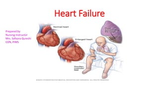
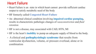
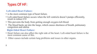
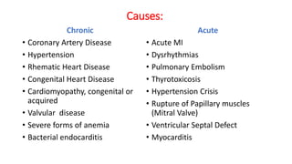
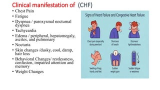
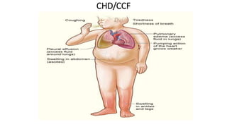
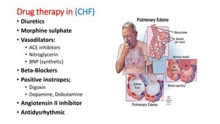
Heart Failure
Prepared by
Nursing Instructor
Mrs. Safoora Qureshi
CON, PIMS
Heart Failure
• Heart Failure is an state in which heart cannot provide sufficient cardiac
output to satisfy metabolic need of the body
• HF formerly called Congested Heart Failure
• An abnormal clinical condition involving impaired cardiac pumping,
results in characteristic pathologic changes of vasoconstriction and fluid
retention
• HF is not a disease, may associated with CAD, HTN, MI
• HF is the heart’s inability to pump an adequate supply of blood to the body.
• A clinical and pathophysiologic syndrome that results from
ventricular dysfunction, volume, or pressure overload, alone or in
combination
Heart Compensatory Mechanism
Reduce Cardiac Out put
carotid baroreceptor stimulation
Renal Perfusion/GFR
Activation of
CNS
HR & Inotropy
Myocardial toxicity
Activation of
RAAS Vasoconstriction
After load
Negative remodeling
Worsens the LV
Symptoms of HF
shortness of breaths, arrythmias,
edema, chest congestion etc
increased
angiotensin -II
From increased
Aldosterone
water retention
Hemodynamic
alteration
Preload
Myocardial injury
Pathophysiology of
Heart Failure
ANP &
BNP
Heart
CHF
Pathology of Ventricular Failure
• Systolic Failure (r-EF); The hallmark of systolic dysfunction is a
decrease in the left ventricular ejection fraction.
• The normal EF is greater than 55% of ventricular volume.
• Systolic failure is caused by
• impaired contractile function(MI)
• increased afterload (hypertension),
• mechanical abnormalities (VHD)
• Diastolic Failure (p-EF):
• is an impaired ability of the ventricles to relax and fill during diastole,
result in decreased in stroke volume and CO.
• High filling pressure due to stiff or noncompliance ventricles, result
in venous engorgement in both pulmonary and systemic circulation.
Types Of HF:
Left-sided Heart Failure:
• is the most common type of heart failure.
• Left-sided heart failure occurs when the left ventricle doesn’t pumps efficiently,
result in reduce CO
• This prevents the body from getting enough oxygen-rich blood.
• The blood backs up into the lungs, which causes shortness of breath, pulmonary
congestion and edema
Right-Sided Heart Failure:
• Heart failure can also affect the right side of the heart. Left-sided heart failure is the
most common cause of this.
• Other causes include certain lung problems and issues in other organs.
z
Causes:
Chronic
• Coronary Artery Disease
• Hypertension
• Rhematic Heart Disease
• Congenital Heart Disease
• Cardiomyopathy, congenital or
acquired
• Valvular disease
• Severe forms of anemia
• Bacterial endocarditis
Acute
• Acute MI
• Dysrhythmias
• Pulmonary Embolism
• Thyrotoxicosis
• Hypertension Crisis
• Rupture of Papillary muscles
(Mitral Valve)
• Ventricular Septal Defect
• Myocarditis
Clinical manifestation of (CHF)
• Chest Pain
• Fatigue
• Dyspnea / paroxysmal nocturnal
dyspnea
• Tachycardia
• Edema / peripheral, hepatomegaly,
ascites, and pulmonary
• Nocturia
• Skin changes /dusky, cool, damp,
hair loss
• Behavioral Changes/ restlessness,
confusion, impaired attention and
memory
• Weight Changes
CHD/CCF
Complication:
• Pleural effusion
• Dysrhythmias
• Left ventricular thrombus
• Hepatomegaly
• Renal failure
Diagnosis:
• History
• Physical Examination
• X-rays
• ECG
• Lab: Cardiac Enzyme BNP
• Serum chemistries such as;
• LFT, RFT, TFT, CBC
• Echocardiogram
• Stress testing
• Cardiac Catheterization
Collaborative Care
• Treatment of underline cause
• Oxygen therapy 2 --- 6 l/min by nasal catheter
• Rest-activity period
• Drug therapy
• Daily weight
• Sodium restricted diet
• Circulatory/ventricular assist device
• (IABP)
• Cardiac resynchronization therapy with
• cardioverter-defibrillator
• Cardiac transplant
ICD IABP
Drug therapy in (CHF)
• Diuretics
• Morphine sulphate
• Vasodilators:
• ACE inhibitors
• Nitroglycerin
• BNP (synthetic)
• Beta-Blockers
• Positive inotropes;
• Digoxin
• Dopamine, Dobutamine
• Angiotensin II inhibitor
• Antidysrhythmic
Nursing Management
Oxygen therapy:
• Administers oxygen as per need by nasal catheter
• Monitor the effectiveness of the therapy to identify hypoxia and establish a
range of oxygen saturation
Position:
• Semi-Fowler position to alleviate dyspnea and pain
Respiratory Monitoring:
• RR, rhythm, depth, and resp.efforts, to evaluate respiratory status
• Auscultate breath sounds to assess congestion
• Monitor dyspnea to observe the improvement of respiratory status
• Note the events that improve or worsen the distress to detect the events,
influence ADLs
Monitor Cardiac status:
• Continue cardiac monitoring to detect arrythmias
 Anxiety reduction:
• Use a reassuring approach to gain client confidence in care provider
• Promote a sense of security by explaining all therapeutic procedures
• Identify source of anxiety/fear and plan anxiety relieving techniques
• Create a atmosphere to facilitate trust
• Instruct the client in use of relaxation techniques (imagery)
• Promote sleep by providing a calm and quiet environment
Energy Management:
• Encourage alternate rest and activity period to reduce cardiac workload
• Provide calming diversionary activities to promote relaxation to reduce
oxygen consumption
• Monitor patient’s oxygen demand during self-care/nursing care activities to
determine the level of activity that can be performed
Fluid /Electrolyte management:
• Daily weight to monitor fluid retention/diuresis and weight reduction
• CVP line monitoring
• Monitor serum electrolyte balance to assess response to treatment
Hypervolemia management;
• Monitor
• respiratory status/distress/SOB to detect pulmonary edema
• hemodynamic status including CVP, MAP to evaluate effective ness of therapy
• renal function and intake output to monitor fluid balance
• effectiveness of diuretics to assess response to treatment
Dietary Management:
• (DASH ) diet is widely used for patients with HF
• Fluid restriction incase of renal insufficiency
• Sodium restricted diet

Heart Failure.pptx

  • 1.
    Heart Failure Prepared by NursingInstructor Mrs. Safoora Qureshi CON, PIMS
  • 2.
    Heart Failure • HeartFailure is an state in which heart cannot provide sufficient cardiac output to satisfy metabolic need of the body • HF formerly called Congested Heart Failure • An abnormal clinical condition involving impaired cardiac pumping, results in characteristic pathologic changes of vasoconstriction and fluid retention • HF is not a disease, may associated with CAD, HTN, MI • HF is the heart’s inability to pump an adequate supply of blood to the body. • A clinical and pathophysiologic syndrome that results from ventricular dysfunction, volume, or pressure overload, alone or in combination
  • 3.
  • 4.
    Reduce Cardiac Output carotid baroreceptor stimulation Renal Perfusion/GFR Activation of CNS HR & Inotropy Myocardial toxicity Activation of RAAS Vasoconstriction After load Negative remodeling Worsens the LV Symptoms of HF shortness of breaths, arrythmias, edema, chest congestion etc increased angiotensin -II From increased Aldosterone water retention Hemodynamic alteration Preload Myocardial injury Pathophysiology of Heart Failure ANP & BNP Heart CHF
  • 5.
    Pathology of VentricularFailure • Systolic Failure (r-EF); The hallmark of systolic dysfunction is a decrease in the left ventricular ejection fraction. • The normal EF is greater than 55% of ventricular volume. • Systolic failure is caused by • impaired contractile function(MI) • increased afterload (hypertension), • mechanical abnormalities (VHD) • Diastolic Failure (p-EF): • is an impaired ability of the ventricles to relax and fill during diastole, result in decreased in stroke volume and CO. • High filling pressure due to stiff or noncompliance ventricles, result in venous engorgement in both pulmonary and systemic circulation.
  • 6.
    Types Of HF: Left-sidedHeart Failure: • is the most common type of heart failure. • Left-sided heart failure occurs when the left ventricle doesn’t pumps efficiently, result in reduce CO • This prevents the body from getting enough oxygen-rich blood. • The blood backs up into the lungs, which causes shortness of breath, pulmonary congestion and edema Right-Sided Heart Failure: • Heart failure can also affect the right side of the heart. Left-sided heart failure is the most common cause of this. • Other causes include certain lung problems and issues in other organs. z
  • 7.
    Causes: Chronic • Coronary ArteryDisease • Hypertension • Rhematic Heart Disease • Congenital Heart Disease • Cardiomyopathy, congenital or acquired • Valvular disease • Severe forms of anemia • Bacterial endocarditis Acute • Acute MI • Dysrhythmias • Pulmonary Embolism • Thyrotoxicosis • Hypertension Crisis • Rupture of Papillary muscles (Mitral Valve) • Ventricular Septal Defect • Myocarditis
  • 8.
    Clinical manifestation of(CHF) • Chest Pain • Fatigue • Dyspnea / paroxysmal nocturnal dyspnea • Tachycardia • Edema / peripheral, hepatomegaly, ascites, and pulmonary • Nocturia • Skin changes /dusky, cool, damp, hair loss • Behavioral Changes/ restlessness, confusion, impaired attention and memory • Weight Changes
  • 9.
  • 10.
    Complication: • Pleural effusion •Dysrhythmias • Left ventricular thrombus • Hepatomegaly • Renal failure
  • 11.
    Diagnosis: • History • PhysicalExamination • X-rays • ECG • Lab: Cardiac Enzyme BNP • Serum chemistries such as; • LFT, RFT, TFT, CBC • Echocardiogram • Stress testing • Cardiac Catheterization
  • 12.
    Collaborative Care • Treatmentof underline cause • Oxygen therapy 2 --- 6 l/min by nasal catheter • Rest-activity period • Drug therapy • Daily weight • Sodium restricted diet • Circulatory/ventricular assist device • (IABP) • Cardiac resynchronization therapy with • cardioverter-defibrillator • Cardiac transplant
  • 13.
  • 14.
    Drug therapy in(CHF) • Diuretics • Morphine sulphate • Vasodilators: • ACE inhibitors • Nitroglycerin • BNP (synthetic) • Beta-Blockers • Positive inotropes; • Digoxin • Dopamine, Dobutamine • Angiotensin II inhibitor • Antidysrhythmic
  • 15.
    Nursing Management Oxygen therapy: •Administers oxygen as per need by nasal catheter • Monitor the effectiveness of the therapy to identify hypoxia and establish a range of oxygen saturation Position: • Semi-Fowler position to alleviate dyspnea and pain Respiratory Monitoring: • RR, rhythm, depth, and resp.efforts, to evaluate respiratory status • Auscultate breath sounds to assess congestion • Monitor dyspnea to observe the improvement of respiratory status • Note the events that improve or worsen the distress to detect the events, influence ADLs
  • 16.
    Monitor Cardiac status: •Continue cardiac monitoring to detect arrythmias  Anxiety reduction: • Use a reassuring approach to gain client confidence in care provider • Promote a sense of security by explaining all therapeutic procedures • Identify source of anxiety/fear and plan anxiety relieving techniques • Create a atmosphere to facilitate trust • Instruct the client in use of relaxation techniques (imagery) • Promote sleep by providing a calm and quiet environment
  • 17.
    Energy Management: • Encouragealternate rest and activity period to reduce cardiac workload • Provide calming diversionary activities to promote relaxation to reduce oxygen consumption • Monitor patient’s oxygen demand during self-care/nursing care activities to determine the level of activity that can be performed Fluid /Electrolyte management: • Daily weight to monitor fluid retention/diuresis and weight reduction • CVP line monitoring • Monitor serum electrolyte balance to assess response to treatment
  • 18.
    Hypervolemia management; • Monitor •respiratory status/distress/SOB to detect pulmonary edema • hemodynamic status including CVP, MAP to evaluate effective ness of therapy • renal function and intake output to monitor fluid balance • effectiveness of diuretics to assess response to treatment Dietary Management: • (DASH ) diet is widely used for patients with HF • Fluid restriction incase of renal insufficiency • Sodium restricted diet