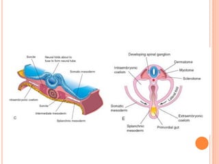
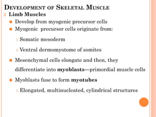
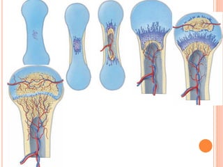
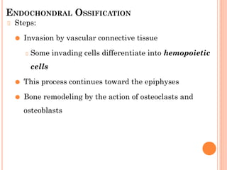
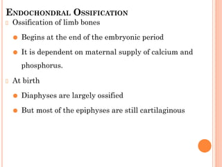
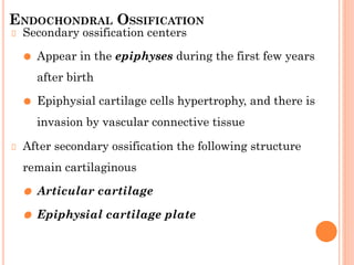
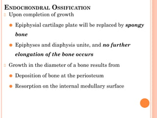
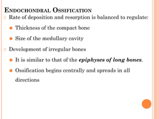
DEVELOPMENT OF SKELETAL MUSCLE,
BONE AND CARTILAGE
Handout for Medical students
Prepared by: Melaku Geletu (MSc. in Anatomy)
DEVELOPMENT OF SKELETAL MUSCLE
Limb Muscles
⚫ Develop from myogenic precursor cells
⚫ Myogenic precursor cells originate from:
Somatic mesoderm
Ventral dermomyotome of somites
⚫ Mesenchymal cells elongate and then, they
differentiate into myoblasts—primordial muscle cells
⚫ Myoblasts fuse to form myotubes
Elongated, multinucleated, cylindrical structures
DEVELOPMENT OF SKELETAL MUSCLE
Myofilaments and myofibrils develop in the cytoplasm of
the myotubes
Myotubes become invested with external laminae
⚫ It segregates them from the surrounding connective
tissue
Fibroblasts produce the perimysium and epimysium layers
Endomysium is formed by the external lamina and
reticular fibers
DEVELOPMENT OF SKELETAL MUSCLE
Most skeletal muscles develop before birth
All remaining muscles develop by the end of first year
Increase in the size of a muscle results of the formation
of more myofilaments
Their ultimate size depends on:
⚫ The amount of exercise that is performed
⚫ Growth of the skeleton
MYOTOMES
Each myotome part of a somite divides into:
⚫ A dorsal epaxial division
Supplied by dorsal primary ramus of spinal n.
⚫ A ventral hypaxial division
Supplied by ventral primary ramus of spinal n.
SKELETAL MUSCLES OF THE TRUNK
Derived from mesenchyme in the myotome regions of the
somites
Intercostal muscles, remain segmentally arranged like
the somites
But most myoblasts migrate away from the myotome and
form nonsegmented muscles.
DERIVATIVES OF EPAXIAL MYOTOMES
Form the extensor muscles of the neck and vertebral
column
Embryonic extensor muscles derived from the sacral and
coccygeal myotomes degenerate
⚫ Their adult derivatives are the dorsal sacrococcygeal
ligaments
DERIVATIVES OF HYPAXIAL MYOTOMES
Cervical myotomes form scalene, prevertebral,
geniohyoid, and infrahyoid muscles
Thoracic myotomes form lateral and ventral flexor
muscles of the vertebral column
Lumbar myotomes form quadratus lumborum muscle
Sacrococcygeal myotomes form muscles of the pelvic
diaphragm, anus and sex organs.
PHARYNGEAL ARCH MUSCLES
Myoblasts from the pharyngeal arches form:
⚫ Muscles of mastication
⚫ Muscles of facial expression
⚫ Muscles of pharynx and larynx
These muscles are innervated by pharyngeal arch
nerves
OCULAR MUSCLES
They are derived from mesenchymal cells near the
prechordal plate
The mesenchyme in this area give rise to three preotic
myotomes
Myoblasts differentiate from mesenchymal cells derived
from these myotomes
Groups of myoblasts, each supplied by its own nerve CN
III, IV, or VI form the extrinsic muscles of the eye
TONGUE MUSCLES
Initially there are four occipital (postotic) myotomes
The first pair disappears
Myoblasts from the remaining three myotomes form
the tongue muscles
They are innervated by hypoglossal nerve (CN XII)
LIMB MUSCLES
Develops from myoblasts surrounding the developing
bones
Precursor myogenic cells originate from the somites in
the ventral part of dermomyotome
They undergo epitheliomesenchymal transformation
The cells then migrate into the primordium of the limb.
CARTILAGE
It develops from mesenchyme
First appears in embryos during the fifth week.
The mesenchyme condenses to form chondrification
centers
The mesenchymal cells differentiate into chondroblasts
Chondroblasts secrete extracellular matrix
Subsequently, collagenous and/or elastic fibers are
deposited in the intercellular matrix
DEVELOPMENT OF BONE
Bones first appear as condensations of mesenchymal
cells that form bone models
Most flat bones develop by intramembranous bone
formation
Most limb bones
⚫ Mesenchymal models transforms into cartilage bone
models
⚫ Later become ossified by endochondral bone
formation
HISTOGENESIS OF BONE
It primarily develops in two types of connective tissue:
⚫ Mesenchyme
⚫ Cartilage
Bone consists of cells and bone matrix
Bone matrix
⚫ An organic intercellular substance
⚫ It comprises collagen fibrils embedded in an
amorphous component.
INTRAMEMBRANOUS OSSIFICATION
Occurs in mesenchyme that has formed a membranous
sheath
The mesenchyme condenses and becomes highly vascular
Some cells differentiate into osteoblasts
⚫ And begin to deposit unmineralized matrix-osteoid
Calcium phosphate is then deposited in osteoid tissue
Osteoblasts are trapped in matrix and become osteocytes
At first, new bone has no organized pattern
Spicules of bone soon become organized and coalesce into
lamellae
INTRAMEMBRANOUS OSSIFICATION
Concentric lamellae develop around blood vessels,
forming osteons (haversian systems)
Some osteoblasts remain at the periphery and continue
to lay down lamellae
⚫ Forming compact bones on the surface
Between the surface plates, the intervening bone
remains spongy
This spongy environment is accentuated by the action of
osteoclasts
Osteoclasts are cells with a hematopoietic origin
INTRAMEMBRANOUS OSSIFICATION
Mesenchyme in the spaces of spongy bone differentiates
into bone marrow
During fetal and postnatal life, bone remodeling
continues
⚫ By coordinated action of osteoclasts and osteoblasts
ENDOCHONDRAL OSSIFICATION
Occurs in preexisting cartilaginous models
Steps:
⚫ Primary center of ossification appears in the diaphysis
⚫ At this center of ossification, chondrocytes increase in
size
⚫ Matrix becomes calcified, and the cells die.
Concurrently, a thin layer of bone is deposited under
the perichondrium
Perichondrium becomes the periosteum.
ENDOCHONDRAL OSSIFICATION
Steps:
⚫ Invasion by vascular connective tissue
Some invading cells differentiate into hemopoietic
cells
⚫ This process continues toward the epiphyses
⚫ Bone remodeling by the action of osteoclasts and
osteoblasts
ENDOCHONDRAL OSSIFICATION
Lengthening of long bones
⚫ Occurs at the diaphysial-epiphysial junction.
⚫ It depends on the epiphysial cartilage plates
(growth plates)
⚫ Cartilage cells in this region proliferate by mitosis
Toward the diaphysis
⚫ Cartilage cells hypertrophy
⚫ Matrix becomes calcified
ENDOCHONDRAL OSSIFICATION
Spicules
⚫ Isolated from each other by vascular invasion from
medullary cavity.
⚫ Bone is deposited within it by osteoblasts
Resorption
⚫ Keeps the spongy bone masses relatively constant in
length
⚫ Enlarges the medullary cavity.
ENDOCHONDRAL OSSIFICATION
Ossification of limb bones
⚫ Begins at the end of the embryonic period
⚫ It is dependent on maternal supply of calcium and
phosphorus.
At birth
⚫ Diaphyses are largely ossified
⚫ But most of the epiphyses are still cartilaginous
ENDOCHONDRAL OSSIFICATION
Secondary ossification centers
⚫ Appear in the epiphyses during the first few years
after birth
⚫ Epiphysial cartilage cells hypertrophy, and there is
invasion by vascular connective tissue
After secondary ossification the following structure
remain cartilaginous
⚫ Articular cartilage
⚫ Epiphysial cartilage plate
ENDOCHONDRAL OSSIFICATION
Upon completion of growth
⚫ Epiphysial cartilage plate will be replaced by spongy
bone
⚫ Epiphyses and diaphysis unite, and no further
elongation of the bone occurs
Growth in the diameter of a bone results from
⚫ Deposition of bone at the periosteum
⚫ Resorption on the internal medullary surface
ENDOCHONDRAL OSSIFICATION
Rate of deposition and resorption is balanced to regulate:
⚫ Thickness of the compact bone
⚫ Size of the medullary cavity
Development of irregular bones
⚫ It is similar to that of the epiphyses of long bones.
⚫ Ossification begins centrally and spreads in all
directions

Development of Skeletal Muscle.pptx.pdf

  • 1.
    DEVELOPMENT OF SKELETALMUSCLE, BONE AND CARTILAGE Handout for Medical students Prepared by: Melaku Geletu (MSc. in Anatomy)
  • 3.
    DEVELOPMENT OF SKELETALMUSCLE Limb Muscles ⚫ Develop from myogenic precursor cells ⚫ Myogenic precursor cells originate from: Somatic mesoderm Ventral dermomyotome of somites ⚫ Mesenchymal cells elongate and then, they differentiate into myoblasts—primordial muscle cells ⚫ Myoblasts fuse to form myotubes Elongated, multinucleated, cylindrical structures
  • 4.
    DEVELOPMENT OF SKELETALMUSCLE Myofilaments and myofibrils develop in the cytoplasm of the myotubes Myotubes become invested with external laminae ⚫ It segregates them from the surrounding connective tissue Fibroblasts produce the perimysium and epimysium layers Endomysium is formed by the external lamina and reticular fibers
  • 5.
    DEVELOPMENT OF SKELETALMUSCLE Most skeletal muscles develop before birth All remaining muscles develop by the end of first year Increase in the size of a muscle results of the formation of more myofilaments Their ultimate size depends on: ⚫ The amount of exercise that is performed ⚫ Growth of the skeleton
  • 6.
    MYOTOMES Each myotome partof a somite divides into: ⚫ A dorsal epaxial division Supplied by dorsal primary ramus of spinal n. ⚫ A ventral hypaxial division Supplied by ventral primary ramus of spinal n.
  • 7.
    SKELETAL MUSCLES OFTHE TRUNK Derived from mesenchyme in the myotome regions of the somites Intercostal muscles, remain segmentally arranged like the somites But most myoblasts migrate away from the myotome and form nonsegmented muscles.
  • 8.
    DERIVATIVES OF EPAXIALMYOTOMES Form the extensor muscles of the neck and vertebral column Embryonic extensor muscles derived from the sacral and coccygeal myotomes degenerate ⚫ Their adult derivatives are the dorsal sacrococcygeal ligaments
  • 9.
    DERIVATIVES OF HYPAXIALMYOTOMES Cervical myotomes form scalene, prevertebral, geniohyoid, and infrahyoid muscles Thoracic myotomes form lateral and ventral flexor muscles of the vertebral column Lumbar myotomes form quadratus lumborum muscle Sacrococcygeal myotomes form muscles of the pelvic diaphragm, anus and sex organs.
  • 10.
    PHARYNGEAL ARCH MUSCLES Myoblastsfrom the pharyngeal arches form: ⚫ Muscles of mastication ⚫ Muscles of facial expression ⚫ Muscles of pharynx and larynx These muscles are innervated by pharyngeal arch nerves
  • 11.
    OCULAR MUSCLES They arederived from mesenchymal cells near the prechordal plate The mesenchyme in this area give rise to three preotic myotomes Myoblasts differentiate from mesenchymal cells derived from these myotomes Groups of myoblasts, each supplied by its own nerve CN III, IV, or VI form the extrinsic muscles of the eye
  • 12.
    TONGUE MUSCLES Initially thereare four occipital (postotic) myotomes The first pair disappears Myoblasts from the remaining three myotomes form the tongue muscles They are innervated by hypoglossal nerve (CN XII)
  • 13.
    LIMB MUSCLES Develops frommyoblasts surrounding the developing bones Precursor myogenic cells originate from the somites in the ventral part of dermomyotome They undergo epitheliomesenchymal transformation The cells then migrate into the primordium of the limb.
  • 14.
    CARTILAGE It develops frommesenchyme First appears in embryos during the fifth week. The mesenchyme condenses to form chondrification centers The mesenchymal cells differentiate into chondroblasts Chondroblasts secrete extracellular matrix Subsequently, collagenous and/or elastic fibers are deposited in the intercellular matrix
  • 15.
    DEVELOPMENT OF BONE Bonesfirst appear as condensations of mesenchymal cells that form bone models Most flat bones develop by intramembranous bone formation Most limb bones ⚫ Mesenchymal models transforms into cartilage bone models ⚫ Later become ossified by endochondral bone formation
  • 16.
    HISTOGENESIS OF BONE Itprimarily develops in two types of connective tissue: ⚫ Mesenchyme ⚫ Cartilage Bone consists of cells and bone matrix Bone matrix ⚫ An organic intercellular substance ⚫ It comprises collagen fibrils embedded in an amorphous component.
  • 17.
    INTRAMEMBRANOUS OSSIFICATION Occurs inmesenchyme that has formed a membranous sheath The mesenchyme condenses and becomes highly vascular Some cells differentiate into osteoblasts ⚫ And begin to deposit unmineralized matrix-osteoid Calcium phosphate is then deposited in osteoid tissue Osteoblasts are trapped in matrix and become osteocytes At first, new bone has no organized pattern Spicules of bone soon become organized and coalesce into lamellae
  • 18.
    INTRAMEMBRANOUS OSSIFICATION Concentric lamellaedevelop around blood vessels, forming osteons (haversian systems) Some osteoblasts remain at the periphery and continue to lay down lamellae ⚫ Forming compact bones on the surface Between the surface plates, the intervening bone remains spongy This spongy environment is accentuated by the action of osteoclasts Osteoclasts are cells with a hematopoietic origin
  • 19.
    INTRAMEMBRANOUS OSSIFICATION Mesenchyme inthe spaces of spongy bone differentiates into bone marrow During fetal and postnatal life, bone remodeling continues ⚫ By coordinated action of osteoclasts and osteoblasts
  • 20.
    ENDOCHONDRAL OSSIFICATION Occurs inpreexisting cartilaginous models Steps: ⚫ Primary center of ossification appears in the diaphysis ⚫ At this center of ossification, chondrocytes increase in size ⚫ Matrix becomes calcified, and the cells die. Concurrently, a thin layer of bone is deposited under the perichondrium Perichondrium becomes the periosteum.
  • 22.
    ENDOCHONDRAL OSSIFICATION Steps: ⚫ Invasionby vascular connective tissue Some invading cells differentiate into hemopoietic cells ⚫ This process continues toward the epiphyses ⚫ Bone remodeling by the action of osteoclasts and osteoblasts
  • 23.
    ENDOCHONDRAL OSSIFICATION Lengthening oflong bones ⚫ Occurs at the diaphysial-epiphysial junction. ⚫ It depends on the epiphysial cartilage plates (growth plates) ⚫ Cartilage cells in this region proliferate by mitosis Toward the diaphysis ⚫ Cartilage cells hypertrophy ⚫ Matrix becomes calcified
  • 24.
    ENDOCHONDRAL OSSIFICATION Spicules ⚫ Isolatedfrom each other by vascular invasion from medullary cavity. ⚫ Bone is deposited within it by osteoblasts Resorption ⚫ Keeps the spongy bone masses relatively constant in length ⚫ Enlarges the medullary cavity.
  • 25.
    ENDOCHONDRAL OSSIFICATION Ossification oflimb bones ⚫ Begins at the end of the embryonic period ⚫ It is dependent on maternal supply of calcium and phosphorus. At birth ⚫ Diaphyses are largely ossified ⚫ But most of the epiphyses are still cartilaginous
  • 26.
    ENDOCHONDRAL OSSIFICATION Secondary ossificationcenters ⚫ Appear in the epiphyses during the first few years after birth ⚫ Epiphysial cartilage cells hypertrophy, and there is invasion by vascular connective tissue After secondary ossification the following structure remain cartilaginous ⚫ Articular cartilage ⚫ Epiphysial cartilage plate
  • 27.
    ENDOCHONDRAL OSSIFICATION Upon completionof growth ⚫ Epiphysial cartilage plate will be replaced by spongy bone ⚫ Epiphyses and diaphysis unite, and no further elongation of the bone occurs Growth in the diameter of a bone results from ⚫ Deposition of bone at the periosteum ⚫ Resorption on the internal medullary surface
  • 28.
    ENDOCHONDRAL OSSIFICATION Rate ofdeposition and resorption is balanced to regulate: ⚫ Thickness of the compact bone ⚫ Size of the medullary cavity Development of irregular bones ⚫ It is similar to that of the epiphyses of long bones. ⚫ Ossification begins centrally and spreads in all directions