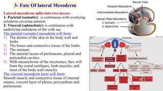
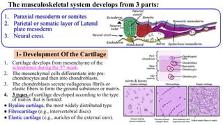
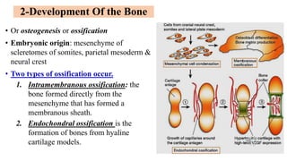
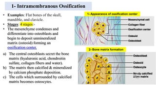
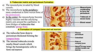
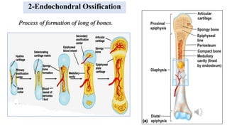
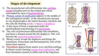
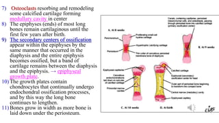
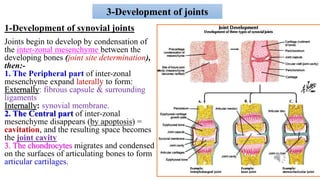
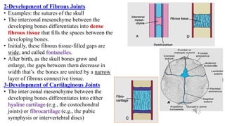
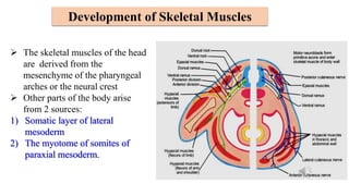
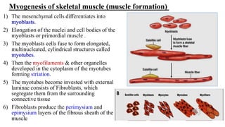
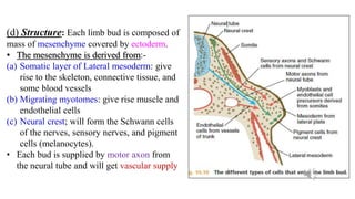
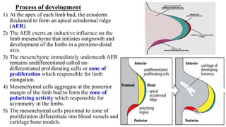
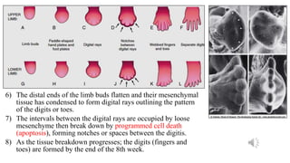
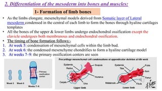
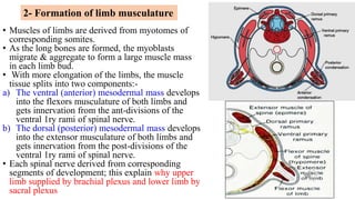
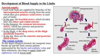
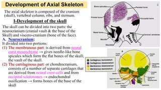
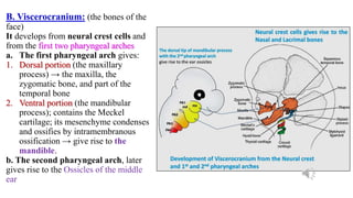
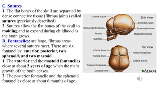
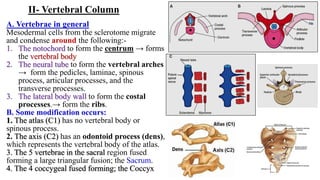
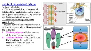
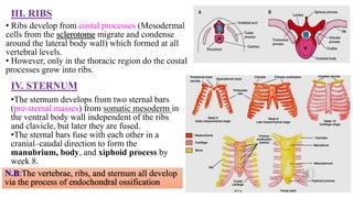
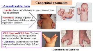
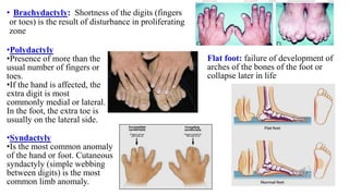
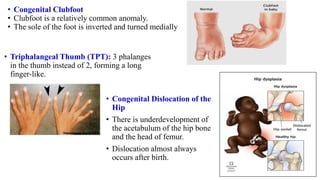
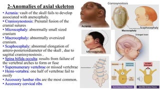
Development Of Musculoskeletal
System
Dr/ Shimaa Antar
Objectives
1. Development of Bone and Cartilage
2. Development of Joints
3. Development of muscles
4. Development of Appendicular Skeleton (limbs)
5. Development of Axial Skeleton
Development of Musculoskeletal System include the followings:-
{
ࣲ
‫ین‬ِ‫ط‬ ‫ن‬ِ‫م‬ ࣲ‫ة‬َ‫ل‬ٰ‫ـ‬َ‫ل‬ُ‫س‬ ‫ن‬ِ‫م‬ َ‫ن‬ٰ‫ـ‬َ‫س‬‫ن‬ِ ۡ
‫ٱۡل‬ ‫َا‬‫ن‬ۡ‫ق‬َ‫ل‬َ‫خ‬ ۡ‫د‬َ‫ق‬َ‫ل‬ َ‫و‬
(
١٢
)
ِ‫ك‬َّ‫م‬ ࣲ
‫ار‬ َ‫ر‬َ‫ق‬ ‫ی‬ِ‫ف‬
ࣰ
‫ة‬َ‫ف‬ۡ‫ط‬ُ‫ن‬ ُ‫ه‬ٰ‫ـ‬َ‫ن‬ۡ‫ل‬َ‫ع‬َ‫ج‬ َّ‫م‬ُ‫ث‬
ࣲ
‫ین‬
(
١٣
)
‫َا‬‫ن‬ۡ‫ق‬َ‫ل‬َ‫خ‬ َّ‫م‬ُ‫ث‬
ۡ‫ض‬ُ‫م‬ۡ‫ٱل‬ ‫َا‬‫ن‬ۡ‫ق‬َ‫ل‬َ‫خ‬َ‫ف‬
ࣰ
‫َة‬‫غ‬ ۡ‫ض‬ُ‫م‬ َ‫ة‬َ‫ق‬َ‫ل‬َ‫ع‬ۡ‫ٱل‬ ‫َا‬‫ن‬ۡ‫ق‬َ‫ل‬َ‫خ‬َ‫ف‬
ࣰ
‫ة‬َ‫ق‬َ‫ل‬َ‫ع‬ َ‫ة‬َ‫ف‬ۡ‫ط‬ُّ‫ن‬‫ٱل‬
َ‫أ‬ َّ‫م‬ُ‫ث‬ ‫ا‬
ࣰ
‫م‬ ۡ
‫ح‬َ‫ل‬ َ‫م‬ٰ‫ـ‬َ‫ظ‬ِ‫ع‬ۡ‫ٱل‬ ‫َا‬‫ن‬ ۡ
‫و‬َ‫س‬َ‫ك‬َ‫ف‬ ‫ا‬
ࣰ
‫م‬ٰ‫ـ‬َ‫ظ‬ِ‫ع‬ َ‫ة‬َ‫غ‬
‫ن‬
ََۚ‫َر‬‫خ‬‫ا‬َ‫ء‬ ‫ا‬ً‫ق‬ۡ‫َل‬‫خ‬ ُ‫ه‬ٰ‫ـ‬َ‫ن‬ۡ‫َأ‬‫ش‬
َ‫ین‬ِ‫ق‬ِ‫ل‬ٰ‫ـ‬َ‫خ‬ۡ‫ٱل‬ ُ‫ن‬َ‫س‬ ۡ
‫ح‬َ‫أ‬ ُ َّ
‫ٱَّلل‬ َ‫ك‬ َ‫ار‬َ‫ب‬َ‫ت‬َ‫ف‬
(
١٤
)
}
Developmental Review
In the 3rd week; The 3 layers (ectoderm, mesoderm &
endoderm) that form the embryo are developed and the
followings occur:
1. The intraembryonic mesoderm differentiates into paraxial,
intermediate and lateral plate
2. The neural folds appeared and form a neural groove which
fused to form neural tube
1.The neural tube will give brain & spinal cord
2. Cells at the lateral border or neural crest cells of the neural fold
dissociate and undergo Epithelial-Mesenchymal transition and
undergo active migration to:-
A. Dorsal pathway: to enter the ectoderm to form melanocytes
and hair follicles
B. Ventral pathway: into the mesoderm to become sensory &
sympathetic ganglia and cells of the adrenal medulla
C. Migrate cranial to neural folds, leaving the neural tube before
closure to contribute to the craniofacial skeleton as well as
neurons for cranial ganglia
1- Fate Of Neural Tube
2- Fate Of Paraxial Mesoderm
 It differentiated into paired blocks of tissue extend in a
craniocaudal direction on each side of the notochord called
somites which appear as bead-like elevations along the
dorsolateral surface of the embryo.
 There are 42–44 pairs of somites (4 occipital, 8 cervical
12 thoracic, 5 lumbar, 5 sacral, and 8 to 10 coccygeal pairs.
 The 1st occipital and the last 5 to 7 coccygeal somites later
disappear.
 The somites then divided into 3 parts which also undergo
Epithelial-Mesenchymal transition.
1-Ventral sclerotome: can migrate and differentiate in many
ways; → forming the bone and cartilage components.
 Cells from the sclerotomes migrates to surround the
notochord and spinal cord, to form axial skeleton including
vertebral column and some bones of the skull.
2- Dermatome: can migrate to form dermis and subcutaneous
tissue of skin.
3- Myotome: can migrate to form all skeletal muscles of body,
head, neck and limbs
Lateral mesoderm splits into two layers:
1- Parietal (somatic): is continuous with overlying
ectoderm covering amnion
2- Visceral (splanchnic): is continuous with
underlying endoderm of the yolk sac
The parietal (somatic) mesoderm will form:
1) The dermis of the skin in the body wall and
limbs
2) The bones and connective tissue of the limbs
3) The sternum
4) The parietal layers of peritoneum, pleural and
pericardial cavities
5) With mesenchyme of the myotomes; they will
form the costal cartilages, limb muscles, and
most of the body wall muscles
The visceral mesoderm layer will form:
Smooth muscle and connective tissue of internal
organs, visceral layer of pleura, pericardium and
peritoneum.
3- Fate Of lateral Mesoderm
The musculoskeletal system develops from 3 parts:
1. Paraxial mesoderm or somites
2. Parietal or somatic layer of Lateral
plate mesoderm
3. Neural crest. Neural crest
1- Development Of the Cartilage
1. Cartilage develops from mesenchyme of the
scleretomes during the 5th week.
2. The mesenchymal cells differentiate into pre-
chondrocytes and then into chondroblasts.
3. The chondroblasts secrete collagenous fibrils or
elastic fibers to form the ground substance or matrix.
4. 3 types of cartilage developed according to the type
of matrix that is formed:
● Hyaline cartilage, the most widely distributed type
● Fibrocartilage (e.g., intervertebral discs)
● Elastic cartilage (e.g., auricles of the external ears).
Joints & bones
• Or osteogenesis or ossification
• Embryonic origin: mesenchyme of
scleretomes of somites, parietal mesoderm &
neural crest
• Two types of ossification occur.
1. Intramembranous ossification: the
bone formed directly from the
mesenchyme that has formed a
membranous sheath.
2. Endochondral ossification is the
formation of bones from hyaline
cartilage models.
2-Development Of the Bone
1- Intramembranous Ossification
• Examples: Flat bones of the skull,
mandible, and clavicle.
• Stages: 4 stages:-
• The mesenchyme condenses and
differentiate into osteoblasts and
begin to deposit unmineralized
matrix (osteoid) forming an
ossification center.
1- Appearance of ossification center
2- Bone matrix formation
a) The central osteoblasts secret the bone
matrix (hyaluronic acid, chondroitin
sulfate, collagen fibers and water).
b) The matrix then calcified & mineralized
by calcium phosphate deposition.
c) The cells which surrounded by calcified
matrix becomes osteocytes.
3- Trabecular matrix and periosteum Formation
a) The mesenchyme invaded by blood
vessels.
b) The mesenchyme in the periphery;
more condensed to form woven bone of
the periosteum.
c) In the center; the mesenchyme become
highly vascular and the calcifying
matrix join around the capillaries to
form bridges of trabeculae that
constitute spongy bone.
4- Formation of compact bone and red marrow
a) The trabecular bone deep to
periosteum thickened forming the
compact bone.
b) The internal trabecula crowds
nearby blood vessels which
brings the hematopoietic cells to
form red marrow
2-Endochondral Ossification
Process of formation of long of bones.
1) The mesenchymal cells differentiate into cartilage
model (chondrocytes) in the central part which
surrounded by layer of perichondrium.
2) Primary center of ossification appears in the middle of
the cartilaginous model; as the chondrocytes increase
in size (hypertrophy), the matrix becomes calcified, and
the cells die forming cavity.→ future of diaphysis
3) A thin layer of bone is deposited under the
perichondrium transforming it to periosteum
4) The cells of periosteum differentiate into osteoblasts,
and bone is formed around the the diaphysis. This will
become the cortical (compact) bone.
5) Blood vessels invade the diaphysis and brings the
osteoblasts, Osteoclasts and haematopoietic cells of the
future bone marrow.
6) Osteoblasts deposit bone matrix over calcified cartilage
& blood vessles forming spongy bone trabeculae, and
bone formation extends to either end of the long bone
Perichondrium
Stages of development
Primary
ossification
center
A B
7) Osteoclasts resorbing and remodeling
some calcified cartilage forming
medullary cavity in center
8) The epiphyses (ends) of most long
bones remain cartilaginous until the
first few years after birth.
9) The secondary centers of ossification
appear within the epiphyses by the
same manner that occurred in the
diaphysis and the entire epiphysis
becomes ossified, but a band of
cartilage remains between the diaphysis
and the epiphysis. → epiphyseal
growth plate.
10) The growth plates contain
chondrocytes that continually undergo
endochondral ossification processes,
and by this way the long bone
continues to lengthen.
11) Bones grow in width as more bone is
laid down under the periosteum.
3-Development of joints
1-Development of synovial joints
Joints begin to develop by condensation of
the inter-zonal mesenchyme between the
developing bones (joint site determination),
then:-
1. The Peripheral part of inter-zonal
mesenchyme expand laterally to form:
Externally: fibrous capsule & surrounding
ligaments
Internally: synovial membrane.
2. The Central part of inter-zonal
mesenchyme disappears (by apoptosis) =
cavitation, and the resulting space becomes
the joint cavity
3. The chondrocytes migrates and condensed
on the surfaces of articulating bones to form
articular cartilages.
2-Development of Fibrous Joints
• Examples: the sutures of the skull
• The interzonal mesenchyme between the
developing bones differentiates into dense
fibrous tissue that fills the spaces between the
developing bones.
• Initially, these fibrous tissue-filled gaps are
wide, and called fontanelles.
• After birth, as the skull bones grow and
enlarge, the gaps between them decrease in
width that’s the bones are united by a narrow
layer of fibrous connective tissue.
3-Development of Cartilaginous Joints
• The inter-zonal mesenchyme between the
developing bones differentiates into either
hyaline cartilage (e.g., the costochondral
joints) or fibrocartilage (e.g., the pubic
symphysis or intervertebral discs)
 The skeletal muscles of the head
are derived from the
mesenchyme of the pharyngeal
arches or the neural crest
 Other parts of the body arise
from 2 sources:
1) Somatic layer of lateral
mesoderm
2) The myotome of somites of
paraxial mesoderm.
Development of Skeletal Muscles
1) The mesenchymal cells differentiates into
myoblasts.
2) Elongation of the nuclei and cell bodies of the
myoblasts or primordial muscle .
3) The myoblasts cells fuse to form elongated,
multinucleated, cylindrical structures called
myotubes.
4) Then the myofilaments & other organelles
developed in the cytoplasm of the myotubes
forming striation.
5) The myotubes become invested with external
laminae consists of Fibroblasts, which
segregate them from the surrounding
connective tissue
6) Fibroblasts produce the perimysium and
epimysium layers of the fibrous sheath of the
muscle
Myogenesis of skeletal muscle (muscle formation)
Development of Appendicular Skeleton (limbs)
(a) Time:
The upper limb buds (forelimb) appear at 28 days;
but the lower limb buds (hindlimb) appeared at 32
days. (1 or 2 days later)
(b) Site:- from the ventrolateral body wall
The U.L. bud develops opposite C4 to T2 somites.
The L.L. bud develops opposite L2 to S3 somites.
(c) Shape:
The upper limb buds appear as paddle
shaped ‫مجداف‬
) ) but the lower limb buds appeared
as flipper-like (‫)زعنفة‬.
• Each bud s flattened, having two borders:
1. Preaxial (cranial) border: marked by the thumb
or big toe.
2. Postaxial (caudal) border: marked by the little
finger or little toe.
Stages of limb development:
1. Development of limb buds:
(d) Structure: Each limb bud is composed of
mass of mesenchyme covered by ectoderm.
• The mesenchyme is derived from:-
(a) Somatic layer of Lateral mesoderm: give
rise to the skeleton, connective tissue, and
some blood vessels
(b) Migrating myotomes: give rise muscle and
endothelial cells
(c) Neural crest; will form the Schwann cells
of the nerves, sensory nerves, and pigment
cells (melanocytes).
• Each bud is supplied by motor axon from
the neural tube and will get vascular supply
1) At the apex of each limb bud, the ectoderm
thickened to form an apical ectodermal ridge
(AER).
2) The AER exerts an inductive influence on the
limb mesenchyme that initiates outgrowth and
development of the limbs in a proximo-distal
axis.
3) The mesenchyme immediately underneath AER
remains undifferentiated called un-
differentiated proliferating cells or zone of
proliferation which responsible for limb
elongation.
4) Mesenchymal cells aggregate at the posterior
margin of the limb bud to form the zone of
polarizing activity which responsible for
asymmetry in the limbs.
5) The mesenchymal cells proximal to zone of
proliferation differentiate into blood vessels and
cartilage bone models.
Process of development
6) The distal ends of the limb buds flatten and their mesenchymal
tissue has condensed to form digital rays outlining the pattern
of the digits or toes.
7) The intervals between the digital rays are occupied by loose
mesenchyme then break down by programmed cell death
(apoptosis), forming notches or spaces between the digitis.
8) As the tissue breakdown progresses; the digits (fingers and
toes) are formed by the end of the 8th week.
• As the limbs elongate, mesenchymal models derived from Somatic layer of Lateral
mesoderm condensed in the central of each limb to form the bones through hyaline cartilages
templates
• All the bones of the upper & lower limbs undergo endochondral ossification except the
clavicle undergoes both membranous and endochondral ossification.
• The timing of bone formation follows:-
1. At week 5: condensation of mesenchymal cells within the limb bud.
2. At week 6: the condensed mesenchyme chondrifies to form a hyaline cartilage model
3. At weeks 7–9: the primary ossification centers are seen
2. Differentiation of the mesoderm into bones and muscles:
1- Formation of limb bones
2- Formation of limb musculature
• Muscles of limbs are derived from myotomes of
corresponding somites.
• As the long bones are formed, the myoblasts
migrate & aggregate to form a large muscle mass
in each limb bud.
• With more elongation of the limbs, the muscle
tissue splits into two components:-
a) The ventral (anterior) mesodermal mass develops
into the flexors musculature of both limbs and
gets innervation from the ant-divisions of the
ventral 1ry rami of spinal nerve.
b) The dorsal (posterior) mesodermal mass develops
into the extensor musculature of both limbs and
gets innervation from the post-divisions of the
ventral 1ry rami of spinal nerve.
• Each spinal nerve derived from corresponding
segments of development; this explain why upper
limb supplied by brachial plexus and lower limb by
sacral plexus
3. Segmentation of the limbs :
• First constriction develops at the base of the paddle, so;
the wrist or ankle areas appear.
• A second constriction on the limb bud appears at the
future site of the elbow or knee.
4. Rotation of the limbs : (7th & 8th week).
a. At first the limbs extend ventrally; the flexor
aspect of the limbs is ventral and the extensor
aspect dorsal.
b. The developing upper and lower limbs rotate in
opposite directions but with same degrees:
c. The upper limbs rotate 90 degrees laterally on
their longitudinal axes; thus, the future elbows
point posterior and the extensor muscles lie on the
lateral and posterior aspects & thumbs faces
laterally.
d. The lower limbs rotate 90 degrees medially; thus,
the future knees face anterior and the extensor
muscles lie on the anterior aspect of the lower limb
& the big toe become medially.
1st constriction 1st constriction
Development of Blood Supply to the Limbs
Arterial supply:
• Each limb buds are supplied by branches of the
intersegmental arteries arising from the aorta
which then give primary axial artery in each
part of limbs.
• In the arm: the brachial artery which divided
later into ulnar and radial arteries
• In the forearm: the common interosseous
artery, which has anterior and posterior
interosseous branches.
• In the thigh: is the deep artery of the thigh
(profunda femoris).
• In the leg: there are the anterior and posterior
tibial arteries.
The venous system:
As the digits (fingers) formed, the marginal sinus
breaks up and the final venous pattern
represented by the basilic and cephalic veins and
their tributaries, develops in upper limb and
saphenous vein in lower limb.
common
interosseous
Development of Axial Skeleton
The axial skeleton is composed of the cranium
(skull), vertebral column, ribs, and sternum.
I-Development of the skull
The skull can be divided into two parts: the
neurocranium (cranial vault & the base of the
Skull) and viscero-cranium (bone of the face).
A. Neurocranium:
It divided into two portions:
(1) The membranous part: is derived from neural
crest mesenchyme → gives needle-like bone
spicules which form the flat bones of the skull;
the vault of the skull.
(2) The cartilaginous part: or chondrocranium,
consists of a number of separate cartilages that
are derived from neural crest cells and from
occipital sclerotomes → endochondral
ossification → forms bones of the base of the
skull
B. Viscerocranium: (the bones of the
face)
It develops from neural crest cells and
from the first two pharyngeal arches
a. The first pharyngeal arch gives:
1. Dorsal portion (the maxillary
process) → the maxilla, the
zygomatic bone, and part of the
temporal bone
2. Ventral portion (the mandibular
process); contains the Meckel
cartilage; its mesenchyme condenses
and ossifies by intramembranous
ossification → give rise to the
mandible.
b. The second pharyngeal arch, later
gives rise to the Ossicles of the middle
ear
C. Sutures
1. The flat bones of the skull are separated by
dense connective tissue (fibrous joints) called
sutures (previously described).
2. Sutures allow the flat bones of the skull to
molding and to expand during childhood as
the brain grows.
D. Fontanelles: are large, fibrous areas
where several sutures meet. There are six
fontanelles: anterior, posterior, two
sphenoid, and two mastoid.
1. The anterior and the mastoid fontanelles
close at about 2 years of age when the main
growth of the brain ceases.
2. The posterior fontanelle and the sphenoid
fontanelles close at about 6 months of age.
II- Vertebral Column
A. Vertebrae in general
Mesodermal cells from the sclerotome migrate
and condense around the following:-
1. The notochord to form the centrum → forms
the vertebral body
2. The neural tube to form the vertebral arches
→ form the pedicles, laminae, spinous
process, articular processes, and the
transverse processes.
3. The lateral body wall to form the costal
processes.→ form the ribs.
B. Some modification occurs:
1. The atlas (C1) has no vertebral body or
spinous process.
2. The axis (C2) has an odontoid process (dens),
which represents the vertebral body of the atlas.
3. The 5 vertebrae in the sacral region fused
forming a large triangular fusion; the Sacrum.
4. The 4 coccygeal fused forming; the Coccyx
Joints of the vertebral column
1. Synovial joints
a. The atlanto-occipital, atlanto-axial
joint and the Facets (between the inferior
and superior articular facets). By the same
mechanism previously described
2. Secondary cartilaginous joints
(symphyses)
The joints between the vertebral bodies in
which the intervertebral disks consists of
the following:
i. Nucleus pulposus: this is a remnant
of the embryonic notochord.
ii. Annulus fibrosus: is an outer rim of
fibrocartilage derived from
mesoderm found between the
vertebral bodies.
III. RIBS
• Ribs develop from costal processes (Mesodermal
cells from the sclerotome migrate and condense
around the lateral body wall) which formed at all
vertebral levels.
• However, only in the thoracic region do the costal
processes grow into ribs.
IV. STERNUM
•The sternum develops from two sternal bars
(pre-sternal masses) from somatic mesoderm in
the ventral body wall independent of the ribs
and clavicle, but later they are fused.
•The sternal bars fuse with each other in a
cranial–caudal direction to form the
manubrium, body, and xiphoid process by
week 8.
N.B:The vertebrae, ribs, and sternum all develop
via the process of endochondral ossification
1-Anomalies of the limbs
•Cleft Hand and Cleft Foot: The hand
or foot is divided into two parts that
oppose each other like lobster claws.
• Cleft Hand; usually absence of third
metacarpal and fusion of digits 1–2 and
4–5
Congenital anomalies
Cleft Hand and Cleft Foot
Amelia
• Amelia: absence of a limb due to suppression of limb
bud development
•Meromelia: absence of part of a
limb: disturbance of differentiation
or growth of the limbs
•Syndactyly
•Is the most common anomaly
of the hand or foot. Cutaneous
syndactyly (simple webbing
between digits) is the most
common limb anomaly.
• Brachydactyly: Shortness of the digits (fingers
or toes) is the result of disturbance in proliferating
zone
•Polydactyly
•Presence of more than the
usual number of fingers or
toes.
•If the hand is affected, the
extra digit is most
commonly medial or lateral.
In the foot, the extra toe is
usually on the lateral side.
Flat foot: failure of development of
arches of the bones of the foot or
collapse later in life
• Congenital Clubfoot
• Clubfoot is a relatively common anomaly.
• The sole of the foot is inverted and turned medially
• Congenital Dislocation of the
Hip
• There is underdevelopment of
the acetabulum of the hip bone
and the head of femur.
• Dislocation almost always
occurs after birth.
• Triphalangeal Thumb (TPT): 3 phalanges
in the thumb instead of 2, forming a long
finger-like.
2-Anomalies of axial skeleton
• Acrania: vault of the skull fails to develop
associated with anencephaly.
• Craniosynostosis: Prenatal fusion of the
cranial sutures
• Microcephaly: abnormally small sized
cranium
• Macrocephaly: abnormally oversized
cranium.
• Scaphocephaly: abnormal elongation of
antero-posteriordiameter of the skull , due to
sagittal craniosynostosis .
• Spina bifida occulta: results from failure of
the vertebral arches to form or fuse
• Supernumerary vertebrae or missed vertebrae
• Hemi-vertabra: one half of vertebrae fail to
ossify
• Accessory lumbar ribs are the most common.
• Accessory cervical ribs
Acrania
Craniosynostosis
Microcephaly
Scaphocephaly
Thank you Allah

Development of Musculoskeletal system.ppsx

  • 1.
  • 2.
    Objectives 1. Development ofBone and Cartilage 2. Development of Joints 3. Development of muscles 4. Development of Appendicular Skeleton (limbs) 5. Development of Axial Skeleton Development of Musculoskeletal System include the followings:- { ࣲ ‫ین‬ِ‫ط‬ ‫ن‬ِ‫م‬ ࣲ‫ة‬َ‫ل‬ٰ‫ـ‬َ‫ل‬ُ‫س‬ ‫ن‬ِ‫م‬ َ‫ن‬ٰ‫ـ‬َ‫س‬‫ن‬ِ ۡ ‫ٱۡل‬ ‫َا‬‫ن‬ۡ‫ق‬َ‫ل‬َ‫خ‬ ۡ‫د‬َ‫ق‬َ‫ل‬ َ‫و‬ ( ١٢ ) ِ‫ك‬َّ‫م‬ ࣲ ‫ار‬ َ‫ر‬َ‫ق‬ ‫ی‬ِ‫ف‬ ࣰ ‫ة‬َ‫ف‬ۡ‫ط‬ُ‫ن‬ ُ‫ه‬ٰ‫ـ‬َ‫ن‬ۡ‫ل‬َ‫ع‬َ‫ج‬ َّ‫م‬ُ‫ث‬ ࣲ ‫ین‬ ( ١٣ ) ‫َا‬‫ن‬ۡ‫ق‬َ‫ل‬َ‫خ‬ َّ‫م‬ُ‫ث‬ ۡ‫ض‬ُ‫م‬ۡ‫ٱل‬ ‫َا‬‫ن‬ۡ‫ق‬َ‫ل‬َ‫خ‬َ‫ف‬ ࣰ ‫َة‬‫غ‬ ۡ‫ض‬ُ‫م‬ َ‫ة‬َ‫ق‬َ‫ل‬َ‫ع‬ۡ‫ٱل‬ ‫َا‬‫ن‬ۡ‫ق‬َ‫ل‬َ‫خ‬َ‫ف‬ ࣰ ‫ة‬َ‫ق‬َ‫ل‬َ‫ع‬ َ‫ة‬َ‫ف‬ۡ‫ط‬ُّ‫ن‬‫ٱل‬ َ‫أ‬ َّ‫م‬ُ‫ث‬ ‫ا‬ ࣰ ‫م‬ ۡ ‫ح‬َ‫ل‬ َ‫م‬ٰ‫ـ‬َ‫ظ‬ِ‫ع‬ۡ‫ٱل‬ ‫َا‬‫ن‬ ۡ ‫و‬َ‫س‬َ‫ك‬َ‫ف‬ ‫ا‬ ࣰ ‫م‬ٰ‫ـ‬َ‫ظ‬ِ‫ع‬ َ‫ة‬َ‫غ‬ ‫ن‬ ََۚ‫َر‬‫خ‬‫ا‬َ‫ء‬ ‫ا‬ً‫ق‬ۡ‫َل‬‫خ‬ ُ‫ه‬ٰ‫ـ‬َ‫ن‬ۡ‫َأ‬‫ش‬ َ‫ین‬ِ‫ق‬ِ‫ل‬ٰ‫ـ‬َ‫خ‬ۡ‫ٱل‬ ُ‫ن‬َ‫س‬ ۡ ‫ح‬َ‫أ‬ ُ َّ ‫ٱَّلل‬ َ‫ك‬ َ‫ار‬َ‫ب‬َ‫ت‬َ‫ف‬ ( ١٤ ) }
  • 3.
    Developmental Review In the3rd week; The 3 layers (ectoderm, mesoderm & endoderm) that form the embryo are developed and the followings occur: 1. The intraembryonic mesoderm differentiates into paraxial, intermediate and lateral plate 2. The neural folds appeared and form a neural groove which fused to form neural tube 1.The neural tube will give brain & spinal cord 2. Cells at the lateral border or neural crest cells of the neural fold dissociate and undergo Epithelial-Mesenchymal transition and undergo active migration to:- A. Dorsal pathway: to enter the ectoderm to form melanocytes and hair follicles B. Ventral pathway: into the mesoderm to become sensory & sympathetic ganglia and cells of the adrenal medulla C. Migrate cranial to neural folds, leaving the neural tube before closure to contribute to the craniofacial skeleton as well as neurons for cranial ganglia 1- Fate Of Neural Tube
  • 4.
    2- Fate OfParaxial Mesoderm  It differentiated into paired blocks of tissue extend in a craniocaudal direction on each side of the notochord called somites which appear as bead-like elevations along the dorsolateral surface of the embryo.  There are 42–44 pairs of somites (4 occipital, 8 cervical 12 thoracic, 5 lumbar, 5 sacral, and 8 to 10 coccygeal pairs.  The 1st occipital and the last 5 to 7 coccygeal somites later disappear.  The somites then divided into 3 parts which also undergo Epithelial-Mesenchymal transition. 1-Ventral sclerotome: can migrate and differentiate in many ways; → forming the bone and cartilage components.  Cells from the sclerotomes migrates to surround the notochord and spinal cord, to form axial skeleton including vertebral column and some bones of the skull. 2- Dermatome: can migrate to form dermis and subcutaneous tissue of skin. 3- Myotome: can migrate to form all skeletal muscles of body, head, neck and limbs
  • 5.
    Lateral mesoderm splitsinto two layers: 1- Parietal (somatic): is continuous with overlying ectoderm covering amnion 2- Visceral (splanchnic): is continuous with underlying endoderm of the yolk sac The parietal (somatic) mesoderm will form: 1) The dermis of the skin in the body wall and limbs 2) The bones and connective tissue of the limbs 3) The sternum 4) The parietal layers of peritoneum, pleural and pericardial cavities 5) With mesenchyme of the myotomes; they will form the costal cartilages, limb muscles, and most of the body wall muscles The visceral mesoderm layer will form: Smooth muscle and connective tissue of internal organs, visceral layer of pleura, pericardium and peritoneum. 3- Fate Of lateral Mesoderm
  • 6.
    The musculoskeletal systemdevelops from 3 parts: 1. Paraxial mesoderm or somites 2. Parietal or somatic layer of Lateral plate mesoderm 3. Neural crest. Neural crest 1- Development Of the Cartilage 1. Cartilage develops from mesenchyme of the scleretomes during the 5th week. 2. The mesenchymal cells differentiate into pre- chondrocytes and then into chondroblasts. 3. The chondroblasts secrete collagenous fibrils or elastic fibers to form the ground substance or matrix. 4. 3 types of cartilage developed according to the type of matrix that is formed: ● Hyaline cartilage, the most widely distributed type ● Fibrocartilage (e.g., intervertebral discs) ● Elastic cartilage (e.g., auricles of the external ears). Joints & bones
  • 7.
    • Or osteogenesisor ossification • Embryonic origin: mesenchyme of scleretomes of somites, parietal mesoderm & neural crest • Two types of ossification occur. 1. Intramembranous ossification: the bone formed directly from the mesenchyme that has formed a membranous sheath. 2. Endochondral ossification is the formation of bones from hyaline cartilage models. 2-Development Of the Bone
  • 8.
    1- Intramembranous Ossification •Examples: Flat bones of the skull, mandible, and clavicle. • Stages: 4 stages:- • The mesenchyme condenses and differentiate into osteoblasts and begin to deposit unmineralized matrix (osteoid) forming an ossification center. 1- Appearance of ossification center 2- Bone matrix formation a) The central osteoblasts secret the bone matrix (hyaluronic acid, chondroitin sulfate, collagen fibers and water). b) The matrix then calcified & mineralized by calcium phosphate deposition. c) The cells which surrounded by calcified matrix becomes osteocytes.
  • 9.
    3- Trabecular matrixand periosteum Formation a) The mesenchyme invaded by blood vessels. b) The mesenchyme in the periphery; more condensed to form woven bone of the periosteum. c) In the center; the mesenchyme become highly vascular and the calcifying matrix join around the capillaries to form bridges of trabeculae that constitute spongy bone. 4- Formation of compact bone and red marrow a) The trabecular bone deep to periosteum thickened forming the compact bone. b) The internal trabecula crowds nearby blood vessels which brings the hematopoietic cells to form red marrow
  • 10.
    2-Endochondral Ossification Process offormation of long of bones.
  • 11.
    1) The mesenchymalcells differentiate into cartilage model (chondrocytes) in the central part which surrounded by layer of perichondrium. 2) Primary center of ossification appears in the middle of the cartilaginous model; as the chondrocytes increase in size (hypertrophy), the matrix becomes calcified, and the cells die forming cavity.→ future of diaphysis 3) A thin layer of bone is deposited under the perichondrium transforming it to periosteum 4) The cells of periosteum differentiate into osteoblasts, and bone is formed around the the diaphysis. This will become the cortical (compact) bone. 5) Blood vessels invade the diaphysis and brings the osteoblasts, Osteoclasts and haematopoietic cells of the future bone marrow. 6) Osteoblasts deposit bone matrix over calcified cartilage & blood vessles forming spongy bone trabeculae, and bone formation extends to either end of the long bone Perichondrium Stages of development Primary ossification center A B
  • 12.
    7) Osteoclasts resorbingand remodeling some calcified cartilage forming medullary cavity in center 8) The epiphyses (ends) of most long bones remain cartilaginous until the first few years after birth. 9) The secondary centers of ossification appear within the epiphyses by the same manner that occurred in the diaphysis and the entire epiphysis becomes ossified, but a band of cartilage remains between the diaphysis and the epiphysis. → epiphyseal growth plate. 10) The growth plates contain chondrocytes that continually undergo endochondral ossification processes, and by this way the long bone continues to lengthen. 11) Bones grow in width as more bone is laid down under the periosteum.
  • 13.
    3-Development of joints 1-Developmentof synovial joints Joints begin to develop by condensation of the inter-zonal mesenchyme between the developing bones (joint site determination), then:- 1. The Peripheral part of inter-zonal mesenchyme expand laterally to form: Externally: fibrous capsule & surrounding ligaments Internally: synovial membrane. 2. The Central part of inter-zonal mesenchyme disappears (by apoptosis) = cavitation, and the resulting space becomes the joint cavity 3. The chondrocytes migrates and condensed on the surfaces of articulating bones to form articular cartilages.
  • 14.
    2-Development of FibrousJoints • Examples: the sutures of the skull • The interzonal mesenchyme between the developing bones differentiates into dense fibrous tissue that fills the spaces between the developing bones. • Initially, these fibrous tissue-filled gaps are wide, and called fontanelles. • After birth, as the skull bones grow and enlarge, the gaps between them decrease in width that’s the bones are united by a narrow layer of fibrous connective tissue. 3-Development of Cartilaginous Joints • The inter-zonal mesenchyme between the developing bones differentiates into either hyaline cartilage (e.g., the costochondral joints) or fibrocartilage (e.g., the pubic symphysis or intervertebral discs)
  • 15.
     The skeletalmuscles of the head are derived from the mesenchyme of the pharyngeal arches or the neural crest  Other parts of the body arise from 2 sources: 1) Somatic layer of lateral mesoderm 2) The myotome of somites of paraxial mesoderm. Development of Skeletal Muscles
  • 16.
    1) The mesenchymalcells differentiates into myoblasts. 2) Elongation of the nuclei and cell bodies of the myoblasts or primordial muscle . 3) The myoblasts cells fuse to form elongated, multinucleated, cylindrical structures called myotubes. 4) Then the myofilaments & other organelles developed in the cytoplasm of the myotubes forming striation. 5) The myotubes become invested with external laminae consists of Fibroblasts, which segregate them from the surrounding connective tissue 6) Fibroblasts produce the perimysium and epimysium layers of the fibrous sheath of the muscle Myogenesis of skeletal muscle (muscle formation)
  • 17.
    Development of AppendicularSkeleton (limbs) (a) Time: The upper limb buds (forelimb) appear at 28 days; but the lower limb buds (hindlimb) appeared at 32 days. (1 or 2 days later) (b) Site:- from the ventrolateral body wall The U.L. bud develops opposite C4 to T2 somites. The L.L. bud develops opposite L2 to S3 somites. (c) Shape: The upper limb buds appear as paddle shaped ‫مجداف‬ ) ) but the lower limb buds appeared as flipper-like (‫)زعنفة‬. • Each bud s flattened, having two borders: 1. Preaxial (cranial) border: marked by the thumb or big toe. 2. Postaxial (caudal) border: marked by the little finger or little toe. Stages of limb development: 1. Development of limb buds:
  • 18.
    (d) Structure: Eachlimb bud is composed of mass of mesenchyme covered by ectoderm. • The mesenchyme is derived from:- (a) Somatic layer of Lateral mesoderm: give rise to the skeleton, connective tissue, and some blood vessels (b) Migrating myotomes: give rise muscle and endothelial cells (c) Neural crest; will form the Schwann cells of the nerves, sensory nerves, and pigment cells (melanocytes). • Each bud is supplied by motor axon from the neural tube and will get vascular supply
  • 19.
    1) At theapex of each limb bud, the ectoderm thickened to form an apical ectodermal ridge (AER). 2) The AER exerts an inductive influence on the limb mesenchyme that initiates outgrowth and development of the limbs in a proximo-distal axis. 3) The mesenchyme immediately underneath AER remains undifferentiated called un- differentiated proliferating cells or zone of proliferation which responsible for limb elongation. 4) Mesenchymal cells aggregate at the posterior margin of the limb bud to form the zone of polarizing activity which responsible for asymmetry in the limbs. 5) The mesenchymal cells proximal to zone of proliferation differentiate into blood vessels and cartilage bone models. Process of development
  • 20.
    6) The distalends of the limb buds flatten and their mesenchymal tissue has condensed to form digital rays outlining the pattern of the digits or toes. 7) The intervals between the digital rays are occupied by loose mesenchyme then break down by programmed cell death (apoptosis), forming notches or spaces between the digitis. 8) As the tissue breakdown progresses; the digits (fingers and toes) are formed by the end of the 8th week.
  • 21.
    • As thelimbs elongate, mesenchymal models derived from Somatic layer of Lateral mesoderm condensed in the central of each limb to form the bones through hyaline cartilages templates • All the bones of the upper & lower limbs undergo endochondral ossification except the clavicle undergoes both membranous and endochondral ossification. • The timing of bone formation follows:- 1. At week 5: condensation of mesenchymal cells within the limb bud. 2. At week 6: the condensed mesenchyme chondrifies to form a hyaline cartilage model 3. At weeks 7–9: the primary ossification centers are seen 2. Differentiation of the mesoderm into bones and muscles: 1- Formation of limb bones
  • 22.
    2- Formation oflimb musculature • Muscles of limbs are derived from myotomes of corresponding somites. • As the long bones are formed, the myoblasts migrate & aggregate to form a large muscle mass in each limb bud. • With more elongation of the limbs, the muscle tissue splits into two components:- a) The ventral (anterior) mesodermal mass develops into the flexors musculature of both limbs and gets innervation from the ant-divisions of the ventral 1ry rami of spinal nerve. b) The dorsal (posterior) mesodermal mass develops into the extensor musculature of both limbs and gets innervation from the post-divisions of the ventral 1ry rami of spinal nerve. • Each spinal nerve derived from corresponding segments of development; this explain why upper limb supplied by brachial plexus and lower limb by sacral plexus
  • 23.
    3. Segmentation ofthe limbs : • First constriction develops at the base of the paddle, so; the wrist or ankle areas appear. • A second constriction on the limb bud appears at the future site of the elbow or knee. 4. Rotation of the limbs : (7th & 8th week). a. At first the limbs extend ventrally; the flexor aspect of the limbs is ventral and the extensor aspect dorsal. b. The developing upper and lower limbs rotate in opposite directions but with same degrees: c. The upper limbs rotate 90 degrees laterally on their longitudinal axes; thus, the future elbows point posterior and the extensor muscles lie on the lateral and posterior aspects & thumbs faces laterally. d. The lower limbs rotate 90 degrees medially; thus, the future knees face anterior and the extensor muscles lie on the anterior aspect of the lower limb & the big toe become medially. 1st constriction 1st constriction
  • 24.
    Development of BloodSupply to the Limbs Arterial supply: • Each limb buds are supplied by branches of the intersegmental arteries arising from the aorta which then give primary axial artery in each part of limbs. • In the arm: the brachial artery which divided later into ulnar and radial arteries • In the forearm: the common interosseous artery, which has anterior and posterior interosseous branches. • In the thigh: is the deep artery of the thigh (profunda femoris). • In the leg: there are the anterior and posterior tibial arteries. The venous system: As the digits (fingers) formed, the marginal sinus breaks up and the final venous pattern represented by the basilic and cephalic veins and their tributaries, develops in upper limb and saphenous vein in lower limb. common interosseous
  • 25.
    Development of AxialSkeleton The axial skeleton is composed of the cranium (skull), vertebral column, ribs, and sternum. I-Development of the skull The skull can be divided into two parts: the neurocranium (cranial vault & the base of the Skull) and viscero-cranium (bone of the face). A. Neurocranium: It divided into two portions: (1) The membranous part: is derived from neural crest mesenchyme → gives needle-like bone spicules which form the flat bones of the skull; the vault of the skull. (2) The cartilaginous part: or chondrocranium, consists of a number of separate cartilages that are derived from neural crest cells and from occipital sclerotomes → endochondral ossification → forms bones of the base of the skull
  • 26.
    B. Viscerocranium: (thebones of the face) It develops from neural crest cells and from the first two pharyngeal arches a. The first pharyngeal arch gives: 1. Dorsal portion (the maxillary process) → the maxilla, the zygomatic bone, and part of the temporal bone 2. Ventral portion (the mandibular process); contains the Meckel cartilage; its mesenchyme condenses and ossifies by intramembranous ossification → give rise to the mandible. b. The second pharyngeal arch, later gives rise to the Ossicles of the middle ear
  • 27.
    C. Sutures 1. Theflat bones of the skull are separated by dense connective tissue (fibrous joints) called sutures (previously described). 2. Sutures allow the flat bones of the skull to molding and to expand during childhood as the brain grows. D. Fontanelles: are large, fibrous areas where several sutures meet. There are six fontanelles: anterior, posterior, two sphenoid, and two mastoid. 1. The anterior and the mastoid fontanelles close at about 2 years of age when the main growth of the brain ceases. 2. The posterior fontanelle and the sphenoid fontanelles close at about 6 months of age.
  • 28.
    II- Vertebral Column A.Vertebrae in general Mesodermal cells from the sclerotome migrate and condense around the following:- 1. The notochord to form the centrum → forms the vertebral body 2. The neural tube to form the vertebral arches → form the pedicles, laminae, spinous process, articular processes, and the transverse processes. 3. The lateral body wall to form the costal processes.→ form the ribs. B. Some modification occurs: 1. The atlas (C1) has no vertebral body or spinous process. 2. The axis (C2) has an odontoid process (dens), which represents the vertebral body of the atlas. 3. The 5 vertebrae in the sacral region fused forming a large triangular fusion; the Sacrum. 4. The 4 coccygeal fused forming; the Coccyx
  • 29.
    Joints of thevertebral column 1. Synovial joints a. The atlanto-occipital, atlanto-axial joint and the Facets (between the inferior and superior articular facets). By the same mechanism previously described 2. Secondary cartilaginous joints (symphyses) The joints between the vertebral bodies in which the intervertebral disks consists of the following: i. Nucleus pulposus: this is a remnant of the embryonic notochord. ii. Annulus fibrosus: is an outer rim of fibrocartilage derived from mesoderm found between the vertebral bodies.
  • 30.
    III. RIBS • Ribsdevelop from costal processes (Mesodermal cells from the sclerotome migrate and condense around the lateral body wall) which formed at all vertebral levels. • However, only in the thoracic region do the costal processes grow into ribs. IV. STERNUM •The sternum develops from two sternal bars (pre-sternal masses) from somatic mesoderm in the ventral body wall independent of the ribs and clavicle, but later they are fused. •The sternal bars fuse with each other in a cranial–caudal direction to form the manubrium, body, and xiphoid process by week 8. N.B:The vertebrae, ribs, and sternum all develop via the process of endochondral ossification
  • 31.
    1-Anomalies of thelimbs •Cleft Hand and Cleft Foot: The hand or foot is divided into two parts that oppose each other like lobster claws. • Cleft Hand; usually absence of third metacarpal and fusion of digits 1–2 and 4–5 Congenital anomalies Cleft Hand and Cleft Foot Amelia • Amelia: absence of a limb due to suppression of limb bud development •Meromelia: absence of part of a limb: disturbance of differentiation or growth of the limbs
  • 32.
    •Syndactyly •Is the mostcommon anomaly of the hand or foot. Cutaneous syndactyly (simple webbing between digits) is the most common limb anomaly. • Brachydactyly: Shortness of the digits (fingers or toes) is the result of disturbance in proliferating zone •Polydactyly •Presence of more than the usual number of fingers or toes. •If the hand is affected, the extra digit is most commonly medial or lateral. In the foot, the extra toe is usually on the lateral side. Flat foot: failure of development of arches of the bones of the foot or collapse later in life
  • 33.
    • Congenital Clubfoot •Clubfoot is a relatively common anomaly. • The sole of the foot is inverted and turned medially • Congenital Dislocation of the Hip • There is underdevelopment of the acetabulum of the hip bone and the head of femur. • Dislocation almost always occurs after birth. • Triphalangeal Thumb (TPT): 3 phalanges in the thumb instead of 2, forming a long finger-like.
  • 34.
    2-Anomalies of axialskeleton • Acrania: vault of the skull fails to develop associated with anencephaly. • Craniosynostosis: Prenatal fusion of the cranial sutures • Microcephaly: abnormally small sized cranium • Macrocephaly: abnormally oversized cranium. • Scaphocephaly: abnormal elongation of antero-posteriordiameter of the skull , due to sagittal craniosynostosis . • Spina bifida occulta: results from failure of the vertebral arches to form or fuse • Supernumerary vertebrae or missed vertebrae • Hemi-vertabra: one half of vertebrae fail to ossify • Accessory lumbar ribs are the most common. • Accessory cervical ribs Acrania Craniosynostosis Microcephaly Scaphocephaly
  • 35.