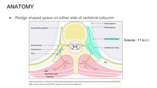
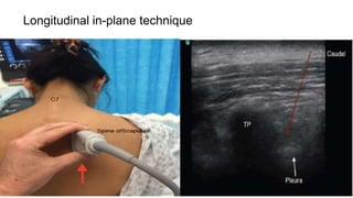
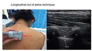
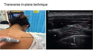
USG-GUIDED THORACIC
PARAVERTEBRAL BLOCK
BACKGROUND
● It is a technique of injecting local anesthetic alongside the thoracic vertebra
close to where the spinal nerves emerge from the intervertebral foramen.
● produces unilateral, segmental, somatic, and sympathetic nerve blockade
BACKGROUND
● Hugo Sellheim of Leipzig (1871–1936) is believed to have pioneered TPVB in
1905.
● Kappis, in 1919, developed the technique of paravertebral injection, which is
comparable to the one in present day use.
● In 1979, Eason and Wyatt re popularized the technique after describing
paravertebral catheter placement.
ANATOMY
● Wedge shaped space on either side of vertebral coloumn
Extends : T1 to L1
BOUNDARIES
● Anterolaterally : parietal pleura
● Base : vertebral body, intervertebral disc, intervertebral foremen with its
contents
● Posteriorly : transverse process and superior costotransverse ligament
● Endothoracic fascia forms the deep border of space and separates the nerve
root from sympathetic ganglia
Endothoracic fascia
● Deep fascia of thorax
● Lies between parietal pleura anteriorly and superior costotransverse ligament
posteriorly
● Medially it is attached to periosteum of vertebral body
● Divides TPVS into two fascial compartments :
○ Anterior extrapleural paravertebral compartment
○ Posterior subendothoracic paravertebral compartment
SAGGITAL SECTION
The SCTL
extends
from the
lower
border of
the
transverse
process
above to
the upper
border of
the
transverse
process
below
● CONTENTS
○ intercostal (spinal) nerve
○ the dorsal ramus
○ intercostal vessels
○ rami communicantes
○ sympathetic chain.
● COMMUNICATIONS
○ Medially : epidural space
○ Laterally : intercostal space
○ PVS of opposite side
Mechanism of block
● It produces ipsilateral somatic and sympathetic nerve blockade due to
○ a direct effect of the local anesthetic on the somatic and sympathetic nerves in the
TPVS
○ extension into the intercostal space laterally, and the epidural space medially
Distribution of anaesthesia
Single injection technique
● Dermatomal distribution -- unpredictable
● Spread both cephalad and caudad to the site of injection to some extent
● Segmental contralateral anesthesia, adjacent to the site of injection, occurs in
approximately 10% of patients
Multiple injection technique
● Small volumes (3–4 mL) of local anesthetic are injected at several contiguous
thoracic levels
● Preferable to single injection technique
INDICATIONS
● Anaesthesia
○ Breast surgery
○ Herniorraphy (thoracolumbar anaesthesia)
○ Chest wound exploration
● Chronic pain management
○ Benign and malignant neuralgia
● Post op analgesia ( as part of balanced analgesic regimen )
○ Thoracotomy
○ Thoracoabdominal thoracic surgery
○ Video-assisted thoracoscopic surgery
○ Cholecystectomy
○ Renal surgery
○ Breast surgery
○ Herniorraphy
○ Liver resection
○ Appendicectomy
○ Minimally invasive cardiac surgery
○ Conventional cardiac surgery (b/l TPVB)
● Miscellaneous
○ Post herpetic neuralgia
○ Relief of pleuritic chest pain
○ Multiple fractured rib
○ Treatment of hyperhydrosis
○ Liver capsule pain after blunt abdominal trauma
CONTRAINDICATIONS
Absolute
● Infection at the site of injection
● Allergy to LA
● Empyema
● Neoplastic mass occupying the paravertebral space
● Coagulopathy
Relative
● Bleeding d/o
● Pt receiving anticoagulants
USG GUIDED TECHNIQUE
● Position : sitting (preferable ) / lateral / prone
● Probe :curved probe (5-8 Mhz) or linear probe oscillating at 10-13 MHz
● Needle : 5-10 cm, 22G blunt needle or 18 G touhy needle
● Local anaesthetic: 10-15 ml of 0.5% ropivacaine /0.5% bupivacaine
Basic USG appearance of key structures
● Bone appears as a hyperechoic line with acoustic shadowing below
● Ribs will appear as more convex structures compared to transverse
processes
● Transverse process will appear more squared off and lie more superficial to
skin compared to ribs
● Pleura appear as a bright hyperechoic line.
● Visual echogenicity of pleura can be accentuated by asking the patient to
breathe deeply. There is no acoustic shadowing seen with pleura
Basic ultrasound appearance of bone and pleura
Various Approaches of thoracic PVB
● The choice of technique to adopt is ultimately down to operator experience
and preference
● The ideal technique should be simple to perform and ensure continuous
needle visualization and avoid the neuraxis.
● Currently, there is no evidence to conclude that one technique is superior to
the other in terms of block success or the ability of the needle tip and injectate
to cross the SCTL
CHOOSING THE LEVEL
● Depends on the dermatomal spread required for blockade.
● Large volume single shot (such as 20 mL of local anaesthetic) PVB supposed
to provide dermatomal spread to 4-5 levels.
● Local anaesthetic spreads in both in the caudad and cephalad directions.
Thus, a PVB placed at a mid-dermatomal level with reference to the surgical
site should suffice for most surgical procedures
● When greater dermatomal spread is desired, the use of multilevel PVB
injections is very effective.
Longitudinal in-plane technique
Longitudinal out of plane technique
Transverse in-plane technique
Duration
● The duration of anesthesia :3–4 h
● Duration of analgesia :8–18 h
● If continuous TPVB is planned : infusion of bupivacaine or levobupivacaine
0.25% or ropivacaine 0.2% at 0.1–0.2 mL/kg/h is started after the initial bolus
injection and continued for 3–4 days or as indicated.
Complications

PARAVERTEBRAL BLOCK.pptx

  • 1.
  • 2.
    BACKGROUND ● It isa technique of injecting local anesthetic alongside the thoracic vertebra close to where the spinal nerves emerge from the intervertebral foramen. ● produces unilateral, segmental, somatic, and sympathetic nerve blockade
  • 3.
    BACKGROUND ● Hugo Sellheimof Leipzig (1871–1936) is believed to have pioneered TPVB in 1905. ● Kappis, in 1919, developed the technique of paravertebral injection, which is comparable to the one in present day use. ● In 1979, Eason and Wyatt re popularized the technique after describing paravertebral catheter placement.
  • 4.
    ANATOMY ● Wedge shapedspace on either side of vertebral coloumn Extends : T1 to L1
  • 5.
    BOUNDARIES ● Anterolaterally :parietal pleura ● Base : vertebral body, intervertebral disc, intervertebral foremen with its contents ● Posteriorly : transverse process and superior costotransverse ligament ● Endothoracic fascia forms the deep border of space and separates the nerve root from sympathetic ganglia
  • 6.
    Endothoracic fascia ● Deepfascia of thorax ● Lies between parietal pleura anteriorly and superior costotransverse ligament posteriorly ● Medially it is attached to periosteum of vertebral body ● Divides TPVS into two fascial compartments : ○ Anterior extrapleural paravertebral compartment ○ Posterior subendothoracic paravertebral compartment
  • 7.
  • 8.
    The SCTL extends from the lower borderof the transverse process above to the upper border of the transverse process below
  • 9.
    ● CONTENTS ○ intercostal(spinal) nerve ○ the dorsal ramus ○ intercostal vessels ○ rami communicantes ○ sympathetic chain. ● COMMUNICATIONS ○ Medially : epidural space ○ Laterally : intercostal space ○ PVS of opposite side
  • 10.
    Mechanism of block ●It produces ipsilateral somatic and sympathetic nerve blockade due to ○ a direct effect of the local anesthetic on the somatic and sympathetic nerves in the TPVS ○ extension into the intercostal space laterally, and the epidural space medially
  • 11.
    Distribution of anaesthesia Singleinjection technique ● Dermatomal distribution -- unpredictable ● Spread both cephalad and caudad to the site of injection to some extent ● Segmental contralateral anesthesia, adjacent to the site of injection, occurs in approximately 10% of patients Multiple injection technique ● Small volumes (3–4 mL) of local anesthetic are injected at several contiguous thoracic levels ● Preferable to single injection technique
  • 12.
    INDICATIONS ● Anaesthesia ○ Breastsurgery ○ Herniorraphy (thoracolumbar anaesthesia) ○ Chest wound exploration ● Chronic pain management ○ Benign and malignant neuralgia ● Post op analgesia ( as part of balanced analgesic regimen ) ○ Thoracotomy ○ Thoracoabdominal thoracic surgery ○ Video-assisted thoracoscopic surgery ○ Cholecystectomy ○ Renal surgery ○ Breast surgery ○ Herniorraphy ○ Liver resection ○ Appendicectomy ○ Minimally invasive cardiac surgery ○ Conventional cardiac surgery (b/l TPVB)
  • 13.
    ● Miscellaneous ○ Postherpetic neuralgia ○ Relief of pleuritic chest pain ○ Multiple fractured rib ○ Treatment of hyperhydrosis ○ Liver capsule pain after blunt abdominal trauma
  • 14.
    CONTRAINDICATIONS Absolute ● Infection atthe site of injection ● Allergy to LA ● Empyema ● Neoplastic mass occupying the paravertebral space ● Coagulopathy Relative ● Bleeding d/o ● Pt receiving anticoagulants
  • 15.
    USG GUIDED TECHNIQUE ●Position : sitting (preferable ) / lateral / prone ● Probe :curved probe (5-8 Mhz) or linear probe oscillating at 10-13 MHz ● Needle : 5-10 cm, 22G blunt needle or 18 G touhy needle ● Local anaesthetic: 10-15 ml of 0.5% ropivacaine /0.5% bupivacaine
  • 16.
    Basic USG appearanceof key structures ● Bone appears as a hyperechoic line with acoustic shadowing below ● Ribs will appear as more convex structures compared to transverse processes ● Transverse process will appear more squared off and lie more superficial to skin compared to ribs ● Pleura appear as a bright hyperechoic line. ● Visual echogenicity of pleura can be accentuated by asking the patient to breathe deeply. There is no acoustic shadowing seen with pleura
  • 17.
    Basic ultrasound appearanceof bone and pleura
  • 18.
  • 19.
    ● The choiceof technique to adopt is ultimately down to operator experience and preference ● The ideal technique should be simple to perform and ensure continuous needle visualization and avoid the neuraxis. ● Currently, there is no evidence to conclude that one technique is superior to the other in terms of block success or the ability of the needle tip and injectate to cross the SCTL
  • 20.
    CHOOSING THE LEVEL ●Depends on the dermatomal spread required for blockade. ● Large volume single shot (such as 20 mL of local anaesthetic) PVB supposed to provide dermatomal spread to 4-5 levels. ● Local anaesthetic spreads in both in the caudad and cephalad directions. Thus, a PVB placed at a mid-dermatomal level with reference to the surgical site should suffice for most surgical procedures ● When greater dermatomal spread is desired, the use of multilevel PVB injections is very effective.
  • 22.
  • 24.
    Longitudinal out ofplane technique
  • 25.
  • 26.
    Duration ● The durationof anesthesia :3–4 h ● Duration of analgesia :8–18 h ● If continuous TPVB is planned : infusion of bupivacaine or levobupivacaine 0.25% or ropivacaine 0.2% at 0.1–0.2 mL/kg/h is started after the initial bolus injection and continued for 3–4 days or as indicated.
  • 27.