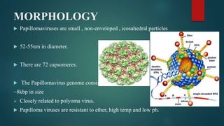
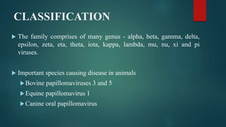
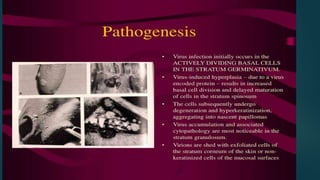
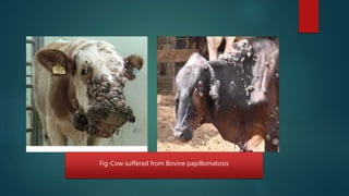
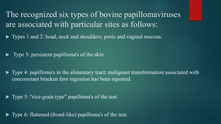
The document discusses papilloma viruses, which are small, non-enveloped viruses that cause wart growths in various animal species. It covers the morphology, classification, replication cycle, and cultivation of papilloma viruses. It also summarizes several specific papilloma virus infections that affect cattle (bovine papillomatosis), horses (equine papillomatosis), and dogs (canine oral papillomatosis).
etiology, local names, definition, transmission, source of infection, epidemiology, pathogenesis, clinical signs, diagnosis, differential diagnosis, treatment prevention and control
local names, definition, etiology,epidemiology lifecycle, pathogenesis, clinical findings, necropsy finding, diagnosis,treatment, control and prevention
etiology, local names, definition, transmission, source of infection, epidemiology, pathogenesis, clinical signs, diagnosis, differential diagnosis, treatment prevention and control
Peste des Petits Ruminants (PPR) in India Epidemiology and ControlBhoj Raj Singh
PPR is endemic in India in sheep & goats. Mainly young stocks are more affected. Disease occurs throughout the year but more common in October & March. Though vaccination is the only method for control & eradication, even the institutes those developed the effective vaccine in India to control the disease fear to use it because many a time outbreaks ensue on vaccination. The other important reason for persistence of disease is undeclared Policy of suppressed reporting of PPR outbreaks.
Bovine tuberculosis epidemiology & control in indiaBhoj Raj Singh
Tuberculosis in India is in hyperendemic state both in human and animals. No DOTS can help in control of human tuberculosis unless tuberculosis is controlled in animals. Control of tuberculosis in animals is a far reacheachable dream in India and thus the Tuberculosis will persist in India till the dooms day.
Blue tongue is a non-contagious, infectious, arthropod-borne viral disease of sheep, goat, cattle and deer, with a worldwide distribution. Initially, the disease was reported in sheep in South Africa in 1881 and it was ascribed as “epizootic catarrh”. In 1905, the disease was renamed as “blue tongue”. In India, the first outbreak of blue tongue disease in sheep and goat was reported by Sapre (1964) from Maharashtra. It is listed under category ‘A’ of disease by OIE. The presence of this disease disrupts international commerce by putting a trade barrier on the movement of animals, their germplasm as well as animal products (OIE Bulletin, 1998).
etiology, local names, definition, transmission, source of infection, epidemiology, pathogenesis, clinical signs, diagnosis, differential diagnosis, treatment prevention and control
Organisms Causing
Systemic Mycoses
Objectives - List two properties that the systemic dimorphics share.
Discuss conversion of the dimorphic yeasts
Identify from cases using colonial, morphological and geographic information, and diseases, the dimorphic fungi.
Differentiate the systemic dimorphic fungi from similar organisms previously discussed.
Describve the diseases, transmission, causative agents, of blastomycosis, paracocciodes, histoplamosis, and cocciomycosis.
Lab objectivesDescribe any additional safety precautions needed when working with the systemic dimorphic fungi.
Using lactophenol cotton blue or tissue stains – identify Blastomyces dermatitidis, Paracocciodes barasilienses, Histoplasma capsulatum, and Cocciodes immitis.
Explain the method from converting these molds to yeast.Describe exoantigen testing
Organisms Histoplasma capsulatumBlastomyces dermatitidisCoccidioides immitisParacoccidioides brasiliensisPneumocytis carinii
Safety considerationsALL handling of specimens and cultures MUST be performed in a biological safety cabinetDo not open plates on benchtopPlates should be sealedIf C. immitis is suspected, use tubed media instead of plates
Transmission and pathogenesisTransmission is by inhalation of airborne conidia which are normally present in soilOrganisms are located in distinct geographical areasDisease severity is dependent upon infective dose and patient’s immune statusCompetent hosts: asymptomatic to mild respiratory diseaseCompromised hosts: dissemination is common
Cultural characteristicsVery slow growingUp to 6 weeks for growth (except C. immitis)Must do direct examinationsExperimental: PCR testing of direct specimensColonial morphology varies with isolation mediaUse both enriched (BHIA) and selective mediaClassic morphology is described on SABHistorically, definitive identification has been made by demonstrating both a yeast or tissue phase (at 370C) and a mold phase (at room temperature)
Methods of identification of
organism grown in cultureExoantigen testingDNA probeMicroscopic morphologyConversion to yeast or tissue phaseSerological testingFour-fold rise in antibody titer between acute and convalescent paired sera
Histoplasma capsulatumCauses histoplasmosis (Darling’s or spelunker’s disease)DistributionOhio and Mississippi River valleys, Appalachian mountainsOrganism is spread by inhalation of airborne conidiaOrganism multiplies in bird droppings and bat guano
Primary disease90-95% are asymptomatic or sub-clinical, with a self-limiting mild respiratory infectionHigh percent of population in endemic areas are skin test positive for organismAcute pulmonary diseaseNight sweats, cough, fever and weight lossSome develop pulmonary cavitary lesions resembling TBOrganism is able to multiply in macrophages (observed as pseudoencapsulated yeasts)Dissemination is rare in the immuno-competent
Disseminated diseaseImmune competent patientsChronic disease of the adrenals, liver, kidn ...
Organisms Causing
Systemic Mycoses
Objectives - List two properties that the systemic dimorphics share.
Discuss conversion of the dimorphic yeasts
Identify from cases using colonial, morphological and geographic information, and diseases, the dimorphic fungi.
Differentiate the systemic dimorphic fungi from similar organisms previously discussed.
Describve the diseases, transmission, causative agents, of blastomycosis, paracocciodes, histoplamosis, and cocciomycosis.
Lab objectivesDescribe any additional safety precautions needed when working with the systemic dimorphic fungi.
Using lactophenol cotton blue or tissue stains – identify Blastomyces dermatitidis, Paracocciodes barasilienses, Histoplasma capsulatum, and Cocciodes immitis.
Explain the method from converting these molds to yeast.Describe exoantigen testing
Organisms Histoplasma capsulatumBlastomyces dermatitidisCoccidioides immitisParacoccidioides brasiliensisPneumocytis carinii
Safety considerationsALL handling of specimens and cultures MUST be performed in a biological safety cabinetDo not open plates on benchtopPlates should be sealedIf C. immitis is suspected, use tubed media instead of plates
Transmission and pathogenesisTransmission is by inhalation of airborne conidia which are normally present in soilOrganisms are located in distinct geographical areasDisease severity is dependent upon infective dose and patient’s immune statusCompetent hosts: asymptomatic to mild respiratory diseaseCompromised hosts: dissemination is common
Cultural characteristicsVery slow growingUp to 6 weeks for growth (except C. immitis)Must do direct examinationsExperimental: PCR testing of direct specimensColonial morphology varies with isolation mediaUse both enriched (BHIA) and selective mediaClassic morphology is described on SABHistorically, definitive identification has been made by demonstrating both a yeast or tissue phase (at 370C) and a mold phase (at room temperature)
Methods of identification of
organism grown in cultureExoantigen testingDNA probeMicroscopic morphologyConversion to yeast or tissue phaseSerological testingFour-fold rise in antibody titer between acute and convalescent paired sera
Histoplasma capsulatumCauses histoplasmosis (Darling’s or spelunker’s disease)DistributionOhio and Mississippi River valleys, Appalachian mountainsOrganism is spread by inhalation of airborne conidiaOrganism multiplies in bird droppings and bat guano
Primary disease90-95% are asymptomatic or sub-clinical, with a self-limiting mild respiratory infectionHigh percent of population in endemic areas are skin test positive for organismAcute pulmonary diseaseNight sweats, cough, fever and weight lossSome develop pulmonary cavitary lesions resembling TBOrganism is able to multiply in macrophages (observed as pseudoencapsulated yeasts)Dissemination is rare in the immuno-competent
Disseminated diseaseImmune competent patientsChronic disease of the adrenals, liver, kidn.
Largest viruses that infect vertebrates
Can be seen under light microscope
Poxvirus diseases are characterized by skin lesions – localized or generalized
Important diseases caused by poxviruses are-
Smallpox
Monkeypox
Cowpox
Tanapox
Molluscum contagiosum
Paratuberculosis is a contagious, chronic and sometimes fatal infection that primarily affects the small intestine of ruminants.
It is caused by the bacterium Mycobacterium avium subspecies paratuberculosis.
Infections normally affect ruminants (mammals that have four compartments of their stomachs, of which the rumen is one),
but have also been seen in a variety of non ruminant species, including rabbits, foxes, and birds. Horses, dogs, and nonhuman primates have been infected experimentally.
Paratuberculosis is found worldwide
DERIVATION OF MODIFIED BERNOULLI EQUATION WITH VISCOUS EFFECTS AND TERMINAL V...Wasswaderrick3
In this book, we use conservation of energy techniques on a fluid element to derive the Modified Bernoulli equation of flow with viscous or friction effects. We derive the general equation of flow/ velocity and then from this we derive the Pouiselle flow equation, the transition flow equation and the turbulent flow equation. In the situations where there are no viscous effects , the equation reduces to the Bernoulli equation. From experimental results, we are able to include other terms in the Bernoulli equation. We also look at cases where pressure gradients exist. We use the Modified Bernoulli equation to derive equations of flow rate for pipes of different cross sectional areas connected together. We also extend our techniques of energy conservation to a sphere falling in a viscous medium under the effect of gravity. We demonstrate Stokes equation of terminal velocity and turbulent flow equation. We look at a way of calculating the time taken for a body to fall in a viscous medium. We also look at the general equation of terminal velocity.
Seminar of U.V. Spectroscopy by SAMIR PANDASAMIR PANDA
Spectroscopy is a branch of science dealing the study of interaction of electromagnetic radiation with matter.
Ultraviolet-visible spectroscopy refers to absorption spectroscopy or reflect spectroscopy in the UV-VIS spectral region.
Ultraviolet-visible spectroscopy is an analytical method that can measure the amount of light received by the analyte.
Deep Behavioral Phenotyping in Systems Neuroscience for Functional Atlasing a...Ana Luísa Pinho
Functional Magnetic Resonance Imaging (fMRI) provides means to characterize brain activations in response to behavior. However, cognitive neuroscience has been limited to group-level effects referring to the performance of specific tasks. To obtain the functional profile of elementary cognitive mechanisms, the combination of brain responses to many tasks is required. Yet, to date, both structural atlases and parcellation-based activations do not fully account for cognitive function and still present several limitations. Further, they do not adapt overall to individual characteristics. In this talk, I will give an account of deep-behavioral phenotyping strategies, namely data-driven methods in large task-fMRI datasets, to optimize functional brain-data collection and improve inference of effects-of-interest related to mental processes. Key to this approach is the employment of fast multi-functional paradigms rich on features that can be well parametrized and, consequently, facilitate the creation of psycho-physiological constructs to be modelled with imaging data. Particular emphasis will be given to music stimuli when studying high-order cognitive mechanisms, due to their ecological nature and quality to enable complex behavior compounded by discrete entities. I will also discuss how deep-behavioral phenotyping and individualized models applied to neuroimaging data can better account for the subject-specific organization of domain-general cognitive systems in the human brain. Finally, the accumulation of functional brain signatures brings the possibility to clarify relationships among tasks and create a univocal link between brain systems and mental functions through: (1) the development of ontologies proposing an organization of cognitive processes; and (2) brain-network taxonomies describing functional specialization. To this end, tools to improve commensurability in cognitive science are necessary, such as public repositories, ontology-based platforms and automated meta-analysis tools. I will thus discuss some brain-atlasing resources currently under development, and their applicability in cognitive as well as clinical neuroscience.
Phenomics assisted breeding in crop improvementIshaGoswami9
As the population is increasing and will reach about 9 billion upto 2050. Also due to climate change, it is difficult to meet the food requirement of such a large population. Facing the challenges presented by resource shortages, climate
change, and increasing global population, crop yield and quality need to be improved in a sustainable way over the coming decades. Genetic improvement by breeding is the best way to increase crop productivity. With the rapid progression of functional
genomics, an increasing number of crop genomes have been sequenced and dozens of genes influencing key agronomic traits have been identified. However, current genome sequence information has not been adequately exploited for understanding
the complex characteristics of multiple gene, owing to a lack of crop phenotypic data. Efficient, automatic, and accurate technologies and platforms that can capture phenotypic data that can
be linked to genomics information for crop improvement at all growth stages have become as important as genotyping. Thus,
high-throughput phenotyping has become the major bottleneck restricting crop breeding. Plant phenomics has been defined as the high-throughput, accurate acquisition and analysis of multi-dimensional phenotypes
during crop growing stages at the organism level, including the cell, tissue, organ, individual plant, plot, and field levels. With the rapid development of novel sensors, imaging technology,
and analysis methods, numerous infrastructure platforms have been developed for phenotyping.
Earliest Galaxies in the JADES Origins Field: Luminosity Function and Cosmic ...Sérgio Sacani
We characterize the earliest galaxy population in the JADES Origins Field (JOF), the deepest
imaging field observed with JWST. We make use of the ancillary Hubble optical images (5 filters
spanning 0.4−0.9µm) and novel JWST images with 14 filters spanning 0.8−5µm, including 7 mediumband filters, and reaching total exposure times of up to 46 hours per filter. We combine all our data
at > 2.3µm to construct an ultradeep image, reaching as deep as ≈ 31.4 AB mag in the stack and
30.3-31.0 AB mag (5σ, r = 0.1” circular aperture) in individual filters. We measure photometric
redshifts and use robust selection criteria to identify a sample of eight galaxy candidates at redshifts
z = 11.5 − 15. These objects show compact half-light radii of R1/2 ∼ 50 − 200pc, stellar masses of
M⋆ ∼ 107−108M⊙, and star-formation rates of SFR ∼ 0.1−1 M⊙ yr−1
. Our search finds no candidates
at 15 < z < 20, placing upper limits at these redshifts. We develop a forward modeling approach to
infer the properties of the evolving luminosity function without binning in redshift or luminosity that
marginalizes over the photometric redshift uncertainty of our candidate galaxies and incorporates the
impact of non-detections. We find a z = 12 luminosity function in good agreement with prior results,
and that the luminosity function normalization and UV luminosity density decline by a factor of ∼ 2.5
from z = 12 to z = 14. We discuss the possible implications of our results in the context of theoretical
models for evolution of the dark matter halo mass function.
The ability to recreate computational results with minimal effort and actionable metrics provides a solid foundation for scientific research and software development. When people can replicate an analysis at the touch of a button using open-source software, open data, and methods to assess and compare proposals, it significantly eases verification of results, engagement with a diverse range of contributors, and progress. However, we have yet to fully achieve this; there are still many sociotechnical frictions.
Inspired by David Donoho's vision, this talk aims to revisit the three crucial pillars of frictionless reproducibility (data sharing, code sharing, and competitive challenges) with the perspective of deep software variability.
Our observation is that multiple layers — hardware, operating systems, third-party libraries, software versions, input data, compile-time options, and parameters — are subject to variability that exacerbates frictions but is also essential for achieving robust, generalizable results and fostering innovation. I will first review the literature, providing evidence of how the complex variability interactions across these layers affect qualitative and quantitative software properties, thereby complicating the reproduction and replication of scientific studies in various fields.
I will then present some software engineering and AI techniques that can support the strategic exploration of variability spaces. These include the use of abstractions and models (e.g., feature models), sampling strategies (e.g., uniform, random), cost-effective measurements (e.g., incremental build of software configurations), and dimensionality reduction methods (e.g., transfer learning, feature selection, software debloating).
I will finally argue that deep variability is both the problem and solution of frictionless reproducibility, calling the software science community to develop new methods and tools to manage variability and foster reproducibility in software systems.
Exposé invité Journées Nationales du GDR GPL 2024
Comparing Evolved Extractive Text Summary Scores of Bidirectional Encoder Rep...University of Maribor
Slides from:
11th International Conference on Electrical, Electronics and Computer Engineering (IcETRAN), Niš, 3-6 June 2024
Track: Artificial Intelligence
https://www.etran.rs/2024/en/home-english/
Nutraceutical market, scope and growth: Herbal drug technologyLokesh Patil
As consumer awareness of health and wellness rises, the nutraceutical market—which includes goods like functional meals, drinks, and dietary supplements that provide health advantages beyond basic nutrition—is growing significantly. As healthcare expenses rise, the population ages, and people want natural and preventative health solutions more and more, this industry is increasing quickly. Further driving market expansion are product formulation innovations and the use of cutting-edge technology for customized nutrition. With its worldwide reach, the nutraceutical industry is expected to keep growing and provide significant chances for research and investment in a number of categories, including vitamins, minerals, probiotics, and herbal supplements.
Nutraceutical market, scope and growth: Herbal drug technology
Papilloma virus
1. PAPILLOMA VIRUS
PRESENTED BY: SURYAADHIKARI
B V SC & AH, 6TH SEM., NPI
CHITWAN , NEPAL
EMAIL- ADHIKARISURYA313@GMAIL.COM
2. INTRODUCTION
Belongs to the family Papillomaviridae.
Papilloma virus--- Papilli Pustule, Oma Tumor
Are oncogenic tumor viruses.
Causes wart in their natural hosts.
Papilloma viruses are strictly species specific & even in experimental condition, do not
infect any host other than natural hosts.
Infect many animals including humans,chimpanzee, monkeys, cattle, deer, dog, horse,
sheep, elephant, elk, opossum, rabbit and birds.
3. MORPHOLOGY
Papillomaviruses are small , non-enveloped , icosahedral particles
52-55nm in diameter.
There are 72 capsomeres.
The Papillomavirus genome consists of circular, d/s DNA
~8kbp in size
Closely related to polyoma virus.
Papilloma viruses are resistant to ether, high temp and low ph.
4. CLASSIFICATION
The family comprises of many genus - alpha, beta, gamma, delta,
epsilon, zeta, eta, theta, iota, kappa, lambda, mu, nu, xi and pi
viruses.
Important species causing disease in animals
Bovine papillomaviruses 3 and 5
Equine papillomavirus 1
Canine oral papillomavirus
5. REPLICATION
Replication is restricted to epithelial cells- mainly squamous, (i.e. skin, and upper
alimentary tract of ruminants), but occasionally in the lower alimentary tract
epithelium.
Replication and virion assembly occur in the nucleus and virion are released by
destruction of the nuclear and cell membranes.
Papillomaviruses replicate in the nucleus and new virions are released with the lysis
of the cell.
Papillomaviruses produce diagnostically significant koilocytotic (vaculated) cells
while replicating.
6. CULTIVATION
Growth on chorioallantoic membrane (CAM) of embryonated hen‘s egg has not been
reported.
Papillomavirus cannot be propagated in cell culture. The virus species induce papillomas in
the host of origin but rarely cause cell transformation or cytopathic effects (CPEs) in cell
culture.
In experimental house, subcutaneous (S/C) inoculation of the virus gives rise to connective
tissue tumors.
Intracerebrally injected calves may develop meningeal fibromoda.
Unweaned hamsters and certain strains of mice (C3H/eB) on subcutaneous inoculation
develop tumor at the inoculation site within 100 days. The tumors are firm, multiple or singe,
measuring about 8-15 nm in diameter and persist for 4-6 months without evidence of
metastasis.
7. BOVINE PAPILLOMATOSIS
(Common warts of cattle)
Cause
Six types of papillomavirus cause bovine papillomatosis.
Occurrence
Bovine papillomatosis occurs frequently worldwide, mainly affecting
young cattle. They
occur with greater frequency in stabled cattle.
8. PATHOGENESIS
The mechanism of tumor induction by papilloma virus is largely unknown.
The virus probably gains entry –
o Directly via abrasion in the skin by direct contact with infected
animals.
o Indirectly by means contaminated inanimate objects like
Halters, syringe and needle, tattooing machine or other equipments used for managemental
practices.
Papillomavirus affect cutaneous and mucosal squamous epithelium and induce benign
circumscribed tumor, which tends to regress spontaneously after a period of time.
9.
10. CLINICAL & PATHOLOGICAL FEATURES
Papilloma develop as small nodular growths of the skin or mucous membrane.
They initially grow slowly, but then more rapidly and eventually become larger, horny,
pendulant and sometimes cauliflower in shape. They ultimately necrose and fall off.
The most common sites affected are the head (particularly around the eyes), neck, and
shoulders.
They may occur on the penis of the bull and in the vaginal mucosa of the female, resulting in
breeding difficulty.
After about a year there is usually spontaneous recovery.
12. The recognized six types of bovine papillomaviruses
are associated with particular sites as follows:
Types 1 and 2: head, neck and shoulders; penis and vaginal mucosa.
Type 3: persistent papilloma's of the skin.
Type 4: papilloma's in the alimentary tract; malignant transformation associated with
concomitant bracken fern ingestion has been reported.
Type 5: "rice-grain type" papilloma's of the teat.
Type 6: flattened (frond-like) papilloma's of the teat.
13. DIAGNOSIS
This is usually based on characteristic gross appearance. Laboratory diagnosis is
not usually sought.
Definitive diagnosis requires histological examination for the presence of
koilocytes.
Although not employed for diagnosis, types 1 and 2 bovine papillomaviruses can
be cultivated in cell cultures and on the chorioallantoic membrane of chicken
embryo.
Confirmed by demonstrating the presence of viral particle in the negatively stained
specimen of wart tissue by electron microscope.
14. PREVENTION
Commercial wart vaccines and autogenous wart vaccines, both consisting of
finely ground warts, are used.
Formalin is often used to kill the virus and a preservative is added.
To prevent spread, affected animals should be isolated.
15. Equine Papillomatosis
(Common warts of horses)
Cause– Equine papillomavirus.
Host- Equidae
Occurrence-Worldwide in horses, mules and donkeys usually up to three
years if age. Warts in older horses persist longer.
MOT- Infection is by direct contact with other infected horses, although
fomites and biting flies may also be involved.
16. CLINICAL FEATURES
Warts generally occur on the nose and lips, vary in size and number and
usually disappear within three months.
Congenital papillomatosis has been reported but occurs rarely.
A different papillomavirus is associated with genital lesions in both male
and female horses.
17. Diagnosis
This is usually based on the characteristic gross lesions.
Histologic examination of affected tissue provides confirmation.
Prevention
Autogenous formalin-inactivated wart vaccines are sometimes administered but
their value is questionable; repeated doses are recommended.
Surgical removal of warts may be helpful.
Equine warts, like the warts of other species, will frequently disappear
spontaneously.
18. Canine Oral Papillomatosis
(Common warts of dogs)
Cause
Three types of papillomavirus cause canine warts, a common worldwide
disease of young dogs
19. CLINICAL & PATHOLOGICAL FEATURE
Numerous papilloma may occur on the mucous membrane of the mouth, lips, tongue,
and pharynx of young dogs.
Infections begin as small, white elevated areas that enlarge to form small cauliflower-
like lesions. Warts may occasionally involve the eyelid.
Skin warts are seen in older dogs and may be caused by a particular type of
papillomavirus.
Warts usually regress spontaneously in several months. Recovered dogs are immune,
and dogs older than two years are usually immune.
20. DIAGNOSIS
The disease is clinically characteristic.
Histologic examination of warts or biopsies is confirmatory.
TREATMENT
Surgical excision may be employed.
Autogenous wart vaccines are considered of questionable value in
treatment.
PREVENTION
Vaccination for prevention is not generally practiced.