Bovine Papillomatosis: Causes, Types, and Clinical Findings
•Download as PPTX, PDF•
7 likes•3,092 views

This document summarizes information about bovine papillomatosis, which is caused by bovine papillomaviruses (BPV). BPV are small DNA viruses that infect the basal layer of epithelium and cause benign proliferative lesions. There are multiple BPV types that cause different lesions in different anatomical locations. The lesions are generally self-limiting but can cause economic impacts. Transmission occurs through direct contact or fomites. While lesions usually regress spontaneously, prevention focuses on disinfection and vaccination.

Recommended
More Related Content
What's hot
What's hot (20)
Similar to Bovine Papillomatosis: Causes, Types, and Clinical Findings
Similar to Bovine Papillomatosis: Causes, Types, and Clinical Findings (20)
More from sivasankar. P
Recently uploaded
Recently uploaded (20)
Bovine Papillomatosis: Causes, Types, and Clinical Findings
- 2. Etiology • Bovine papillomaviruses (BPV) belong to the Family papillomaviridae genus Papillomavirus (PV), 16 genera present. • These are small DNA viruses infecting humans as well as many domestic and wild animal species, including birds, causing benign hyper proliferative lesions of both mucosal and cutaneous epithelia, infects almost all species • These tumours are benign, self-limiting and, in most cases, spontaneously regress. • Differentiation of types is based on the histological features of the lesion and DNA identification by hybridization or PCR • Papillomaviruses cannot be grown in conventional cell cultures, but their genomes readily can be sequenced with molecular methods, without need for culture. • Although a single BPV type is detected in an individual papilloma, a single animal may have papillomas at different sites associated with different BPV types. (Garcia-Perez A et al., 2003) • Papillomaviruses are resistant to diverse environmental insults, infectivity survives lipid solvents and detergents, low pH, and high temperatures.
- 3. Structure and genetic organization • BPVs are small non-enveloped viruses with an icosahedral capsid around 50–60 nm in diameter. • Double stranded circular DNA, genome of 7.3-8.0 kb • Covalently closed , genome is infectious • L1-L2 protein forms capsid • E1-E8 protein are non strutural protein • E5 oncoprotein- pivotal role in cell transformation. • Infects epithelium and fibroblast • Cutaneous warts are most common in younger animals (under 2 years) and usually spontaneously regress due to the animal's immune response without significant scarring. • The duration of infection is very variable (from one month to over a year) and recurrence is possible • This viruses are host specific, lesion specific and site specific • Individual strains of bovine papillomavirus differ, with capability of inducing neoplasms
- 4. types
- 5. Genotypes • In cattle 14 types have been identified: BPV-1, 2 and BPV-5 which cause fibropapillomas and BPV-3,4,6,9 and 10 which cause true epithelial papillomas • BPV-1 and BPV-2 can also induce sarcomas and fibrosarcomas in other mammals, including equids (equine sarcoid) and, experimentally rabbits, hamsters and mice. These two causes cross infections • BPV- 1 - frond fibropapillomas of teat skin and penile fibropapilloma • BPV- 1 and BPV-2 - fibropapilloma of the skin of the anteroventral part of the body including the forehead, neck and back, the common cutaneous wart • BPV-2 - cauliflower-like fibropapillomas of the anogenital and ventral abdominal skin
- 6. Genotypes • BPV-2 - associated with bladder cancer in cattle in association with the ingestion of bracken fern (Pteridium spp) • BPV-3 - cutaneous papilloma • BPV-4 - papilloma of the esophagus, esophageal groove, fore stomachs and small intestine; this is capable of becoming malignant, particularly in animals fed bracken fern. • BPV-4 has site specificity to the upper alimentary tract • BPV-5 - rice grain fibropapilloma on the udder. BPV-5 has also been demonstrated in cutaneous skin warts • BPV-6 - frond epithelial papillomas of the bovine udder and teats •
- 7. Epidemiology • Distribution is worldwide and it occurs in cattle, goat, horse ,rabbit, fish and human. Oral papillomatosis occurs in dogs and rabbits but the disease is uncommon in sheep and pigs • Bovine papiloma virus, ovine papilloma virus, deer fibroma virus will cause fibroblastic tumour in cats • Common in young animals and immune compromised animals • The skin surfaces of the neck, legs, back and abdomen are the more usual sites. The infective virus concentrated in the outer keratinized epithelium of the papilloma and when shed, can readily contaminate fomites such as fences, stanchions and boards of the stable. These fomites with virus readily transmit the disease to susceptible cattle by causing skin wound. • The disease is more common in housed cattle than in cattle on pasture. Campo et al.,1994
- 8. Transmission • The method of spread is by direct contact with infected animals, infection gaining entry through cutaneous abrasions or by fomites • Virus can also persist on inanimate objects in live stock buildings and infect animals rubbing against them • The calf can contract infection through direct contact during suckling (Blood et al., 2002) • Crops of warts sometimes occur around ear tags, at branding sites or along scratches made by barbed wire, and can be spread by tattooing implements, dehorning shears and by procedures such as tuberculin testing • An extensive outbreak of perianal warts is recorded in beef heifers, the infection reported to be spread by rectal examination for pregnancy • Congenital infection is recorded in the foal and calf, but is rare (Desrochers et al., 1994) • Papillomaviruses also cause transmissible fibropapillomas of the genitalia of young cattle, with venereal spread analogous to that of human genital warts
- 9. Risk factors • All species may be affected but the disease is commonest in cattle and horses • Age: Cutaneous papillomas of the head and neck occur predominantly in young animals, the lack of susceptibility of adults to natural infection being ascribed to immunity acquired by apparent or inapparent infection when young. • Many of the viruses are associated with teat and udder lesions, probably as a result of transmission during milking. • The occurrence of cutaneous warts and their severity can be influenced by factors that induce immunosuppression and latent infection has converted to clinical disease with the administration of immunosuppressive agents Olson et al.,1992
- 10. Economic importance • Papillomatosis affected animals are usually otherwise healthy, and there are normally no systemic effects. • Cutaneous warts are quite common in young cattle, especially when they are housed, but ordinarily they cause little harm and regress spontaneously. • In purebred animals they may interfere with sales and shows because of their unsightly appearance. • Animals with extensive lesions may lose condition, and secondary bacterial invasion of traumatized warts may cause concern • Warts on the teats of dairy cows often cause interference with milking. • In horses, the lesions are usually small and cause little inconvenience, but they are esthetically unattractive. • In all species, the development of warts on the genitalia requires immediate treatment.
- 11. Pathogenesis • The virus infects the basal keratinocytes, replicating its genome in the diferentiating spinous and granular layers causing the excessive growth that is characteristic of wart formation. • Papillomas are the result of basal cell hyperplasia without viral antigen production.(Levkutova et al.,1998) • Expression of the late structural proteins of the virus is limited to the differentiated cells of the squamous layer where the new virus particles are encapsulated and shed into the environment as the cells die. • The tumor contains epithelial and connective tissues and can be a papilloma or a fibropapilloma, depending on the relative proportions of epithelial and connective tissue present • papillomas contain little connective tissue, and fibropapillomas are mostly fibrous tissue, with very little epithelial tissue. • fibropapillomas are uncommon in horses, but are the common in cattle, sheep, and wild ruminants.
- 12. Pathogenesis
- 13. Pathology • Warts caused by the Xipapillomavirus group have a cauliflower-like appearance and can attain the size of a fist; most common on the head, neck and shoulders, they may also occur in other locations. • Cutaneous fibropapillomas caused by Deltapapillomavirus group have a nodular appearance • Papillomavirus has been observed in squamous cell carcinoma of bovine eyes . (Rutten et al.,1992) • Equine sarcoid are associated with BPV-1 and BPV-2
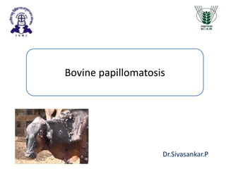
- 14. Clinical findings • The incubation period for cutaneous warts produced by BPV is about 30 days, and the duration of naturally and experimentally produced fibropapillomas ranges from 1-12 months before regression • The solid outgrowths of epidermis may be sessile or pedunculated. • Most common type in cattle occurs on head and neck and has cauliflower- like appearance, but lesion site and appearance varies with papilloma type. • In the horse, lesions are on the face and lips • Warts are occur anywhere on the body but are most commonly seen in the head and neck area. Less common but often more important sites include teats and scrotum. Warts vary greatly in shape from almost flat pea-sized lumps to large orange sized balls on stalks.
- 15. Clinical findings • Warts on the teats manifest with different forms depending on the papillomavirus type and may show an increasing frequency with age. The frond form have filiform projections • Other forms are a flat, round type which is usually multiple, always sessile and up to 2 cm in diameter. • The third form has an elongated structure appearing like a rice grain. • Teat warts may regress during the dry period and recur with the next lactation. • Genital warts on the vulva and penis make mating impracticable because the lesions are of large size, are friable, and bleed easily. They commonly become infected and flyblown.
- 16. Clinical findings • Alimentary tract warts are rarely observed clinically in farm animals in most countries, Papillomas occur on the lateral and dorsal aspects of the tongue, the soft palate, oropharynx, esophagus, esophageal groove and rumen. • Paillomas occurring in the esophageal groove and in the reticulum are a cause of chronic ruminal tympany. • Less common manifestations of papillomatosis in cattle include lesions in the urinary bladder, which cause no clinical signs but may predispose to enzootic hematuria. BPV-4 papillomas in the upper alimentary tract of cattle being fed bracken fern are the focus for transformation to squamous cell carcinomas. • Cattle fed bracken fern are immunosuppressed which promotes the persistence and spread of the papilloma virus, and mutagens in bracken fern cause neoplastic transformation of papilloma cells.
- 17. Association with cancer • BPV-4 causes squamous cell carcinomas of the alimentary tract and BPV- 1,2 causes carcinomas and haemangioendotheliomas of the urinary bladder, in both cases in animals that have fed on bracken (Pteridium aquilinum (Jarret et al.,1978;Campo, 2006) • Bracken contains several immunosuppressants and mutagens, including quercetin and ptaquiloside. • Bladder cancer leads to enzootic hematuria
- 18. Diagnostic confirmation • In most cases the diagnosis is obvious. • History and clinical sign • Histology • DNA identification by PCR • Biopsy or tissue scraping • Immunohistochemical staining
- 19. Treatment • There is no completely effective treatment, particularly for severe cases. • Simple surgical removal or if the warts has a significant stalk using a rubber ring • Cryosurgery with liquid nitrogen has recently come into use and should be very effective when the cutaneous papillomas are not too large. • Autogenous vaccine reported to be successful (Khursheed et al.,2008) • Recovery in 3-6 weeks is recorded in 80-85% cases where the warts are on the body surface or penis of cattle but in only 33% when the warts are on the teats. • Binary ethylenimine inactivated virus vaccine of BPV -1,2 reported to be effective for prevention and for therapeutics ( Pangty et al.,2010)
- 20. Treatment • Autogenous vaccine was reported to be effective (Sreeparvathy et al.,2011; Kursheed et al., 2008) • Terziev et al .,2015 reported autogenous vacine not effective wart > 3 cm • Autohemotherpy reported to be effective ( Arun et al.,2017; Ganesh et al., 211; Mathi et al., 2016) • Kavitha et al., 2011 reported autogenous vaccine 92 %, anthiomaline 81% thuja 70%, topical thuja ointment 57% was effective • Ivermectin showed 100% reovery in calves (Jana et al., 2013) • Peptide therapy reported to be arrest growth in early growth of wart by BPV-1,2 ( Pangty et al.,2011)
- 21. Case reports • 11 teat sample, rice grain type from UP – 2 sample showed mixed infection of BPV-1,2 (Kumar et al.,2013) • 20 cases of wart reported in UP in buffaloe with BPV-2 and successfully transmitted to golden syrian hamster( Vidya et al., 2010) • 6 cases of enzootic hematuria was reported in mukteshwar infected with BPV-2(Leisangthem et al., 2006) • 27 cases of GI tract papilloma was reported in IVRI, Bareilley slaughter house infected with BPV-1,2,5 is the first world report of GI papilloma in buffaloe (Kumar et al.,2012) ( Vidya et al., 2010)
- 22. Prevention • Disinfection with formaldehyde of stalls, fence posts and other environmental virus reservoirs can prevent transmission. • Tattoo or tagging pliers can be disinfected between use on calves, with 2 to 4% solution of formaldehyde • Maintain two sets of the instruments and alternate them in use there by providing adequate time in the formaldehyde to inactivate the virus.
- 23. Control • Vaccination has been shown experimentally to be an effective prevention and gives complete protection in cattle • The vaccine must contain all serotypes of the papillomavirus because they are very type-specific.(Jarett et al., 1990) • Avoidance of close contact between infected and uninfected animals should be encouraged • And the use of communal equipment between affected and unaffected animals should be avoided.
- 24. THANK YOU