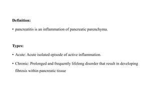
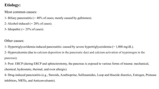
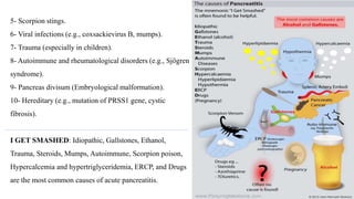
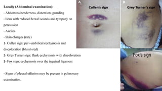
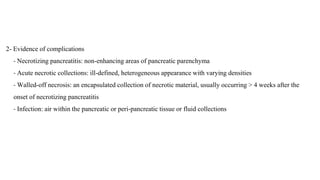
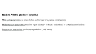
Pancreatitis
Mohammed Alqahtani
PGY1
Definition:
• pancreatitis is an inflammation of pancreatic parenchyma.
Types:
• Acute: Acute isolated episode of active inflammation.
• Chronic: Prolonged and frequently lifelong disorder that result in developing
fibrosis within pancreatic tissue
Etiology:
Most common causes:
1- Biliary pancreatitis (∼ 40% of cases; mostly caused by gallstones).
2- Alcohol-induced (∼ 20% of cases).
3- Idiopathic (∼ 25% of cases).
Other causes:
1- Hypertriglyceridemia-induced pancreatitis: caused by severe hypertriglyceridemia (> 1,000 mg/dL).
2- Hypercalcemia (due to calcium deposition in the pancreatic duct and calcium activation of trypsinogen in the
pancreas).
3- Post- ERCP (during ERCP and sphincterotomy, the pancreas is exposed to various forms of trauma: mechanical,
chemical, hydrostatic, thermal, and even allergic).
4- Drug-induced pancreatitis (e.g., Steroids, Azathioprine, Sulfonamides, Loop and thiazide diuretics, Estrogen, Protease
inhibitors, NRTIs, and Anticonvulsants).
5- Scorpion stings.
6- Viral infections (e.g., coxsackievirus B, mumps).
7- Trauma (especially in children).
8- Autoimmune and rheumatological disorders (e.g., Sjögren
syndrome).
9- Pancreas divisum (Embryological malformation).
10- Hereditary (e.g., mutation of PRSS1 gene, cystic
fibrosis).
I GET SMASHED: Idiopathic, Gallstones, Ethanol,
Trauma, Steroids, Mumps, Autoimmune, Scorpion poison,
Hypercalcemia and hypertriglyceridemia, ERCP, and Drugs
are the most common causes of acute pancreatitis.
Clinical features:
Symptoms:
- Constant, sever epigastric pain, classically radiating towards the back, become worse after meals and when
supine and improves on leaning forward
- Nausea, vomiting
- Fever
- If pulmonary complications are present: chest pain, dyspnea
Examination findings:
General:
- Signs of shock: tachycardia, hypotension, oliguria/anuria
- Possibly jaundice in patients with biliary pancreatitis
Locally (Abdominal examination):
- Abdominal tenderness, distention, guarding
- Ileus with reduced bowel sounds and tympany on
percussion
- Ascites
- Skin changes (rare)
1- Cullen sign: peri-umbilical ecchymosis and
discoloration (bluish-red)
2- Grey Turner sign: flank ecchymosis with discoloration
3- Fox sign: ecchymosis over the inguinal ligament
- Signs of pleural effusion may be present in pulmonary
examination.
Diagnosis:
Criteria:
Two of the three following criteria should be met for a diagnosis of acute pancreatitis:
Clinically > Characteristic abdominal pain.
Laboratory > ↑ Serum pancreatic enzymes: lipase or amylase ≥ 3× ULN.
Imaging > Characteristic findings of acute pancreatitis on cross-sectional imaging.
Laboratory tests:
- CBC (↑ WBC and Hematocrit)
- Renal panel
- Liver panel (↑ liver enzymes and bilirubin in biliary pancreatitis)
- Pancreatic enzymes [Amylase and Lipase (more sensitive and specific)] (often normal in chronic pancreatitis).
Note: not necessarily correlate with severity of inflammation
- ESR, CRP and LDH (severity marker)
- Triglyceride
- Calcium level
Imaging:
Ultrasound abdomen:
Indications: first-line imaging modality for all patients.
Findings:
1- Features of acute pancreatitis (visible in 20% of cases):
- Enlarged hypoechoic pancreas (pancreatic edema)
- Peri-pancreatic fluid and/or ascites
2- Features of biliary pancreatitis:
- Cholelithiasis and/or gallbladder sludge
- Dilated biliary tree
3- Evidence of complications: pancreatic pseudocysts, walled-off necrosis (typically > 4 weeks from symptom
onset).
CT abdomen and pelvis with IV contrast:
Indications:
1- Diagnostic uncertainty (e.g., typical clinical features in a patient with moderately elevated pancreatic enzymes)
2- Severe pancreatitis : optimally performed > 5–7 days after symptom onset
3- Lack of improvement (after > 7 days) or sudden acute deterioration
4- To evaluate for underlying etiology if routine diagnostic studies are negative
Findings:
1- Features of acute pancreatitis
- Pancreatic parenchyma enlargement with edema
- Indistinct pancreatic margins with surrounding fat stranding
- Peri-pancreatic free fluid
2- Evidence of complications
- Necrotizing pancreatitis: non-enhancing areas of pancreatic parenchyma
- Acute necrotic collections: ill-defined, heterogeneous appearance with varying densities
- Walled-off necrosis: an encapsulated collection of necrotic material, usually occurring > 4 weeks after the
onset of necrotizing pancreatitis
- Infection: air within the pancreatic or peri-pancreatic tissue or fluid collections
Magnetic Resonance Cholangiopancreatography (MRCP)
Indication: prior to therapeutic ERCP in suspected biliary pancreatitis
Diagnostic Endoscopic Retrograde Cholangiopancreatography (ERCP)
Indications:
1- Suspected choledocholithiasis (if MRCP or MRI are not feasible)
2- To evaluate for sphincter of Oddi dysfunction in patients with recurrent pancreatitis and normal or
inconclusive EUS and MRCP
Severity index:
There are several scores used to assess the
severity and prognosis of acute pancreatitis:
Ranson criteria:
is one of the oldest predictive models used
to estimate severity and prognosis of biliary and
nonbiliary pancreatitis, but full assessment is
only possible after 48 hours, and sensitivity for
predicting severity and outcome can be as low as
70%.
Bedside Index of Severity of Acute
Pancreatitis (BISAP):
- Used to estimate in-hospital mortality due
to pancreatitis
Revised Atlanta grades of severity:
Mild acute pancreatitis: no organ failure and no local or systemic complications
Moderate acute pancreatitis: transient organ failure (< 48 hours) and/or local or systemic complications
Severe acute pancreatitis: persistent organ failure (> 48 hours)
Management:
Mainly supportive
Hospital admission is usually required
IV fluid:
- Intravenous fluid resuscitation in the first 12–24 hours has the greatest impact on the clinical outcome of patients
with acute pancreatitis.
- Crystalloids are preferred (e.g., NS or RL), with infusion rate 5-10 mL/kg/h or roughly 250-500 mL/h (NS
preferred in patient with Hypercalcemia).
- Adequate fluid replacement can be assessed by an improvement in vital signs (goal heart rate <120 and MAP
between 65 to 85 mmHg), urine output (>0.5 to 1 cc/kg/hour) and reduction in hematocrit (goal 35 to 44 %).
- Fluid resuscitation mainly limited to the first 24 to 48 hours after onset of the disease. Continued aggressive fluid
resuscitation after 48 hours may associated with an increased need for intubation and increased risk of abdominal
compartment syndrome.
Analgesia:
- Opioid (Meperidine superior to morphine in pancreatitis because studies showed that morphine caused an increase
in sphincter of Oddi pressure).
- NSAID (e.g., diclofenac or ibuprofen), contraindicated in patient with renal impairment.
Antiemetic as needed (e.g., metoclopramide or ondanserton).
Monitoring:
- Patients with acute pancreatitis should be monitored closely in the first 24 to 48 hours.
- Vital signs including oxygen saturation should be monitored and supplemental oxygen administered to maintain
arterial oxygen saturation of greater than 95 %.
- Blood gas analysis should be performed if oxygen saturation is less than 90 %.
- Urine output should be measured hourly and fluids should be titrated to maintain urine output (> 0.5 to 1
cc/kg/hour).
- Serum glucose levels should be monitored hourly in patients with severe pancreatitis and hyperglycemia (blood
glucose greater than 180 to 200 mg/dL) should be treated as it can increase the risk of secondary pancreatic
infections.
Electrolytes replacement as needed.
Nutrition:
- Bowel rest is no longer routinely recommended.
- In the absence of ileus, nausea or vomiting, oral feeding can be initiated early (within 24 hours) as tolerated. We
usually start with a clear, low fat, soft diet.
- Enteral feeding rather than parenteral nutrition is recommended in patients with moderately severe and severe
acute pancreatitis who cannot tolerate oral feeding. We also initiate enteral feeding when it becomes clear that the
patient will not be able to consume nourishment by mouth.
- Parenteral nutrition should be initiated only in patients who do not tolerate enteral feeding or if the target rate of
enteral feeding is not achieved within 48 to 72 hours.
Antibiotics:
- Up to 20 percent of patients with acute pancreatitis develop an extrapancreatic infection (eg, bloodstream
infections, pneumonia, and urinary tract infections). Extrapancreatic infections are associated with an increase in
mortality. When an infection is suspected, antibiotics should be started while the source of the infection is being
determined. However, if cultures are negative and no source of infection is identified, antibiotics should be
discontinued.
- Prophylactic antibiotics are not recommended in patients with acute pancreatitis, regardless of the type (interstitial
or necrotizing) or disease severity (mild, moderately severe, or severe).
Management of the underlying cause:
- Biliary pancreatitis > therapeutic ERCP (indicated within 24 hours if there is concurrent cholangitis) and
cholecystectomy.
- Alcohol-induced pancreatitis > check mg and phosphorus level and replete as needed, alcohol cessation and
vitamin supplementation (B1,B6, B9 and B12).
Acute pancreatitis during pregnancy:
- Most commonly affects multiparous women, and the majority of cases occur in the third trimester.
- Physiologic changes during pregnancy such as altered estrogen and progesterone levels increase the rick of
choledocholithiasis.
- Ultrasound and MRI as imaging modalities are preferred during pregnancy.
Complications:
Necrotizing pancreatitis:
Definition: necrosis of pancreatic and peri-pancreatic tissue
Diagnosis: non-enhancing areas of pancreatic parenchyma on CT abdomen with contrast.
Treatment: Usually can be managed conservatively.
Infected necrotizing pancreatitis:
Definition: bacterial superinfection of necrotic pancreatic parenchyma.
Diagnosis:
- Laboratory studies: persistent or worsening leukocytosis, bacteremia, increasing inflammatory markers.
- CT abdomen with contrast: gas within the pancreas and/or peri-pancreatic tissue or fluid collections.
- Fine-needle aspiration of necrotic areas: not routinely recommended.
Treatment of Infected necrotizing pancreatitis:
1- Broad-spectrum empiric antibiotics with good tissue penetration (e.g., carbapenems) for 4 weeks.
2- Drainage of infected material if there is clinical deterioration or persistence of symptoms despite antibiotic
therapy:
- Operative pancreatic debridement (necrosectomy) should ideally be performed at least 2–4 weeks after initial
presentation. [14]
- Minimally invasive procedures (e.g., image-guided percutaneous drainage) can be performed in the first 2
weeks in seriously ill patients.
Walled-off necrosis:
Definition:
- An encapsulated collection of sterile necrotic material, usually occurring 4 weeks after the onset of necrotizing
pancreatitis.
- Previously known as pancreatic abscess.
Diagnosis: CT abdomen with IV contrast showing an encapsulated heterogeneous collection containing fluid and
debris.
Treatment (of symptomatic walled-off necrosis): percutaneous drainage or transmural endoscopic necrosectomy.
Other localized complications:
- Pancreatic pseudocyst.
- Abdominal compartment syndrome.
- Pancreatic hemorrhage.
Systemic complications:
- Shock, SIRS, sepsis, DIC
- Pneumonia, respiratory failure, ARDS
- Pleural effusion
- Prerenal failure due to volume depletion
- Hypocalcemia
- Paralytic ileus
- Pancreatic ascites
Mortality:
- In patients without organ failure: < 1%
- In patients with organ failure: ∼ 30%
- Higher mortality in patients with biliary pancreatitis than in patients with alcohol-
induced pancreatitis (21.5% vs 16.8%).
References:
- Amboss
- Uptodate
- MKSAP

pancreatitis

  • 1.
  • 2.
    Definition: • pancreatitis isan inflammation of pancreatic parenchyma. Types: • Acute: Acute isolated episode of active inflammation. • Chronic: Prolonged and frequently lifelong disorder that result in developing fibrosis within pancreatic tissue
  • 3.
    Etiology: Most common causes: 1-Biliary pancreatitis (∼ 40% of cases; mostly caused by gallstones). 2- Alcohol-induced (∼ 20% of cases). 3- Idiopathic (∼ 25% of cases). Other causes: 1- Hypertriglyceridemia-induced pancreatitis: caused by severe hypertriglyceridemia (> 1,000 mg/dL). 2- Hypercalcemia (due to calcium deposition in the pancreatic duct and calcium activation of trypsinogen in the pancreas). 3- Post- ERCP (during ERCP and sphincterotomy, the pancreas is exposed to various forms of trauma: mechanical, chemical, hydrostatic, thermal, and even allergic). 4- Drug-induced pancreatitis (e.g., Steroids, Azathioprine, Sulfonamides, Loop and thiazide diuretics, Estrogen, Protease inhibitors, NRTIs, and Anticonvulsants).
  • 4.
    5- Scorpion stings. 6-Viral infections (e.g., coxsackievirus B, mumps). 7- Trauma (especially in children). 8- Autoimmune and rheumatological disorders (e.g., Sjögren syndrome). 9- Pancreas divisum (Embryological malformation). 10- Hereditary (e.g., mutation of PRSS1 gene, cystic fibrosis). I GET SMASHED: Idiopathic, Gallstones, Ethanol, Trauma, Steroids, Mumps, Autoimmune, Scorpion poison, Hypercalcemia and hypertriglyceridemia, ERCP, and Drugs are the most common causes of acute pancreatitis.
  • 5.
    Clinical features: Symptoms: - Constant,sever epigastric pain, classically radiating towards the back, become worse after meals and when supine and improves on leaning forward - Nausea, vomiting - Fever - If pulmonary complications are present: chest pain, dyspnea Examination findings: General: - Signs of shock: tachycardia, hypotension, oliguria/anuria - Possibly jaundice in patients with biliary pancreatitis
  • 6.
    Locally (Abdominal examination): -Abdominal tenderness, distention, guarding - Ileus with reduced bowel sounds and tympany on percussion - Ascites - Skin changes (rare) 1- Cullen sign: peri-umbilical ecchymosis and discoloration (bluish-red) 2- Grey Turner sign: flank ecchymosis with discoloration 3- Fox sign: ecchymosis over the inguinal ligament - Signs of pleural effusion may be present in pulmonary examination.
  • 7.
    Diagnosis: Criteria: Two of thethree following criteria should be met for a diagnosis of acute pancreatitis: Clinically > Characteristic abdominal pain. Laboratory > ↑ Serum pancreatic enzymes: lipase or amylase ≥ 3× ULN. Imaging > Characteristic findings of acute pancreatitis on cross-sectional imaging.
  • 8.
    Laboratory tests: - CBC(↑ WBC and Hematocrit) - Renal panel - Liver panel (↑ liver enzymes and bilirubin in biliary pancreatitis) - Pancreatic enzymes [Amylase and Lipase (more sensitive and specific)] (often normal in chronic pancreatitis). Note: not necessarily correlate with severity of inflammation - ESR, CRP and LDH (severity marker) - Triglyceride - Calcium level
  • 9.
    Imaging: Ultrasound abdomen: Indications: first-lineimaging modality for all patients. Findings: 1- Features of acute pancreatitis (visible in 20% of cases): - Enlarged hypoechoic pancreas (pancreatic edema) - Peri-pancreatic fluid and/or ascites 2- Features of biliary pancreatitis: - Cholelithiasis and/or gallbladder sludge - Dilated biliary tree 3- Evidence of complications: pancreatic pseudocysts, walled-off necrosis (typically > 4 weeks from symptom onset).
  • 10.
    CT abdomen andpelvis with IV contrast: Indications: 1- Diagnostic uncertainty (e.g., typical clinical features in a patient with moderately elevated pancreatic enzymes) 2- Severe pancreatitis : optimally performed > 5–7 days after symptom onset 3- Lack of improvement (after > 7 days) or sudden acute deterioration 4- To evaluate for underlying etiology if routine diagnostic studies are negative Findings: 1- Features of acute pancreatitis - Pancreatic parenchyma enlargement with edema - Indistinct pancreatic margins with surrounding fat stranding - Peri-pancreatic free fluid
  • 11.
    2- Evidence ofcomplications - Necrotizing pancreatitis: non-enhancing areas of pancreatic parenchyma - Acute necrotic collections: ill-defined, heterogeneous appearance with varying densities - Walled-off necrosis: an encapsulated collection of necrotic material, usually occurring > 4 weeks after the onset of necrotizing pancreatitis - Infection: air within the pancreatic or peri-pancreatic tissue or fluid collections
  • 12.
    Magnetic Resonance Cholangiopancreatography(MRCP) Indication: prior to therapeutic ERCP in suspected biliary pancreatitis Diagnostic Endoscopic Retrograde Cholangiopancreatography (ERCP) Indications: 1- Suspected choledocholithiasis (if MRCP or MRI are not feasible) 2- To evaluate for sphincter of Oddi dysfunction in patients with recurrent pancreatitis and normal or inconclusive EUS and MRCP
  • 13.
    Severity index: There areseveral scores used to assess the severity and prognosis of acute pancreatitis: Ranson criteria: is one of the oldest predictive models used to estimate severity and prognosis of biliary and nonbiliary pancreatitis, but full assessment is only possible after 48 hours, and sensitivity for predicting severity and outcome can be as low as 70%.
  • 14.
    Bedside Index ofSeverity of Acute Pancreatitis (BISAP): - Used to estimate in-hospital mortality due to pancreatitis
  • 15.
    Revised Atlanta gradesof severity: Mild acute pancreatitis: no organ failure and no local or systemic complications Moderate acute pancreatitis: transient organ failure (< 48 hours) and/or local or systemic complications Severe acute pancreatitis: persistent organ failure (> 48 hours)
  • 16.
  • 17.
    IV fluid: - Intravenousfluid resuscitation in the first 12–24 hours has the greatest impact on the clinical outcome of patients with acute pancreatitis. - Crystalloids are preferred (e.g., NS or RL), with infusion rate 5-10 mL/kg/h or roughly 250-500 mL/h (NS preferred in patient with Hypercalcemia). - Adequate fluid replacement can be assessed by an improvement in vital signs (goal heart rate <120 and MAP between 65 to 85 mmHg), urine output (>0.5 to 1 cc/kg/hour) and reduction in hematocrit (goal 35 to 44 %). - Fluid resuscitation mainly limited to the first 24 to 48 hours after onset of the disease. Continued aggressive fluid resuscitation after 48 hours may associated with an increased need for intubation and increased risk of abdominal compartment syndrome.
  • 18.
    Analgesia: - Opioid (Meperidinesuperior to morphine in pancreatitis because studies showed that morphine caused an increase in sphincter of Oddi pressure). - NSAID (e.g., diclofenac or ibuprofen), contraindicated in patient with renal impairment. Antiemetic as needed (e.g., metoclopramide or ondanserton).
  • 19.
    Monitoring: - Patients withacute pancreatitis should be monitored closely in the first 24 to 48 hours. - Vital signs including oxygen saturation should be monitored and supplemental oxygen administered to maintain arterial oxygen saturation of greater than 95 %. - Blood gas analysis should be performed if oxygen saturation is less than 90 %. - Urine output should be measured hourly and fluids should be titrated to maintain urine output (> 0.5 to 1 cc/kg/hour). - Serum glucose levels should be monitored hourly in patients with severe pancreatitis and hyperglycemia (blood glucose greater than 180 to 200 mg/dL) should be treated as it can increase the risk of secondary pancreatic infections.
  • 20.
    Electrolytes replacement asneeded. Nutrition: - Bowel rest is no longer routinely recommended. - In the absence of ileus, nausea or vomiting, oral feeding can be initiated early (within 24 hours) as tolerated. We usually start with a clear, low fat, soft diet. - Enteral feeding rather than parenteral nutrition is recommended in patients with moderately severe and severe acute pancreatitis who cannot tolerate oral feeding. We also initiate enteral feeding when it becomes clear that the patient will not be able to consume nourishment by mouth. - Parenteral nutrition should be initiated only in patients who do not tolerate enteral feeding or if the target rate of enteral feeding is not achieved within 48 to 72 hours.
  • 21.
    Antibiotics: - Up to20 percent of patients with acute pancreatitis develop an extrapancreatic infection (eg, bloodstream infections, pneumonia, and urinary tract infections). Extrapancreatic infections are associated with an increase in mortality. When an infection is suspected, antibiotics should be started while the source of the infection is being determined. However, if cultures are negative and no source of infection is identified, antibiotics should be discontinued. - Prophylactic antibiotics are not recommended in patients with acute pancreatitis, regardless of the type (interstitial or necrotizing) or disease severity (mild, moderately severe, or severe).
  • 22.
    Management of theunderlying cause: - Biliary pancreatitis > therapeutic ERCP (indicated within 24 hours if there is concurrent cholangitis) and cholecystectomy. - Alcohol-induced pancreatitis > check mg and phosphorus level and replete as needed, alcohol cessation and vitamin supplementation (B1,B6, B9 and B12).
  • 23.
    Acute pancreatitis duringpregnancy: - Most commonly affects multiparous women, and the majority of cases occur in the third trimester. - Physiologic changes during pregnancy such as altered estrogen and progesterone levels increase the rick of choledocholithiasis. - Ultrasound and MRI as imaging modalities are preferred during pregnancy.
  • 24.
    Complications: Necrotizing pancreatitis: Definition: necrosisof pancreatic and peri-pancreatic tissue Diagnosis: non-enhancing areas of pancreatic parenchyma on CT abdomen with contrast. Treatment: Usually can be managed conservatively. Infected necrotizing pancreatitis: Definition: bacterial superinfection of necrotic pancreatic parenchyma. Diagnosis: - Laboratory studies: persistent or worsening leukocytosis, bacteremia, increasing inflammatory markers. - CT abdomen with contrast: gas within the pancreas and/or peri-pancreatic tissue or fluid collections. - Fine-needle aspiration of necrotic areas: not routinely recommended.
  • 25.
    Treatment of Infectednecrotizing pancreatitis: 1- Broad-spectrum empiric antibiotics with good tissue penetration (e.g., carbapenems) for 4 weeks. 2- Drainage of infected material if there is clinical deterioration or persistence of symptoms despite antibiotic therapy: - Operative pancreatic debridement (necrosectomy) should ideally be performed at least 2–4 weeks after initial presentation. [14] - Minimally invasive procedures (e.g., image-guided percutaneous drainage) can be performed in the first 2 weeks in seriously ill patients.
  • 26.
    Walled-off necrosis: Definition: - Anencapsulated collection of sterile necrotic material, usually occurring 4 weeks after the onset of necrotizing pancreatitis. - Previously known as pancreatic abscess. Diagnosis: CT abdomen with IV contrast showing an encapsulated heterogeneous collection containing fluid and debris. Treatment (of symptomatic walled-off necrosis): percutaneous drainage or transmural endoscopic necrosectomy.
  • 27.
    Other localized complications: -Pancreatic pseudocyst. - Abdominal compartment syndrome. - Pancreatic hemorrhage. Systemic complications: - Shock, SIRS, sepsis, DIC - Pneumonia, respiratory failure, ARDS - Pleural effusion - Prerenal failure due to volume depletion - Hypocalcemia - Paralytic ileus - Pancreatic ascites
  • 28.
    Mortality: - In patientswithout organ failure: < 1% - In patients with organ failure: ∼ 30% - Higher mortality in patients with biliary pancreatitis than in patients with alcohol- induced pancreatitis (21.5% vs 16.8%).
  • 29.