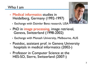
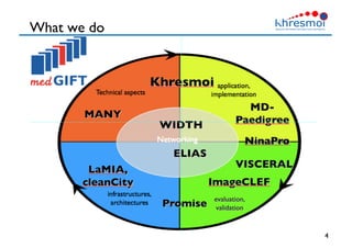
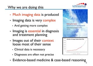
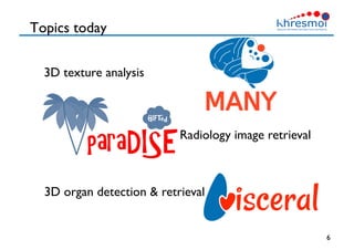
Medical 3D data retrieval
Henning Müller
Where we are
2
Who I am 
•  Medical informatics studies in 
Heidelberg, Germany (1992-1997)
•  Exchange with Daimler Benz research, USA
•  PhD in image processing, image retrieval,
Geneva, Switzerland (1998-2002)
•  Exchange with Monash University, Melbourne, AUS
•  Postdoc, assistant prof. in Geneva University
hospitals in medical informatics (2002-)
•  Professor in Computer Science at the 
HES-SO, Sierre, Switzerland (2007-)
3
What we do
4
Why we are doing this
•  Much imaging data is produced
•  Imaging data is very complex
•  And getting more complex
•  Imaging is essential in diagnosis 
and treatment planning
•  Images out of their context
loose most of their sense
•  Clinical data is necessary
•  Diagnoses are often not precise
•  Evidence-based medicine  case-based reasoning
5
Topics today
6
3D texture analysis
Radiology image retrieval
3D organ detection  retrieval
Diagnosis aid
7
8
3D texture
3D texture vs. 3D objects
•  Not the outer
shape of structures
but the inner 
structure
•  Hard to visualize, 
often in several views
•  Borders are important because texture needs
to be compared inside the same objects
•  Exact place can be important, shape of organs as well
•  Variations among subjects are very high
•  Particularly for healthy tissue, linked to other factors
 9
Viewing 3D texture
•  Multi-planar rendering
•  3D views
•  Semi-transparency
•  MDs need to get used to it
10
A short video
11
4D texture, pulmonary embolism
•  4D dual energy CT
•  Other 4D data include time components
•  Goal is to measure tissue perfusion
•  Find whether and which parts of the lung are blocked
•  Emergency radiology has very little time for decisions
•  Do things that physicians can not do (quantifying)
12
Material Attenuation Coefficient vs keV
0.1
1
10
100
40
 50
 60
 70
 80
 90
 100
 110
 120
 130
 140
Photon Energy (keV)
m(E)(cm2/mg)
Iodine
Water
80 keV 140 keV
Texture characteristics
•  Localized characterizations of scales and local
orientations are important for textures 
•  Often choices for characterization are arbitrary
•  Riesz transform implements nth order directional
derivatives at multiple scales
•  Linear combinations of the filters allows to create texture
templates to detect specific textures
13
local orientation
scale
Combining filters for detection
14
Texture templates
15
Scale and orientation modeling
Templates for lung textures
16
Signatures allow for visually checking the
modeled information
3D texture templates
17
•  Vertical planes
•  3D checkerboard
•  3D wiggled
checkerboard
18
Radiology image retrieval
Khresmoi application
•  Mixing multilingual data from many resources
and semantic information for medical retrieval
•  LinkedLifeData
19
The informed patient
20
Identifying user requirements
•  Surveys among several radiologists
•  Also GPs and patients
•  Observing diagnosis processes
•  Analyzing search log files
•  Eye tracking on a radiology viewing station
•  What are information needs and what are
tasks that are hard and where help is needed?
•  Test the developed systems in user studies
•  Analyze feedback
•  Record the system use for understanding problems
21
Eye tracking
22
Recording user tests
23
Data used for ParaDISE
•  Scientific data of the biomedical literature 
•  600’000 articles and 1.6 mio figures of the open access
literature (4 mio images if separating compound figures)
•  Public data source but only 2D data
•  Clinical data form the Vienna Medical University
image archive
•  5TB of data of two consecutive months
•  Radiology reports for each case
•  Private data source, so access only with password
•  Link medical cases with similar cases from the
literature based on image data and text
 24
Connecting different data levels
25
EHR, PACS
Context is important (25 yo vs. 88 yo)!
26
ParaDISE architecture
27
Interfaces for search: radiology
28
Searching text and images
29
Playful interfaces
30
Semantics in radiology
•  Map text to ontologies such as RadLex
•  LinkedLifeData is a huge knowledge source
•  Permits synonyms, hypernyms, several languages, etc.
•  Use structure and links
•  Anatomic regions are linked with modalities
•  Specific findings (such as fibrosis or micro nodules) are
linked with anatomic regions and also with modalities
•  Use visual information and semantics to extract
information from the images
•  Detecting modality and anatomic region from the images
31
Visual words in 3D
•  Visual features can depend strongly on the
application domain
•  Many benchmarks show that using a large feature set
and then reducing it is often performing best
•  Early vs. late fusion is not absolutely clear
•  Modeling the feature space based on what is
actually present in the images
•  Use techniques known in text retrieval
•  Removing stop words
•  Latent semantic analysis, synonyms, etc.
32
Visual words and region detection
33
34
3D organ detection 
and retrieval
Goals
•  Organize benchmarks on image analysis in
very large data sets 10 TB
•  Identifying anatomic structures
•  Finding similar cases
•  Many challenges
•  10 TB can not be downloaded and also sending hard
disks to participants is not easy
•  How can we obtain a solid ground truth to compare
algorithms on a large scale
•  Scalability is a major factor, so efficiency of algorithms
35
Cloud-based benchmarking
36
Test
Creation of the database
37
Annotated data (20-55 organs)
38
Gold and silver corpus
•  Training data will be fully annotated manually
•  Part of the test data will also be annotated
manually (gold corpus)
•  Does not scale to 10 TB
•  Based on results and evaluation a larger silver
corpus will be created
•  Based on high quality systems on gold corpus via
majority voting
•  Annotate organs where there are strong differences
between participating systems
•  Maximize the gain of manual annotation
39
Participation is open!!
•  Data will be released this summer (August 1)
•  http://www.visceral.eu/benchmark-1/
•  Registration system is almost finished
•  Participants will receive a virtual machine they
can configure to their needs in the Azure cloud
•  Linux, Windows, …
•  First phase on training data (can be downloaded if
really needed), access via Azure API
•  Then the organizers will take over the virtual
machine and run things on test data
•  Detailed protocol is being defined
40
MICCAI workshop
•  http://www.medicalcomputervision.org/
•  Linked to the MICCAI conference
•  Medical Image Computing for Computer Assisted Intervention
•  Scientific part of the workshop and part related
to VISCERAL
•  Big data in medical image analysis
•  Discuss the challenges and orient the work of the
project towards real challenges
•  Community effort 
41
Conclusions
•  Medical imaging offers many interesting
opportunities in multidimensional data analysis
•  Challenges remain such as data confidentiality,
3D/4D visualization, small regions of interest
•  Images should never be regarded out of their context
•  Application-driven approaches can really help
physicians and get support from them
•  Creating (large) data sets for developing tools
requires much effort
•  Open data sets are really important to advance sciece
42
Contact and more information
•  More information can be found at 
•  http://khresmoi.eu/
•  http://visceral.eu/
•  http://medgift.hevs.ch/
•  http://publications.hevs.ch/
•  Contact:
•  Henning.mueller@hevs.ch
43

Medical 3D data retrieval

  • 1.
    Medical 3D dataretrieval Henning Müller
  • 2.
  • 3.
    Who I am •  Medical informatics studies in Heidelberg, Germany (1992-1997) •  Exchange with Daimler Benz research, USA •  PhD in image processing, image retrieval, Geneva, Switzerland (1998-2002) •  Exchange with Monash University, Melbourne, AUS •  Postdoc, assistant prof. in Geneva University hospitals in medical informatics (2002-) •  Professor in Computer Science at the HES-SO, Sierre, Switzerland (2007-) 3
  • 4.
  • 5.
    Why we aredoing this •  Much imaging data is produced •  Imaging data is very complex •  And getting more complex •  Imaging is essential in diagnosis and treatment planning •  Images out of their context loose most of their sense •  Clinical data is necessary •  Diagnoses are often not precise •  Evidence-based medicine case-based reasoning 5
  • 6.
    Topics today 6 3D textureanalysis Radiology image retrieval 3D organ detection retrieval
  • 7.
  • 8.
  • 9.
    3D texture vs.3D objects •  Not the outer shape of structures but the inner structure •  Hard to visualize, often in several views •  Borders are important because texture needs to be compared inside the same objects •  Exact place can be important, shape of organs as well •  Variations among subjects are very high •  Particularly for healthy tissue, linked to other factors 9
  • 10.
    Viewing 3D texture • Multi-planar rendering •  3D views •  Semi-transparency •  MDs need to get used to it 10
  • 11.
  • 12.
    4D texture, pulmonaryembolism •  4D dual energy CT •  Other 4D data include time components •  Goal is to measure tissue perfusion •  Find whether and which parts of the lung are blocked •  Emergency radiology has very little time for decisions •  Do things that physicians can not do (quantifying) 12 Material Attenuation Coefficient vs keV 0.1 1 10 100 40 50 60 70 80 90 100 110 120 130 140 Photon Energy (keV) m(E)(cm2/mg) Iodine Water 80 keV 140 keV
  • 13.
    Texture characteristics •  Localizedcharacterizations of scales and local orientations are important for textures •  Often choices for characterization are arbitrary •  Riesz transform implements nth order directional derivatives at multiple scales •  Linear combinations of the filters allows to create texture templates to detect specific textures 13 local orientation scale
  • 14.
  • 15.
    Texture templates 15 Scale andorientation modeling
  • 16.
    Templates for lungtextures 16 Signatures allow for visually checking the modeled information
  • 17.
    3D texture templates 17 • Vertical planes •  3D checkerboard •  3D wiggled checkerboard
  • 18.
  • 19.
    Khresmoi application •  Mixingmultilingual data from many resources and semantic information for medical retrieval •  LinkedLifeData 19
  • 20.
  • 21.
    Identifying user requirements • Surveys among several radiologists •  Also GPs and patients •  Observing diagnosis processes •  Analyzing search log files •  Eye tracking on a radiology viewing station •  What are information needs and what are tasks that are hard and where help is needed? •  Test the developed systems in user studies •  Analyze feedback •  Record the system use for understanding problems 21
  • 22.
  • 23.
  • 24.
    Data used forParaDISE •  Scientific data of the biomedical literature •  600’000 articles and 1.6 mio figures of the open access literature (4 mio images if separating compound figures) •  Public data source but only 2D data •  Clinical data form the Vienna Medical University image archive •  5TB of data of two consecutive months •  Radiology reports for each case •  Private data source, so access only with password •  Link medical cases with similar cases from the literature based on image data and text 24
  • 25.
    Connecting different datalevels 25 EHR, PACS
  • 26.
    Context is important(25 yo vs. 88 yo)! 26
  • 27.
  • 28.
  • 29.
  • 30.
  • 31.
    Semantics in radiology • Map text to ontologies such as RadLex •  LinkedLifeData is a huge knowledge source •  Permits synonyms, hypernyms, several languages, etc. •  Use structure and links •  Anatomic regions are linked with modalities •  Specific findings (such as fibrosis or micro nodules) are linked with anatomic regions and also with modalities •  Use visual information and semantics to extract information from the images •  Detecting modality and anatomic region from the images 31
  • 32.
    Visual words in3D •  Visual features can depend strongly on the application domain •  Many benchmarks show that using a large feature set and then reducing it is often performing best •  Early vs. late fusion is not absolutely clear •  Modeling the feature space based on what is actually present in the images •  Use techniques known in text retrieval •  Removing stop words •  Latent semantic analysis, synonyms, etc. 32
  • 33.
    Visual words andregion detection 33
  • 34.
    34 3D organ detection and retrieval
  • 35.
    Goals •  Organize benchmarkson image analysis in very large data sets 10 TB •  Identifying anatomic structures •  Finding similar cases •  Many challenges •  10 TB can not be downloaded and also sending hard disks to participants is not easy •  How can we obtain a solid ground truth to compare algorithms on a large scale •  Scalability is a major factor, so efficiency of algorithms 35
  • 36.
  • 37.
    Creation of thedatabase 37
  • 38.
  • 39.
    Gold and silvercorpus •  Training data will be fully annotated manually •  Part of the test data will also be annotated manually (gold corpus) •  Does not scale to 10 TB •  Based on results and evaluation a larger silver corpus will be created •  Based on high quality systems on gold corpus via majority voting •  Annotate organs where there are strong differences between participating systems •  Maximize the gain of manual annotation 39
  • 40.
    Participation is open!! • Data will be released this summer (August 1) •  http://www.visceral.eu/benchmark-1/ •  Registration system is almost finished •  Participants will receive a virtual machine they can configure to their needs in the Azure cloud •  Linux, Windows, … •  First phase on training data (can be downloaded if really needed), access via Azure API •  Then the organizers will take over the virtual machine and run things on test data •  Detailed protocol is being defined 40
  • 41.
    MICCAI workshop •  http://www.medicalcomputervision.org/ • Linked to the MICCAI conference •  Medical Image Computing for Computer Assisted Intervention •  Scientific part of the workshop and part related to VISCERAL •  Big data in medical image analysis •  Discuss the challenges and orient the work of the project towards real challenges •  Community effort 41
  • 42.
    Conclusions •  Medical imagingoffers many interesting opportunities in multidimensional data analysis •  Challenges remain such as data confidentiality, 3D/4D visualization, small regions of interest •  Images should never be regarded out of their context •  Application-driven approaches can really help physicians and get support from them •  Creating (large) data sets for developing tools requires much effort •  Open data sets are really important to advance sciece 42
  • 43.
    Contact and moreinformation •  More information can be found at •  http://khresmoi.eu/ •  http://visceral.eu/ •  http://medgift.hevs.ch/ •  http://publications.hevs.ch/ •  Contact: •  Henning.mueller@hevs.ch 43