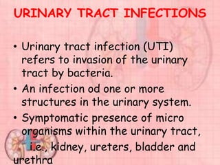
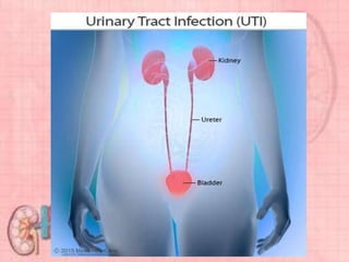
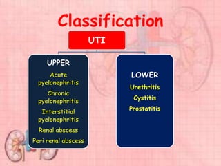
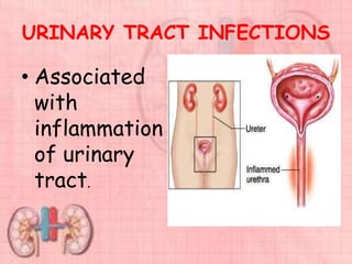
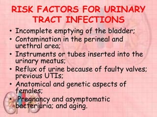
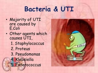
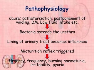
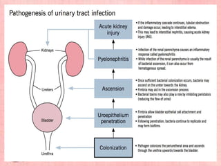
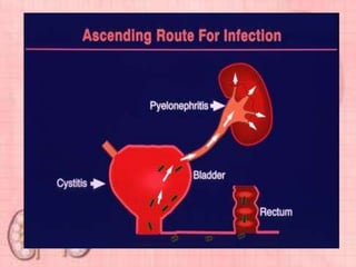
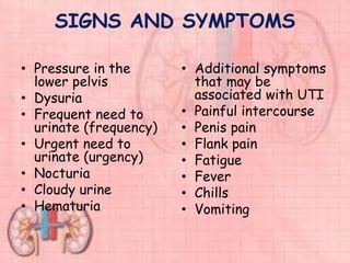
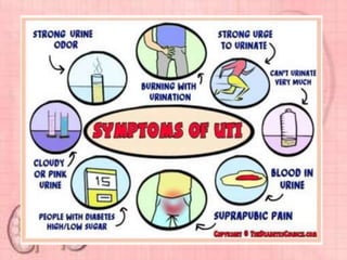
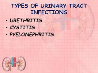
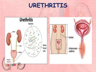
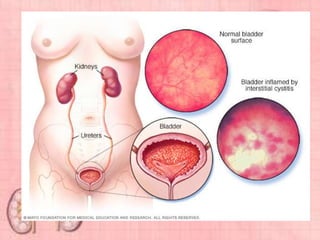
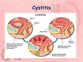
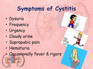
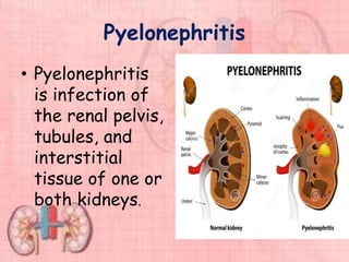
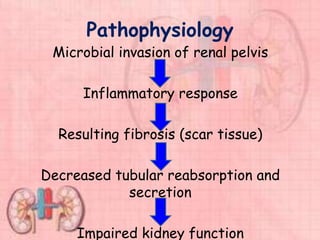
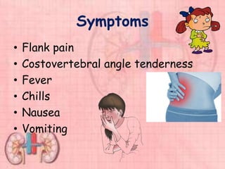
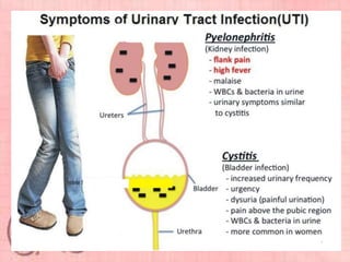
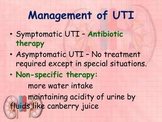
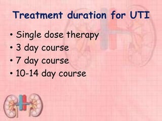
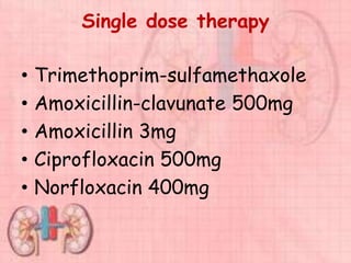
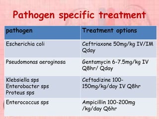
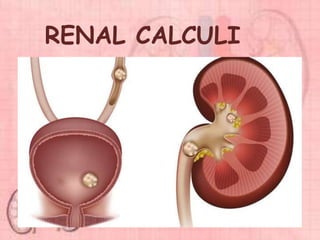
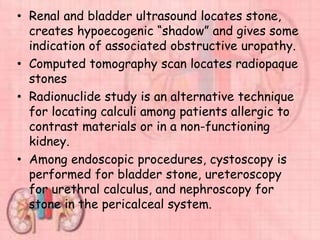
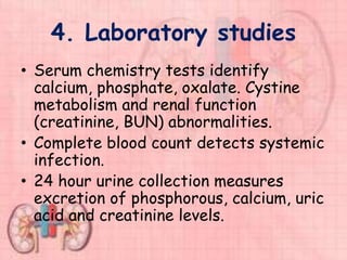
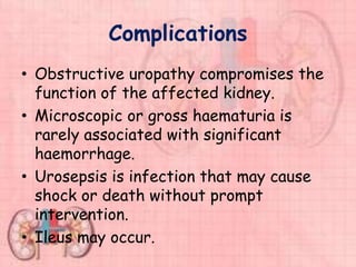
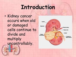
URINARY TRACT
INFECTION
PRESENTED BY
BLESSY SUSAN BABU
FIRST YEAR MSC NURSING
KINS, KARAD
INTRODUCTION
• Urinary tract infection (UTI) is a
common and painful human illness
that, fortunately, is rapidly
responsive to modern antibiotic
therapy.
• UTI is the second most common
bacterial disease.
• In the hospital, UTIs are the most
common hospital acquired infection.
URINARY TRACT INFECTIONS
• Urinary tract infection (UTI)
refers to invasion of the urinary
tract by bacteria.
• An infection od one or more
structures in the urinary system.
• Symptomatic presence of micro
organisms within the urinary tract,
i.e., kidney, ureters, bladder and
urethra
Classification
UTI
UPPER
Acute
pyelonephritis
Chronic
pyelonephritis
Interstitial
pyelonephritis
Renal abscess
Peri renal abscess
LOWER
Urethritis
Cystitis
Prostatitis
URINARY TRACT INFECTIONS
• Associated
with
inflammation
of urinary
tract.
RISK FACTORS FOR URINARY
TRACT INFECTIONS
• Incomplete emptying of the bladder;
• Contamination in the perineal and
urethral area;
• Instruments or tubes inserted into the
urinary meatus;
• Reflux of urine because of faulty valves;
previous UTIs;
• Anatomical and genetic aspects of
females;
• Pregnancy and asymptomatic
bacteriuria; and aging.
Why women are at more risk
for UTIs?
• Women tend to
have urinary tract
infections more
often than men do
because the
urethra is shorter
in women than in
men, so bacteria
have a shorter
distance to travel.
Bacteria & UTI
• Majority of UTI
are caused by
E.Coli
• Other agents which
causes UTI,
1. Staphylococcus
2. Proteus
3. Pseudomonas
4. Klebsiella
5. Enterococcus
Pathophysiology
Cause: catheterization, postponement of
voiding, DM, Low fluid intake etc.
Bacteria ascends the urethra
Lining of urinary tract becomes inflammed
Micturition reflex triggered
Urgency, frequency, burning haematuria,
irritability, pyuria
SIGNS AND SYMPTOMS
• Pressure in the
lower pelvis
• Dysuria
• Frequent need to
urinate (frequency)
• Urgent need to
urinate (urgency)
• Nocturia
• Cloudy urine
• Hematuria
• Additional symptoms
that may be
associated with UTI
• Painful intercourse
• Penis pain
• Flank pain
• Fatigue
• Fever
• Chills
• Vomiting
TYPES OF URINARY TRACT
INFECTIONS
• URETHRITIS
• CYSTITIS
• PYELONEPHRITIS
URETHRITIS
URETHRITIS
• Urethritis is inflammation of the
urethra that may result from a
chemical irritant, bacterial
infection, trauma, or exposure to a
sexually transmitted infection
(STI).
• Post-traumatic urethritis can occur
with intermittent catheterization
or instrumentation of the urethra.
URETHRITIS
• Urethritis can also be caused by
spermicidal agents.
• Gonorrhea and chlamydiosis are
STIs that can cause urethritis in
men.
• It is common to have some degree
of urethritis in association with
bladder or prostatic infections.
URETHRITIS
• It categorized into one of two
forms, based on etiology;
1. Gonococcal urethritis
2. Nongonococcal urethritis
(Chlamydia trachomatis)
Symptoms of urethritis
• Urinary frequency
• Urgency
• Dysuria
• Discharge from the penis
• Itching
• Stomach pain
• Fever and chills
Cystitis
• Cystitis is inflammation and infection
of the bladder wall.
• It can be due to bacteria, viruses,
fungi, or parasites.
• About 90% of UTIs are caused by
Escherichia coli.
• In most cases, the causative organisms
first grow in the perineal area and then
ascend into the bladder.
Cystitis
Symptoms of Cystitis
• Dysuria
• Frequency
• Urgency
• Cloudy urine
• Suprapubic pain
• Hematuria
• Occasionally fever & rigors
Pyelonephritis
• Pyelonephritis
is infection of
the renal pelvis,
tubules, and
interstitial
tissue of one or
both kidneys.
Pathophysiology
Microbial invasion of renal pelvis
Inflammatory response
Resulting fibrosis (scar tissue)
Decreased tubular reabsorption and
secretion
Impaired kidney function
Symptoms
• Flank pain
• Costovertebral angle tenderness
• Fever
• Chills
• Nausea
• Vomiting
Diagnostic tests for UTI
• History
• Physical examination
• Lab parameters
• Imaging studies
Diagnostic tests for UTI
• Urinalysis test: urine is examined
for white and red blood cells and
bacteria, also assess pyuria.
• Urine culture and sensitivity:
bacteria are grown in a culture and
tested against different antibiotics
to see which drug best destroys
the bacteria.
Diagnostic tests for UTI
• Blood test: WBC with
differential, leukocytosis and
increased number of neutrophils
• CBC, ESR, CRP and Blood culture
• Imaging: ultrasonography,
intravenous pyelogram,
cystoscopy.
UTI
URINALYS
IS
URINE MICROSCOPY
AND CULTURE
ADULT
FEMALE
LOWER UTI
Treat without
further
investigation
MALE ANY
UTI
Ultrasound
cystoscopy
PYELONEPHR
ITIS
COMPLICATE
D
Blood
cultures
CT Scan
Check renal
function
Management of UTI
• Symptomatic UTI – Antibiotic
therapy
• Asymptomatic UTI – No treatment
required except in special situations.
• Non-specific therapy:
more water intake
maintaining acidity of urine by
fluids like canberry juice
Anti-microbial therapy
• Goals of therapy:
Elimination of infection
Relief of acute symptoms
Prevention of reoccurrence and
long term complications
Treatment duration for UTI
• Single dose therapy
• 3 day course
• 7 day course
• 10-14 day course
Single dose therapy
• Trimethoprim-sulfamethaxole
• Amoxicillin-clavunate 500mg
• Amoxicillin 3mg
• Ciprofloxacin 500mg
• Norfloxacin 400mg
7 day therapy
• Used less for uncomplicated UTI
• Useful in
recurrent cases
pregnancy
UTI with other risk factors
14 day therapy
• For complicated UTI
• High risk of mortality and
morbidity
Pathogen specific treatment
pathogen Treatment options
Escherichia coli Ceftriaxone 50mg/kg IV/IM
Qday
Pseudomonas aeroginosa Gentamycin 6-7.5mg/kg IV
Q8hr/ Qday
Klebsiella sps
Enterobacter sps
Proteus sps
Ceftadizine 100-
150mg/kg/day IV Q8hr
Enterococcus sps Ampicillin 100-200mg
/kg/day Q6hr
Catheter associated UTI
• Asymptomatic UTI develop in
catheterized patients after 10-14
days.
• Antibiotic treatment – eradicate
organism but high chance of
relapse.
• Catheter removal before treatment
is beneficial.
Nursing Management
• Acute Pain related to inflammation
of the urethra, bladder, and other
urinary structures
• Impaired Urinary Elimination
related to frequency, nocturia,
dysuria, and incontinence.
Patient Education
• To drink more fluid
• Keep perineum clean and dry.
• In female clean the perineum from
front to back.
• Empty bladder soon after intercourse.
• Avoid use of any chemical products.
• If any burning micturition or dysuria,
consult a doctor as early as possible.
Prevention
• Void frequently—at least every 3 hours while
awake.
• Drink up to 3000 mL of fluid a day if there are
no fluid restrictions from the HCP. Preferably
drink water.
• Take showers; avoid tub baths.
• Wipe perineum from the front to the back after
toileting.
• Take medication prescribed for urinary tract
infection (UTI) until it is all gone.
• If UTI is associated with another source of
infection, such as vaginitis or prostatitis, ensure
that both infections are treated.
RENAL CALCULI
Renal calculi
• Definition
Nephrolithiasis refers to renal
stone disease; urolithiasis refers to
the presence of stones in the urinary
system. Stones, or calculi are formed
in the urinary tract from the kidney
to bladder by the crystallization of
substances excreted in the urine.
Incidence
• Urinary calculi are more common in men
than in women.
• Incidence of urinary calculi peaks
between the 3rd and 5th decades of life.
• 50% reoccurrence with in 5-10 years.
• There is seasonal variation with stones
occurring more often in the summer
months suspecting the role of
dehydration in this process.
Etiology & Risk Factors
• Metabolic
Abnormalities that result in increased
urine levels of calcium, oxaluric acid, uric
acid or citric acid.
• Climate
Warm climate that causes increased
fluid loss. Low urine volume and increased
solute concentration in urine.
Etiology & Risk Factors
• Diet
 large intake of dietary proteins that
increases uric acid excretion.
 excessive amounts of tea or fruit
juices that elevate urinary oxalate
level.
 large intake of calcium and oxalate.
 low fluid intake that increases
concentration
Etiology & Risk Factors
• Genetic factors
family history of stones formation,
cystinuria, gout or renal acidosis.
• Life style
sedentary occupation and immobility
• A major pre-disposing factor is the
presence of UTI.
Continued…
• Infection increases the presence of
organic matter around which minerals
can precipitate and increases the
alkalinity of the urine by the production
of ammonia. This results in precipitation
of calcium phosphate and magnesium
ammonium phosphate.
• Stasis of urine also permits
• Drug-induced stones (Indinavir
and Nelfinavir Stones)
These agents are excreted as
urinary crystals that may result
in crystal deposition or stone
formation.
Pathophysiology
Slow urine flow, resulting in super saturation
of the urine with the particular element that
first become crystallized and later become
stone.
Damage to the lining of the urinary tract
Decreased inhibitor substances in the urine
that would otherwise prevent super
saturation and crystalline aggregation.
Types of stones
• Calcium phosphate
• Calcium oxalate
• Uric acid
• Cystine
• Struvite
Clinical Manifestations
• Severe
abdominal or
flank pain
• Frequency and
dysuria
• Oliguria and
anuria in
obstruction
Clinical Manifestations
• Heamaturia
• Renal colic
• Nausea
• Hydronephrosis
Clinical Manifestations
• Sharp, severe pain
Most characteristic manifestation of
renal or urethral calculi
Caused by movement of the calculus
and consequent irritation
Renal colic originates deep in the
lumbar region and radiates around
the side and down toward the testicle
in the male and the bladder in the
female
Urethral colic radiates toward the
genitalia and thigh
Continued…
When the pain is severe, the client
usually has nausea, vomiting, pallor,
grunting respirations, elevated
blood pressure and pulse,
diaphoresis.
Urinary tract infection
Other manifestations of calculi include
infection with an elevated temperature
and white blood cell (WBC) count and
urine obstruction that causes hydro
ureter, hydronephrosis or both
Haematuria
Pain resulting from the passage of a
calculus down the ureter is intense and
colicky. The patient may be in mild
shock with cool, moist skin.
Diagnostic evaluation
Assessment
1. History
Prior stone formation
Renal or bladder colic type pain without
objective evidence of calculi formation
Risk factors
Location, character and duration of
current pain
Current and previous radiation patterns
(indicates possible location and
movement of calculus through the
urinary system)
2. Physical Examination
Vital signs include increased pulse,
respirations and blood pressure
associated with colicky pain;
Fever indicates serious infection
Hyperactive bowel sound occur with
nausea and vomiting; hypoactive or
absent bowel sounds occur with
ileus.
3. Diagnostic studies
Urinalysis, urine culture and sensitivity
testing determine the presence of
urinary tract infection, haematuria or
urine crystals.
Radioactive studies
 ninety percent of calculi are visible on
radio graphic images.
 Calcium phosphate stones are brightest
on radiograph; uric stones are least
visible (radiolucent)
 KUB using plain abdominal film detects
larger, radiopaque stones.
 Intravenous urography (IVU) locates
radiopaque stones, allowing evaluation of
associated obstructive uropathy and crude
evaluation of renal function (ability to
concentrate and excrete contrast material)
 It is a standard method for examining the
urinary tract for obstruction in cases of
renal colic
 Tomograms locate stones in the pericalceal
system. They are performed in combination
with IVP
• Renal and bladder ultrasound locates stone,
creates hypoecogenic “shadow” and gives some
indication of associated obstructive uropathy.
• Computed tomography scan locates radiopaque
stones
• Radionuclide study is an alternative technique
for locating calculi among patients allergic to
contrast materials or in a non-functioning
kidney.
• Among endoscopic procedures, cystoscopy is
performed for bladder stone, ureteroscopy
for urethral calculus, and nephroscopy for
stone in the pericalceal system.
4. Laboratory studies
• Serum chemistry tests identify
calcium, phosphate, oxalate. Cystine
metabolism and renal function
(creatinine, BUN) abnormalities.
• Complete blood count detects systemic
infection.
• 24 hour urine collection measures
excretion of phosphorous, calcium, uric
acid and creatinine levels.
Complications
• Obstructive uropathy compromises the
function of the affected kidney.
• Microscopic or gross haematuria is
rarely associated with significant
haemorrhage.
• Urosepsis is infection that may cause
shock or death without prompt
intervention.
• Ileus may occur.
Calcium oxalate
• Increased hydration
• Reduce dietary oxalate
• Give thiazide diuretics
• Give cellulose phosphate to cholate
calcium and prevent GI absorption
• Give cholestyramine to bind oxalate
• Give calcium lactate to precipitate
oxalate in GI tract.
Calcium phosphate
• Treat underlying causes and other
stones
• Administer antimicrobial agents,
acetohydroxamic acid and
antibiotics
• Use surgical intervention to remove
stone
• Take measure to acidify urine
Uric acid stones
• Reduce urinary concentration of
uric acid
• Alkalinize urine with potassium
citrate
• Administer allopurinol
• Reduce dietary purines
Cystine
• Increase hydration
• Give alpha-penicillamine and
tiopronin to prevent cystin
crystallization
• Give pottassium citrate to
maintain alkaline urine.
Struvite stones
• Complete removal of the stone with
subsequent sterilization of the urinary
tract is the treatment of choice for
patients who can tolerate the
procedures.
• Percutaneous nephrolithotomy is the
preferred surgical approach for most
patients.
• At times, extracorporeal lithotripsy
may be used in combination with a
percutaneous approach. Open surgery is
rarely required.
Continued..
• Antimicrobial treatment is best
reserved for dealing with acute
infection and for maintenance of sterile
urine after surgery.
• Urine culture and culture of stone
fragments removed at surgery should
guide the choice of antibiotic.
• For patients who are not candidates for
surgical removal of stone,
acetohydroxamic acid, an inhibitor of
urease can be used.
1. Ureteroscopy
• Involves first visualizing the stone and then
destroying it.
• Access to the stone is accomplished by
inserting a ureteroscope into the ureter and
then inserting a laser, electrohydraulic
lithotriptor or ultrasound device through the
ureteroscope to fragment and remove the
stones.
• A stent may be inserted and left in place for
48 hours or more after the procedure to keep
the ureter patent.
• Hospital stays are generally brief, and some
patients can be treated as outpatients.
2. Lithotripsy
• Laser Lithrotripsy. A newer
treatment for calculi is laser
lithotripsy. Laser are used
together with a uretero-scope
to remove or loosen impacted
stones. Constant water
irrigation of the ureter is
required to dissipate the heat.
3.Extracorporeal shock wave
lithotripsy (ESWL)
• ESWL is a non-invasive procedure used to
break up stones in the calyx of the kidney.
• In ESWL, a high energy amplitude of
pressure, or shock wave, is generated by the
abrupt release of energy and transmitted
through water and soft tissues. When the
shock wave encounters a substance of
different intensity (a renal stone), a
compression wave causes the surface of the
stone to fragment. Repeated shock waves
focused on the stone eventually reduce it to
many small pieces.
Continued…
• These small pieces are excreted
in the urine usually without
difficulty. The fragment may be
passed up to 3 months after the
procedure.
• Stone size should be 1.5-2cm.
4. Percutaneous lithotripsy
• Percutaneous lithotripsy involves
the insertion of guide
percutaneously (through the skin)
under fluroscopy near the area of
the stone. An ultrasonic wave is
aimed at the stone to break it into
fragments.
• Stone size should be >2.5cm
Post operative complications
• Immediate
Pain
Urinary infection
Obstructive uropathy
Haematuria
Urinoma- urinoma happens as a result of
urethral tear which allows the entry of
free fluid into the retro peritoneum
Renal and perirenal haematuria
Surrounding organ injury
Continued..
• Delayed
Renal functional loss
Hypertension
Residual calculi
Recurrent calculi
Open surgical procedures
• If the stone is too large or if
endourologic and lithotripsy procedures
fail to remove it, an open surgical
procedure is performed
• Ureterolithotomy is the surgical
removal of a stone from the ureter
through a flank incision for higher
stones or an abdominal incision for lower
ones. A Penrose drain and ureteral
catheter are usually placed
postoperatively for healing and drainage
of urine.
Continued..
• Cystolithotomy, removal of bladder
calculi through a suprapubic incision, is
used only when stones cannot be
crushed and removed transurethrally.
Stricture (abnormal narrowing) is the
most common postoperative
complication.
• A stone is removed from the renal
pelvis by pyelo-lithotomy and from the
renal calyx by a nephrolithotomy
Medications
• Lortab (500)mg one tab by mouth
every 6 hours as needed for pain
• Percocet (325) mg one tab by
mouth every 6 hours as needed for
pain
• Pyridium (100, 200) mg one tab per
mouth every 8 hours for dysuria
(burning)
• Cipro (250, 500) mg one tab per
mouth twice a day
Prognosis
• Despite advances in the treatment of
urinary calculi, it is often impossible to
remove all stone fragments completely.
From 5 to 30 percent of patients have
residual stone burden requiring on going
treatment.
• Recurrence rate is approximately 30
percent within years
• ESWL and endoscopic stone removal
techniques have significantly improved
long term prognosis of renal function
after calculus removal.
Nursing diagnosis
• Acute Pain related to the presence
of, obstruction, or movement of a
stone within the urinary system
• Altered urinary elimination related
to presence of urinary calculi
• Risk for infection related to
obstructing Risk for infection
related to obstructing urinary
calculus
Nursing intervention
• Adequate hydration, dietary sodium
restrictions, and the use of medications
minimise stone formation
• High fluid intake at least 3000 ml per
day is recommended
• Dietary intervention may be important
in the management of formation
urolithiasis
• Nutritional management should include
limiting oxalate – foods and there by
reducing oxalate excretion.
Cancer of bladder
Introduction
• Rapid un controlled growth of
abnormal cells in the bladder.
• Begins in the lining of the bladder &
spread through the lining into the
muscular wall of the bladder.
• Invasive bladder cancer: spread to
lymph nodes, other organs in the
pelvis or other organs (liver and
lungs)
Incidence
• In India bladder cancer is the fifth
most common cancer in men.
• Incidence is much lower in females
1.5 cases/1,00,000
• Male to female ratio = 8.6:1
Risk factors
• Smoking
• Environmental carcinogens
• Recurrent/chronic bacterial
infections
• Bladder stones
• High urinary pH
• High cholesterol intake
• Pelvic radiation therapy
• Cancers arising from prostate,
colon & rectum
Risk
factors
Classification
90% of bladder cancers are transitional cell
carcinoma. The other 10% are squamous
cell carcinoma, adenocarcinoma, sarcoma,
small call carcinoma
Stage CIS : Flat cancer limited to the innermost lining of the bladder.
It is high grade
Stage T1 : Cancer penetrated into the sub mucosal tissue.
stageT2 : Cancer penetrated through muscular bladder wall.
Stage T3 : Cancer penetrated through muscular bladder wall into the
surrounding fat.
Stage T4 : Cancer penetrated into the adjacent structures (prostate,
uterus or vagina). Regional lymph nodes not involved yet.
Stage T1-4N1-2M1-2 : Cancer spread out of abdomen/pelvic wall to
lymph nodes or distant organs like liver, lungs or bones.
Signs & symptoms
• Haematuria
• Urinary tract infection
• Pain with metastasis
• Any change in voiding/ urine
• Frequency
• Urgency
• Dysuria
Investigations
Physical Examination
Urinalysis
Urine Cytology
Cystoscopy
CT Scan
Pyelography
Biopsy
Ultrasound
•Transurethral
resection
•Chemotherapy
•Radiotherapy
Trimodality
Therapy
TREATMENT
• Treatment depends on the
stage of the cancer
• Surgery
• Radiation therapy
• Chemotherapy (combination of
drugs)
• Immunotherapy
Surgical management
Transurethral
resection
Cystectomy
Surgery
• In early stage cancer, the tumour is
removed with a laser or high-energy
electricity
• Advanced cancer may require complete
or partial removal of the bladder
(cystectomy) and nearby lymph nodes,
tissues and organs.
• If the bladder is removed, a new way
for the body to divert urine outside the
body is made,
Continent urinary diversion
• Use of intestinal segment to
bypass/ reconstruct/ replace the
normal urinary tract.
• Goals:
Storage of urine without absorption
Prevent reflux of urine back to the
kidneys
Urinary diversions
• Cutaneous ureterostomy
• ileal conduit
• Continent ileostomy
• Orthotopic neobladder
Radiation therapy
• The use of high-energy x-rays to
destroy cancer cells.
• External beam: outside the body
• May be used to treat bladder
cancer before or after surgery.
• Side effects may include fatigue,
mild skin reactions, upset stomach,
loose bowel movements, bladder
irritation and bleeding from the
bladder.
Chemotherapy
• Use of drugs to kill cancer cells.
• Earlier stage cancers are more likely to
be treated with intravesical (local)
chemotherapy
• Advanced cancers are more likely to be
treated with systemic (whole body)
chemotherapy.
• Standard systemic treatment is MVAC,
a four drug combination : methotrexate,
vinblastine, doxorubicin and cisplastin.
Immunotherapy
• Uses materials made by the body or in a
laboratory to boost patient’s natural
defences against cancer.
• BCG (Bacillus Calmette Guerin) is the
most common immunotherapy drug for
bladder cancer; it is given using
intravesical therapy.
• BCG irritates the inside of the bladder,
affecting the patient’s immune cells to
the bladder to fight the tumour
Introduction
• Kidney cancer
occurs when old
or damaged
cells continue to
divide and
multiply
uncontrollably.
• Kidney cancer is most treatable and
curable if caught in the earliest
stage of the disease. Untreated or
advanced kidney cancer can spread
from kidney into surrounding
tissues and into the lymph nodes,
lungs, liver, bones and brain, where
it can form another cancerous
tumour this is called metastasis.
• Kidney cancer is more common in
men than in women.
Causes
• Age and Gender as Risk factors
• The genetic connection
• Smoking
• Obesity
• Hypertension
• Exposure to radiation
Signs and Symptoms
• classic symptoms
Haematuria
dull pain in the flank area
a mass in the area
Other symptoms
• Anaemia
• Fatigue
• Fever
• Weight loss
• swelling in the legs
• Anorexia
• Constipation
Diagnostic Tests
• Physical examination
• lab tests:
complete blood count
urinalysis
serum calcium
• Imaging studies:
ultrasound abdomen
abdominal CT scan
MRI Scan
PET Scan
renal angiography
intravenous pyelogram
biopsy
Staging
N categories for kidney cancer:
• N0 : No spread to nearby lymph nodes
• N1 : Tumour has spread to nearby lymph
nodes
M categories for kidney cancer:
• M0 : There is no spread to distant
lymph nodes or other organs.
• M1 : Distant metastasis is present,
distant lymph nodes and to organs like
lungs, bone, brain and liver
Partial
nephrectomy
:
• for treating small
renal
tumours(<4cm)
• Bilateral renal cell
carcinoma
• It can be done via
laparoscopic
techniques:
Radical
nephrectomy
• Surgical removal of kidney
along with adrenal gland,
retroperitoneal lymph
nodes and perinephric fat
• In cases where the tumour
has spread into the renal
vein, IVC and right atrium,
this portion of tumour can
be surgically removed as
well.
• Medications like tryosine
kinase inhibitors including
nexaver and repamycin
have shown to improve the
prognosis for advanced
Nursing Assessment
Risk
factors
Haematu
ria
Fatigue
Weight
loss
Irritativ
e voiding
syndrome
Assessment
Feelings
about
impotence
Knowledge
of disease
Coping
ability
Signs of
metastasi
s
Nursing diagnosis
• Impaired urinary elimination
related to haematuria and
transurethral surgery.
• Acute pain related to irritative
voiding symptoms and catheter
related discomfort.
• Anxiety related to diagnosis of
cancer
Nursing interventions
• Maintaining urinary elimination
after TUS
• Controlling pain.
• Relieving anxiety
• Patient teaching:
Preoperative care
• Stop cigarette smoking
• Physiotherapy assessment
• Elimination – a baseline assessment of
renal function, urinalysis and the
pattern and characteristics of the
urinary output
• Educate the patient on the following
postoperative care:
• Breathing and coughing exercises
• Use of an incentive spirometer
Preoperative care
• Splinting of the wound
• Pain control
• Urinary catheter
• Wound incision line – for both open and
laparoscopic procedures
• The possible presence of a wound drain
with an open procedure
• Leg exercises and use of anti-thrombosis
stockings and anticoagulants
• Fasting, intravenous therapy, oral fluids
and then a light diet
Postoperative care
• Monitor:
–Respiration
–Colour
–Pulse oximetry for oxygen saturation
levels
–Signs of respiratory complications,
e.g. spontaneous pneumothorax or
atelectasis (changes in
respiration and oxygen saturation
levels)
• Administer prescribed oxygen
• Place in a semi-Fowler’s position
• Ensure regular use of an incentive
spirometer
• Use pillows to splint the wound when
conducting breathing and coughing
exercises
• Monitor for signs of hypovolaemic shock
due to haemorrhage:
– Vital signs
– Colour
– Skin
– Wound dressing (check that no blood has seeped round
to the back)
– Wound drainage
• Monitor pain:
• Assess the level of pain (open versus
laparoscopic): following a open
nephrectomy, the patient will have a
large surgical wound (with or without a
wound drain), and the pain will be more
intense than in patients whose procedure
was carried out via a laparoscopic
approach
• With laparoscopic procedures, shoulder
pain and a bloated abdomen may occur
and can be alleviated by pain medication
and mobilisation
• Administer analgesia (PCA) and
assess its effectiveness
• Monitor:
• Urinary output, and record this on the
fluid balance chart
• The colour and odour of the urine
• The administration of prophylactic
intravenous antibiotics prior to removal of
the urinary catheter
• The postoperative voiding pattern
• Renal function, e.g. serum creatinine level
• Bowel sounds
• Possible paralytic ileus
• Commence oral fluids followed by a light
diet
• Prevent constipation:
• Early mobilisation
• A high-fibre diet and oral fluids
• The wound:
– Monitor for signs of infection
– Remove the wound drain (if present), usually at 48–72
hours
• Remove sutures/staples (if present) at 7–
10 days – the patient can return to the
outpatients department or attend a
community healthcare provider for this
Discharge advice
• Monitor urinary output and signs of UTI
• Ensure a fluid intake of 2 L/day
• Care of the incision site(s)
• Follow-up care in relation to renal function
• Avoid strenuous exercises in the initial
recovery phase
• Avoid lifting weights for 6–8 weeks
• As the patient has only one functioning
kidney, they are advised to avoid contact
sports in the future
• Details of medication and its impact on
kidney function
•
Urinary disorders

Urinary disorders

  • 1.
    URINARY TRACT INFECTION PRESENTED BY BLESSYSUSAN BABU FIRST YEAR MSC NURSING KINS, KARAD
  • 3.
    INTRODUCTION • Urinary tractinfection (UTI) is a common and painful human illness that, fortunately, is rapidly responsive to modern antibiotic therapy. • UTI is the second most common bacterial disease. • In the hospital, UTIs are the most common hospital acquired infection.
  • 4.
    URINARY TRACT INFECTIONS •Urinary tract infection (UTI) refers to invasion of the urinary tract by bacteria. • An infection od one or more structures in the urinary system. • Symptomatic presence of micro organisms within the urinary tract, i.e., kidney, ureters, bladder and urethra
  • 6.
  • 7.
    URINARY TRACT INFECTIONS •Associated with inflammation of urinary tract.
  • 8.
    RISK FACTORS FORURINARY TRACT INFECTIONS • Incomplete emptying of the bladder; • Contamination in the perineal and urethral area; • Instruments or tubes inserted into the urinary meatus; • Reflux of urine because of faulty valves; previous UTIs; • Anatomical and genetic aspects of females; • Pregnancy and asymptomatic bacteriuria; and aging.
  • 9.
    Why women areat more risk for UTIs? • Women tend to have urinary tract infections more often than men do because the urethra is shorter in women than in men, so bacteria have a shorter distance to travel.
  • 10.
    Bacteria & UTI •Majority of UTI are caused by E.Coli • Other agents which causes UTI, 1. Staphylococcus 2. Proteus 3. Pseudomonas 4. Klebsiella 5. Enterococcus
  • 11.
    Pathophysiology Cause: catheterization, postponementof voiding, DM, Low fluid intake etc. Bacteria ascends the urethra Lining of urinary tract becomes inflammed Micturition reflex triggered Urgency, frequency, burning haematuria, irritability, pyuria
  • 14.
    SIGNS AND SYMPTOMS •Pressure in the lower pelvis • Dysuria • Frequent need to urinate (frequency) • Urgent need to urinate (urgency) • Nocturia • Cloudy urine • Hematuria • Additional symptoms that may be associated with UTI • Painful intercourse • Penis pain • Flank pain • Fatigue • Fever • Chills • Vomiting
  • 16.
    TYPES OF URINARYTRACT INFECTIONS • URETHRITIS • CYSTITIS • PYELONEPHRITIS
  • 17.
  • 18.
    URETHRITIS • Urethritis isinflammation of the urethra that may result from a chemical irritant, bacterial infection, trauma, or exposure to a sexually transmitted infection (STI). • Post-traumatic urethritis can occur with intermittent catheterization or instrumentation of the urethra.
  • 19.
    URETHRITIS • Urethritis canalso be caused by spermicidal agents. • Gonorrhea and chlamydiosis are STIs that can cause urethritis in men. • It is common to have some degree of urethritis in association with bladder or prostatic infections.
  • 20.
    URETHRITIS • It categorizedinto one of two forms, based on etiology; 1. Gonococcal urethritis 2. Nongonococcal urethritis (Chlamydia trachomatis)
  • 21.
    Symptoms of urethritis •Urinary frequency • Urgency • Dysuria • Discharge from the penis • Itching • Stomach pain • Fever and chills
  • 22.
    Cystitis • Cystitis isinflammation and infection of the bladder wall. • It can be due to bacteria, viruses, fungi, or parasites. • About 90% of UTIs are caused by Escherichia coli. • In most cases, the causative organisms first grow in the perineal area and then ascend into the bladder.
  • 24.
  • 25.
    Symptoms of Cystitis •Dysuria • Frequency • Urgency • Cloudy urine • Suprapubic pain • Hematuria • Occasionally fever & rigors
  • 26.
    Pyelonephritis • Pyelonephritis is infectionof the renal pelvis, tubules, and interstitial tissue of one or both kidneys.
  • 27.
    Pathophysiology Microbial invasion ofrenal pelvis Inflammatory response Resulting fibrosis (scar tissue) Decreased tubular reabsorption and secretion Impaired kidney function
  • 28.
    Symptoms • Flank pain •Costovertebral angle tenderness • Fever • Chills • Nausea • Vomiting
  • 31.
    Diagnostic tests forUTI • History • Physical examination • Lab parameters • Imaging studies
  • 32.
    Diagnostic tests forUTI • Urinalysis test: urine is examined for white and red blood cells and bacteria, also assess pyuria. • Urine culture and sensitivity: bacteria are grown in a culture and tested against different antibiotics to see which drug best destroys the bacteria.
  • 33.
    Diagnostic tests forUTI • Blood test: WBC with differential, leukocytosis and increased number of neutrophils • CBC, ESR, CRP and Blood culture • Imaging: ultrasonography, intravenous pyelogram, cystoscopy.
  • 34.
    UTI URINALYS IS URINE MICROSCOPY AND CULTURE ADULT FEMALE LOWERUTI Treat without further investigation MALE ANY UTI Ultrasound cystoscopy PYELONEPHR ITIS COMPLICATE D Blood cultures CT Scan Check renal function
  • 35.
    Management of UTI •Symptomatic UTI – Antibiotic therapy • Asymptomatic UTI – No treatment required except in special situations. • Non-specific therapy: more water intake maintaining acidity of urine by fluids like canberry juice
  • 36.
    Anti-microbial therapy • Goalsof therapy: Elimination of infection Relief of acute symptoms Prevention of reoccurrence and long term complications
  • 37.
    Treatment duration forUTI • Single dose therapy • 3 day course • 7 day course • 10-14 day course
  • 38.
    Single dose therapy •Trimethoprim-sulfamethaxole • Amoxicillin-clavunate 500mg • Amoxicillin 3mg • Ciprofloxacin 500mg • Norfloxacin 400mg
  • 39.
    7 day therapy •Used less for uncomplicated UTI • Useful in recurrent cases pregnancy UTI with other risk factors 14 day therapy • For complicated UTI • High risk of mortality and morbidity
  • 40.
    Pathogen specific treatment pathogenTreatment options Escherichia coli Ceftriaxone 50mg/kg IV/IM Qday Pseudomonas aeroginosa Gentamycin 6-7.5mg/kg IV Q8hr/ Qday Klebsiella sps Enterobacter sps Proteus sps Ceftadizine 100- 150mg/kg/day IV Q8hr Enterococcus sps Ampicillin 100-200mg /kg/day Q6hr
  • 41.
    Catheter associated UTI •Asymptomatic UTI develop in catheterized patients after 10-14 days. • Antibiotic treatment – eradicate organism but high chance of relapse. • Catheter removal before treatment is beneficial.
  • 42.
    Nursing Management • AcutePain related to inflammation of the urethra, bladder, and other urinary structures • Impaired Urinary Elimination related to frequency, nocturia, dysuria, and incontinence.
  • 43.
    Patient Education • Todrink more fluid • Keep perineum clean and dry. • In female clean the perineum from front to back. • Empty bladder soon after intercourse. • Avoid use of any chemical products. • If any burning micturition or dysuria, consult a doctor as early as possible.
  • 44.
    Prevention • Void frequently—atleast every 3 hours while awake. • Drink up to 3000 mL of fluid a day if there are no fluid restrictions from the HCP. Preferably drink water. • Take showers; avoid tub baths. • Wipe perineum from the front to the back after toileting. • Take medication prescribed for urinary tract infection (UTI) until it is all gone. • If UTI is associated with another source of infection, such as vaginitis or prostatitis, ensure that both infections are treated.
  • 47.
  • 48.
    Renal calculi • Definition Nephrolithiasisrefers to renal stone disease; urolithiasis refers to the presence of stones in the urinary system. Stones, or calculi are formed in the urinary tract from the kidney to bladder by the crystallization of substances excreted in the urine.
  • 49.
    Incidence • Urinary calculiare more common in men than in women. • Incidence of urinary calculi peaks between the 3rd and 5th decades of life. • 50% reoccurrence with in 5-10 years. • There is seasonal variation with stones occurring more often in the summer months suspecting the role of dehydration in this process.
  • 50.
    Etiology & RiskFactors • Metabolic Abnormalities that result in increased urine levels of calcium, oxaluric acid, uric acid or citric acid. • Climate Warm climate that causes increased fluid loss. Low urine volume and increased solute concentration in urine.
  • 51.
    Etiology & RiskFactors • Diet  large intake of dietary proteins that increases uric acid excretion.  excessive amounts of tea or fruit juices that elevate urinary oxalate level.  large intake of calcium and oxalate.  low fluid intake that increases concentration
  • 52.
    Etiology & RiskFactors • Genetic factors family history of stones formation, cystinuria, gout or renal acidosis. • Life style sedentary occupation and immobility • A major pre-disposing factor is the presence of UTI.
  • 53.
    Continued… • Infection increasesthe presence of organic matter around which minerals can precipitate and increases the alkalinity of the urine by the production of ammonia. This results in precipitation of calcium phosphate and magnesium ammonium phosphate. • Stasis of urine also permits
  • 54.
    • Drug-induced stones(Indinavir and Nelfinavir Stones) These agents are excreted as urinary crystals that may result in crystal deposition or stone formation.
  • 55.
    Pathophysiology Slow urine flow,resulting in super saturation of the urine with the particular element that first become crystallized and later become stone. Damage to the lining of the urinary tract Decreased inhibitor substances in the urine that would otherwise prevent super saturation and crystalline aggregation.
  • 56.
    Types of stones •Calcium phosphate • Calcium oxalate • Uric acid • Cystine • Struvite
  • 57.
    Clinical Manifestations • Severe abdominalor flank pain • Frequency and dysuria • Oliguria and anuria in obstruction
  • 58.
    Clinical Manifestations • Heamaturia •Renal colic • Nausea • Hydronephrosis
  • 59.
  • 60.
    Most characteristic manifestationof renal or urethral calculi Caused by movement of the calculus and consequent irritation Renal colic originates deep in the lumbar region and radiates around the side and down toward the testicle in the male and the bladder in the female Urethral colic radiates toward the genitalia and thigh
  • 61.
    Continued… When the painis severe, the client usually has nausea, vomiting, pallor, grunting respirations, elevated blood pressure and pulse, diaphoresis.
  • 62.
    Urinary tract infection Othermanifestations of calculi include infection with an elevated temperature and white blood cell (WBC) count and urine obstruction that causes hydro ureter, hydronephrosis or both Haematuria Pain resulting from the passage of a calculus down the ureter is intense and colicky. The patient may be in mild shock with cool, moist skin.
  • 63.
  • 64.
    1. History Prior stoneformation Renal or bladder colic type pain without objective evidence of calculi formation Risk factors Location, character and duration of current pain Current and previous radiation patterns (indicates possible location and movement of calculus through the urinary system)
  • 65.
    2. Physical Examination Vitalsigns include increased pulse, respirations and blood pressure associated with colicky pain; Fever indicates serious infection Hyperactive bowel sound occur with nausea and vomiting; hypoactive or absent bowel sounds occur with ileus.
  • 66.
    3. Diagnostic studies Urinalysis,urine culture and sensitivity testing determine the presence of urinary tract infection, haematuria or urine crystals. Radioactive studies  ninety percent of calculi are visible on radio graphic images.  Calcium phosphate stones are brightest on radiograph; uric stones are least visible (radiolucent)
  • 67.
     KUB usingplain abdominal film detects larger, radiopaque stones.  Intravenous urography (IVU) locates radiopaque stones, allowing evaluation of associated obstructive uropathy and crude evaluation of renal function (ability to concentrate and excrete contrast material)  It is a standard method for examining the urinary tract for obstruction in cases of renal colic  Tomograms locate stones in the pericalceal system. They are performed in combination with IVP
  • 68.
    • Renal andbladder ultrasound locates stone, creates hypoecogenic “shadow” and gives some indication of associated obstructive uropathy. • Computed tomography scan locates radiopaque stones • Radionuclide study is an alternative technique for locating calculi among patients allergic to contrast materials or in a non-functioning kidney. • Among endoscopic procedures, cystoscopy is performed for bladder stone, ureteroscopy for urethral calculus, and nephroscopy for stone in the pericalceal system.
  • 69.
    4. Laboratory studies •Serum chemistry tests identify calcium, phosphate, oxalate. Cystine metabolism and renal function (creatinine, BUN) abnormalities. • Complete blood count detects systemic infection. • 24 hour urine collection measures excretion of phosphorous, calcium, uric acid and creatinine levels.
  • 70.
    Complications • Obstructive uropathycompromises the function of the affected kidney. • Microscopic or gross haematuria is rarely associated with significant haemorrhage. • Urosepsis is infection that may cause shock or death without prompt intervention. • Ileus may occur.
  • 72.
    Calcium oxalate • Increasedhydration • Reduce dietary oxalate • Give thiazide diuretics • Give cellulose phosphate to cholate calcium and prevent GI absorption • Give cholestyramine to bind oxalate • Give calcium lactate to precipitate oxalate in GI tract.
  • 73.
    Calcium phosphate • Treatunderlying causes and other stones • Administer antimicrobial agents, acetohydroxamic acid and antibiotics • Use surgical intervention to remove stone • Take measure to acidify urine
  • 74.
    Uric acid stones •Reduce urinary concentration of uric acid • Alkalinize urine with potassium citrate • Administer allopurinol • Reduce dietary purines
  • 75.
    Cystine • Increase hydration •Give alpha-penicillamine and tiopronin to prevent cystin crystallization • Give pottassium citrate to maintain alkaline urine.
  • 76.
    Struvite stones • Completeremoval of the stone with subsequent sterilization of the urinary tract is the treatment of choice for patients who can tolerate the procedures. • Percutaneous nephrolithotomy is the preferred surgical approach for most patients. • At times, extracorporeal lithotripsy may be used in combination with a percutaneous approach. Open surgery is rarely required.
  • 77.
    Continued.. • Antimicrobial treatmentis best reserved for dealing with acute infection and for maintenance of sterile urine after surgery. • Urine culture and culture of stone fragments removed at surgery should guide the choice of antibiotic. • For patients who are not candidates for surgical removal of stone, acetohydroxamic acid, an inhibitor of urease can be used.
  • 79.
    1. Ureteroscopy • Involvesfirst visualizing the stone and then destroying it. • Access to the stone is accomplished by inserting a ureteroscope into the ureter and then inserting a laser, electrohydraulic lithotriptor or ultrasound device through the ureteroscope to fragment and remove the stones. • A stent may be inserted and left in place for 48 hours or more after the procedure to keep the ureter patent. • Hospital stays are generally brief, and some patients can be treated as outpatients.
  • 80.
    2. Lithotripsy • LaserLithrotripsy. A newer treatment for calculi is laser lithotripsy. Laser are used together with a uretero-scope to remove or loosen impacted stones. Constant water irrigation of the ureter is required to dissipate the heat.
  • 82.
    3.Extracorporeal shock wave lithotripsy(ESWL) • ESWL is a non-invasive procedure used to break up stones in the calyx of the kidney. • In ESWL, a high energy amplitude of pressure, or shock wave, is generated by the abrupt release of energy and transmitted through water and soft tissues. When the shock wave encounters a substance of different intensity (a renal stone), a compression wave causes the surface of the stone to fragment. Repeated shock waves focused on the stone eventually reduce it to many small pieces.
  • 84.
    Continued… • These smallpieces are excreted in the urine usually without difficulty. The fragment may be passed up to 3 months after the procedure. • Stone size should be 1.5-2cm.
  • 85.
    4. Percutaneous lithotripsy •Percutaneous lithotripsy involves the insertion of guide percutaneously (through the skin) under fluroscopy near the area of the stone. An ultrasonic wave is aimed at the stone to break it into fragments. • Stone size should be >2.5cm
  • 87.
    Post operative complications •Immediate Pain Urinary infection Obstructive uropathy Haematuria Urinoma- urinoma happens as a result of urethral tear which allows the entry of free fluid into the retro peritoneum Renal and perirenal haematuria Surrounding organ injury
  • 88.
    Continued.. • Delayed Renal functionalloss Hypertension Residual calculi Recurrent calculi
  • 89.
    Open surgical procedures •If the stone is too large or if endourologic and lithotripsy procedures fail to remove it, an open surgical procedure is performed • Ureterolithotomy is the surgical removal of a stone from the ureter through a flank incision for higher stones or an abdominal incision for lower ones. A Penrose drain and ureteral catheter are usually placed postoperatively for healing and drainage of urine.
  • 90.
    Continued.. • Cystolithotomy, removalof bladder calculi through a suprapubic incision, is used only when stones cannot be crushed and removed transurethrally. Stricture (abnormal narrowing) is the most common postoperative complication. • A stone is removed from the renal pelvis by pyelo-lithotomy and from the renal calyx by a nephrolithotomy
  • 91.
    Medications • Lortab (500)mgone tab by mouth every 6 hours as needed for pain • Percocet (325) mg one tab by mouth every 6 hours as needed for pain • Pyridium (100, 200) mg one tab per mouth every 8 hours for dysuria (burning) • Cipro (250, 500) mg one tab per mouth twice a day
  • 92.
    Prognosis • Despite advancesin the treatment of urinary calculi, it is often impossible to remove all stone fragments completely. From 5 to 30 percent of patients have residual stone burden requiring on going treatment. • Recurrence rate is approximately 30 percent within years • ESWL and endoscopic stone removal techniques have significantly improved long term prognosis of renal function after calculus removal.
  • 93.
    Nursing diagnosis • AcutePain related to the presence of, obstruction, or movement of a stone within the urinary system • Altered urinary elimination related to presence of urinary calculi • Risk for infection related to obstructing Risk for infection related to obstructing urinary calculus
  • 94.
    Nursing intervention • Adequatehydration, dietary sodium restrictions, and the use of medications minimise stone formation • High fluid intake at least 3000 ml per day is recommended • Dietary intervention may be important in the management of formation urolithiasis • Nutritional management should include limiting oxalate – foods and there by reducing oxalate excretion.
  • 95.
  • 96.
    Introduction • Rapid uncontrolled growth of abnormal cells in the bladder. • Begins in the lining of the bladder & spread through the lining into the muscular wall of the bladder. • Invasive bladder cancer: spread to lymph nodes, other organs in the pelvis or other organs (liver and lungs)
  • 97.
    Incidence • In Indiabladder cancer is the fifth most common cancer in men. • Incidence is much lower in females 1.5 cases/1,00,000 • Male to female ratio = 8.6:1
  • 98.
    Risk factors • Smoking •Environmental carcinogens • Recurrent/chronic bacterial infections • Bladder stones
  • 99.
    • High urinarypH • High cholesterol intake • Pelvic radiation therapy • Cancers arising from prostate, colon & rectum Risk factors
  • 100.
    Classification 90% of bladdercancers are transitional cell carcinoma. The other 10% are squamous cell carcinoma, adenocarcinoma, sarcoma, small call carcinoma
  • 102.
    Stage CIS :Flat cancer limited to the innermost lining of the bladder. It is high grade Stage T1 : Cancer penetrated into the sub mucosal tissue. stageT2 : Cancer penetrated through muscular bladder wall. Stage T3 : Cancer penetrated through muscular bladder wall into the surrounding fat. Stage T4 : Cancer penetrated into the adjacent structures (prostate, uterus or vagina). Regional lymph nodes not involved yet. Stage T1-4N1-2M1-2 : Cancer spread out of abdomen/pelvic wall to lymph nodes or distant organs like liver, lungs or bones.
  • 103.
    Signs & symptoms •Haematuria • Urinary tract infection • Pain with metastasis • Any change in voiding/ urine
  • 105.
  • 106.
  • 107.
  • 108.
  • 109.
    TREATMENT • Treatment dependson the stage of the cancer • Surgery • Radiation therapy • Chemotherapy (combination of drugs) • Immunotherapy
  • 110.
  • 111.
    Surgery • In earlystage cancer, the tumour is removed with a laser or high-energy electricity • Advanced cancer may require complete or partial removal of the bladder (cystectomy) and nearby lymph nodes, tissues and organs. • If the bladder is removed, a new way for the body to divert urine outside the body is made,
  • 112.
    Continent urinary diversion •Use of intestinal segment to bypass/ reconstruct/ replace the normal urinary tract. • Goals: Storage of urine without absorption Prevent reflux of urine back to the kidneys
  • 113.
    Urinary diversions • Cutaneousureterostomy • ileal conduit • Continent ileostomy • Orthotopic neobladder
  • 114.
    Radiation therapy • Theuse of high-energy x-rays to destroy cancer cells. • External beam: outside the body • May be used to treat bladder cancer before or after surgery. • Side effects may include fatigue, mild skin reactions, upset stomach, loose bowel movements, bladder irritation and bleeding from the bladder.
  • 115.
    Chemotherapy • Use ofdrugs to kill cancer cells. • Earlier stage cancers are more likely to be treated with intravesical (local) chemotherapy • Advanced cancers are more likely to be treated with systemic (whole body) chemotherapy. • Standard systemic treatment is MVAC, a four drug combination : methotrexate, vinblastine, doxorubicin and cisplastin.
  • 116.
    Immunotherapy • Uses materialsmade by the body or in a laboratory to boost patient’s natural defences against cancer. • BCG (Bacillus Calmette Guerin) is the most common immunotherapy drug for bladder cancer; it is given using intravesical therapy. • BCG irritates the inside of the bladder, affecting the patient’s immune cells to the bladder to fight the tumour
  • 118.
    Introduction • Kidney cancer occurswhen old or damaged cells continue to divide and multiply uncontrollably.
  • 119.
    • Kidney canceris most treatable and curable if caught in the earliest stage of the disease. Untreated or advanced kidney cancer can spread from kidney into surrounding tissues and into the lymph nodes, lungs, liver, bones and brain, where it can form another cancerous tumour this is called metastasis. • Kidney cancer is more common in men than in women.
  • 120.
    Causes • Age andGender as Risk factors • The genetic connection • Smoking • Obesity • Hypertension • Exposure to radiation
  • 121.
    Signs and Symptoms •classic symptoms Haematuria dull pain in the flank area a mass in the area
  • 122.
    Other symptoms • Anaemia •Fatigue • Fever • Weight loss • swelling in the legs • Anorexia • Constipation
  • 123.
    Diagnostic Tests • Physicalexamination • lab tests: complete blood count urinalysis serum calcium
  • 124.
    • Imaging studies: ultrasoundabdomen abdominal CT scan MRI Scan PET Scan renal angiography intravenous pyelogram biopsy
  • 125.
    Staging N categories forkidney cancer: • N0 : No spread to nearby lymph nodes • N1 : Tumour has spread to nearby lymph nodes M categories for kidney cancer: • M0 : There is no spread to distant lymph nodes or other organs. • M1 : Distant metastasis is present, distant lymph nodes and to organs like lungs, bone, brain and liver
  • 127.
    Partial nephrectomy : • for treatingsmall renal tumours(<4cm) • Bilateral renal cell carcinoma • It can be done via laparoscopic techniques:
  • 128.
    Radical nephrectomy • Surgical removalof kidney along with adrenal gland, retroperitoneal lymph nodes and perinephric fat • In cases where the tumour has spread into the renal vein, IVC and right atrium, this portion of tumour can be surgically removed as well. • Medications like tryosine kinase inhibitors including nexaver and repamycin have shown to improve the prognosis for advanced
  • 129.
  • 130.
  • 131.
    Nursing diagnosis • Impairedurinary elimination related to haematuria and transurethral surgery. • Acute pain related to irritative voiding symptoms and catheter related discomfort. • Anxiety related to diagnosis of cancer
  • 132.
    Nursing interventions • Maintainingurinary elimination after TUS • Controlling pain. • Relieving anxiety • Patient teaching:
  • 134.
    Preoperative care • Stopcigarette smoking • Physiotherapy assessment • Elimination – a baseline assessment of renal function, urinalysis and the pattern and characteristics of the urinary output • Educate the patient on the following postoperative care: • Breathing and coughing exercises • Use of an incentive spirometer
  • 135.
    Preoperative care • Splintingof the wound • Pain control • Urinary catheter • Wound incision line – for both open and laparoscopic procedures • The possible presence of a wound drain with an open procedure • Leg exercises and use of anti-thrombosis stockings and anticoagulants • Fasting, intravenous therapy, oral fluids and then a light diet
  • 136.
    Postoperative care • Monitor: –Respiration –Colour –Pulseoximetry for oxygen saturation levels –Signs of respiratory complications, e.g. spontaneous pneumothorax or atelectasis (changes in respiration and oxygen saturation levels)
  • 137.
    • Administer prescribedoxygen • Place in a semi-Fowler’s position • Ensure regular use of an incentive spirometer • Use pillows to splint the wound when conducting breathing and coughing exercises • Monitor for signs of hypovolaemic shock due to haemorrhage: – Vital signs – Colour – Skin – Wound dressing (check that no blood has seeped round to the back) – Wound drainage
  • 138.
    • Monitor pain: •Assess the level of pain (open versus laparoscopic): following a open nephrectomy, the patient will have a large surgical wound (with or without a wound drain), and the pain will be more intense than in patients whose procedure was carried out via a laparoscopic approach • With laparoscopic procedures, shoulder pain and a bloated abdomen may occur and can be alleviated by pain medication and mobilisation
  • 139.
    • Administer analgesia(PCA) and assess its effectiveness • Monitor: • Urinary output, and record this on the fluid balance chart • The colour and odour of the urine • The administration of prophylactic intravenous antibiotics prior to removal of the urinary catheter • The postoperative voiding pattern • Renal function, e.g. serum creatinine level • Bowel sounds • Possible paralytic ileus
  • 140.
    • Commence oralfluids followed by a light diet • Prevent constipation: • Early mobilisation • A high-fibre diet and oral fluids • The wound: – Monitor for signs of infection – Remove the wound drain (if present), usually at 48–72 hours • Remove sutures/staples (if present) at 7– 10 days – the patient can return to the outpatients department or attend a community healthcare provider for this
  • 141.
    Discharge advice • Monitorurinary output and signs of UTI • Ensure a fluid intake of 2 L/day • Care of the incision site(s) • Follow-up care in relation to renal function • Avoid strenuous exercises in the initial recovery phase • Avoid lifting weights for 6–8 weeks • As the patient has only one functioning kidney, they are advised to avoid contact sports in the future • Details of medication and its impact on kidney function •