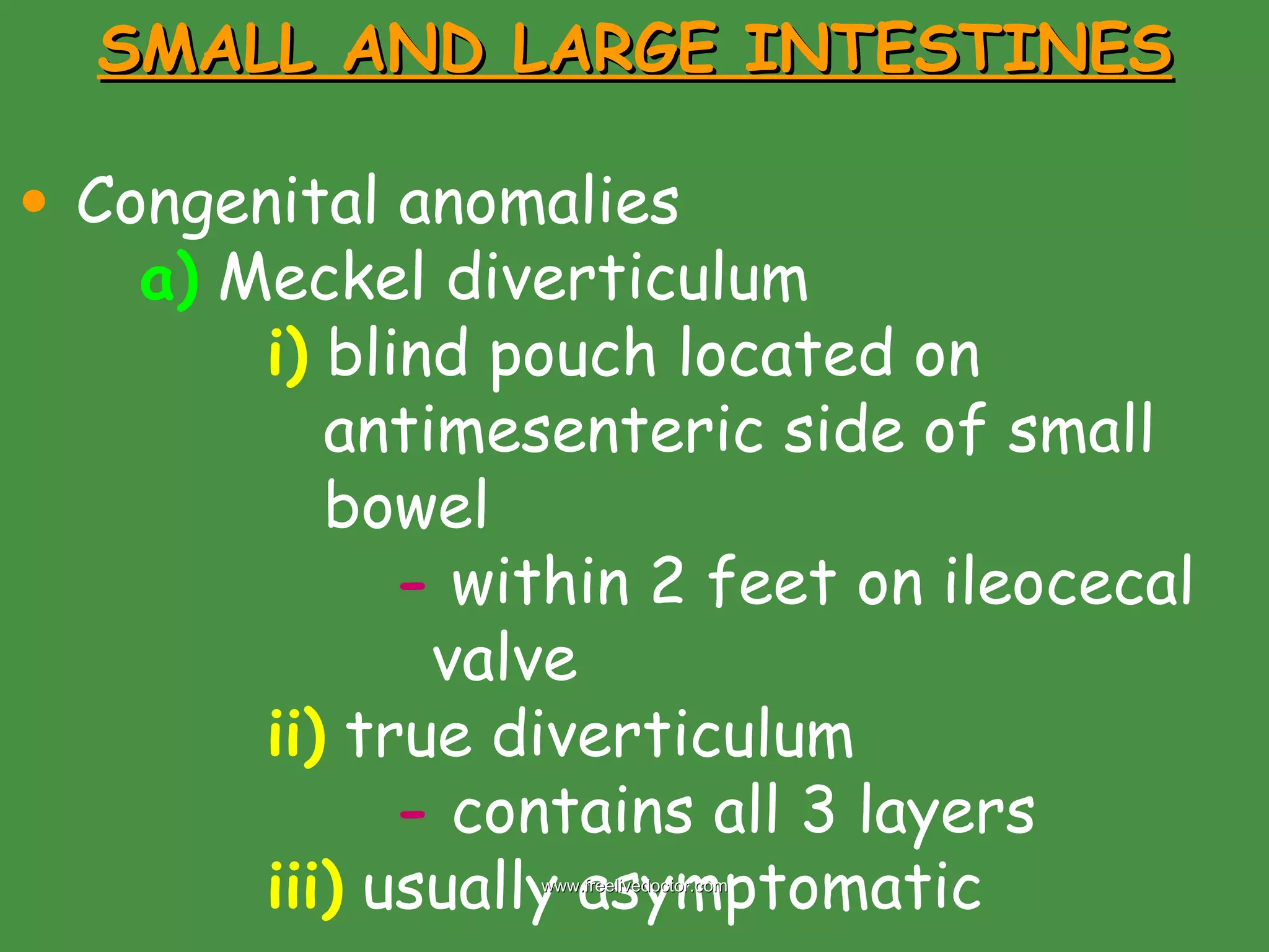
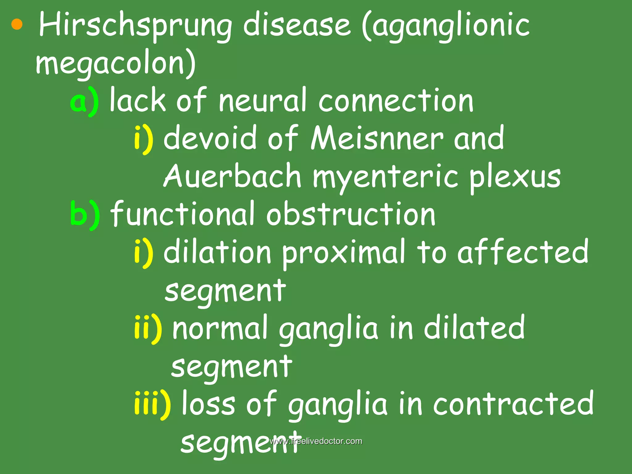
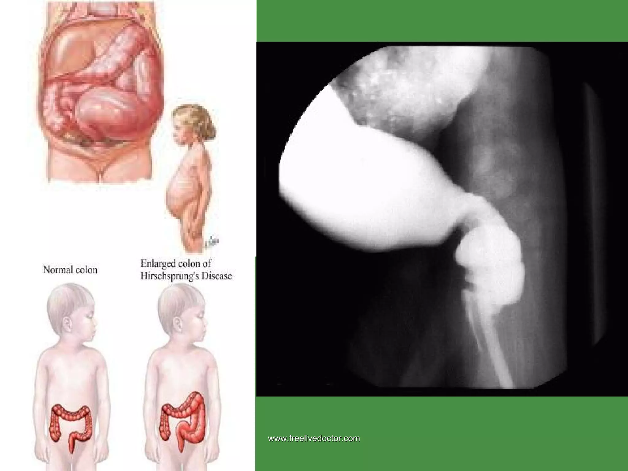
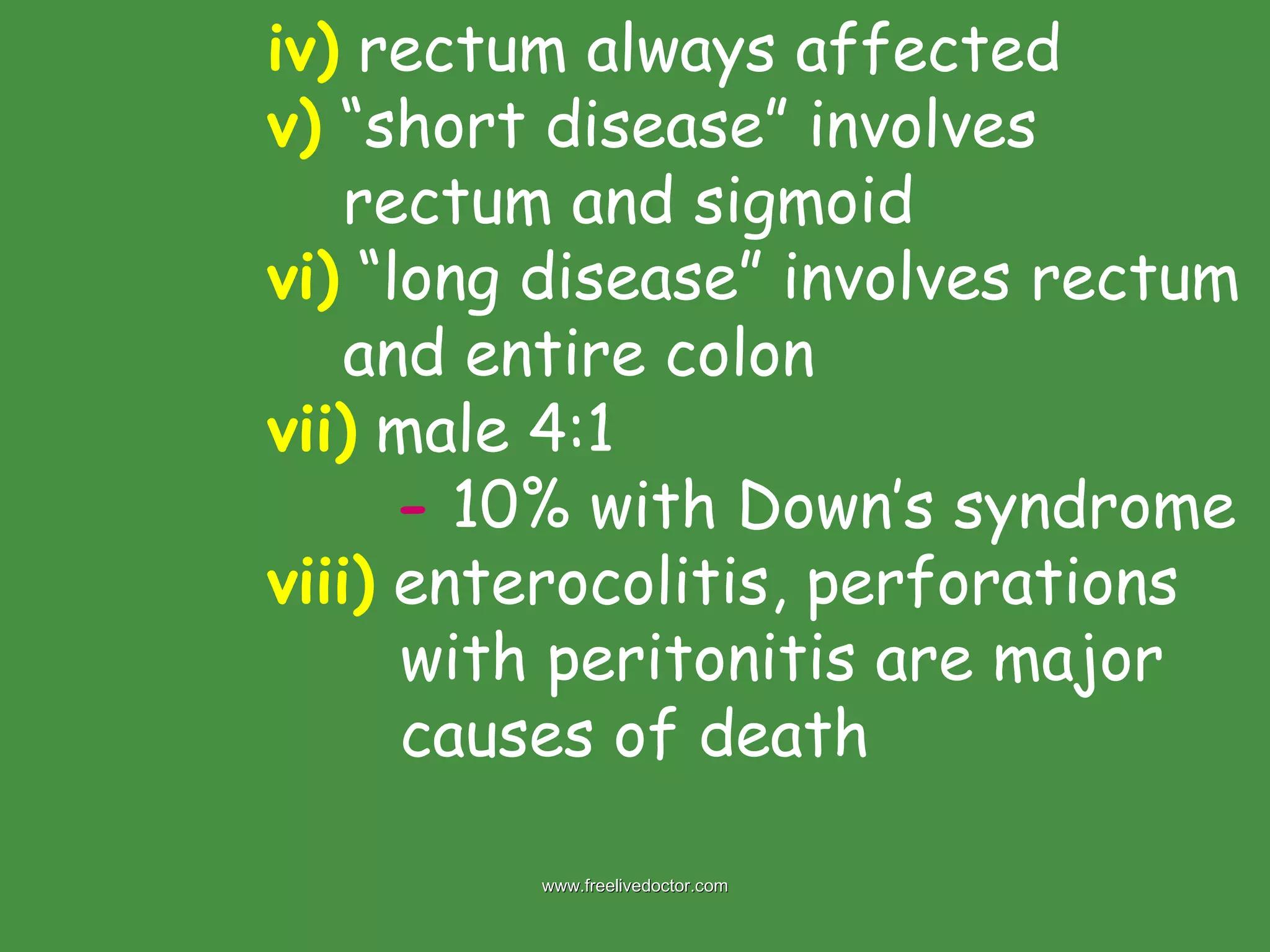
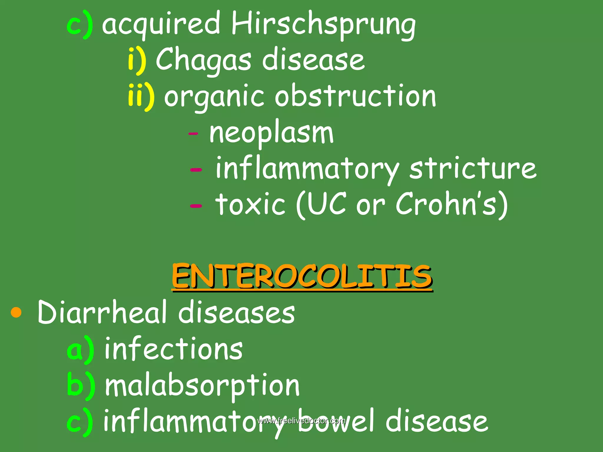
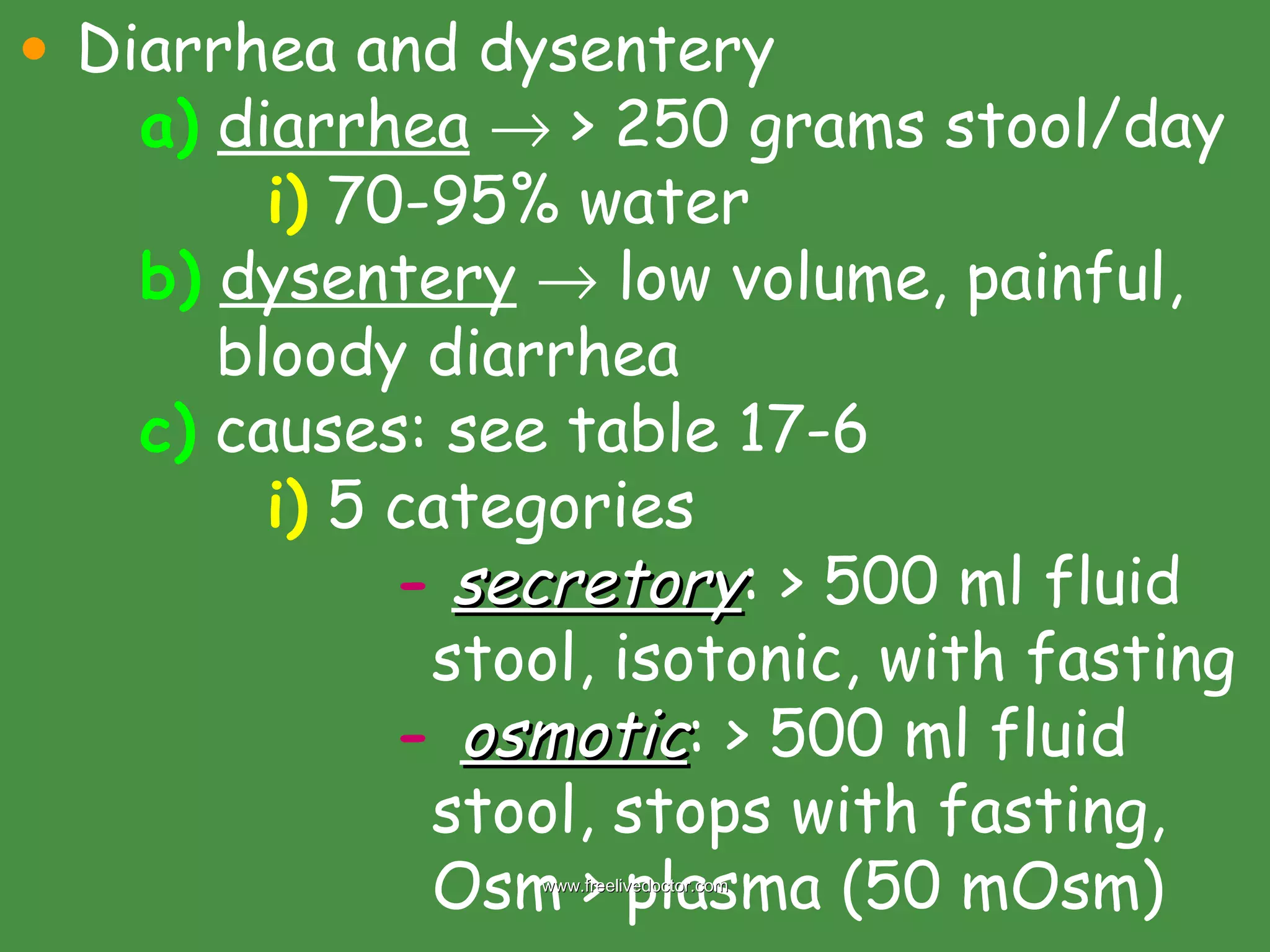
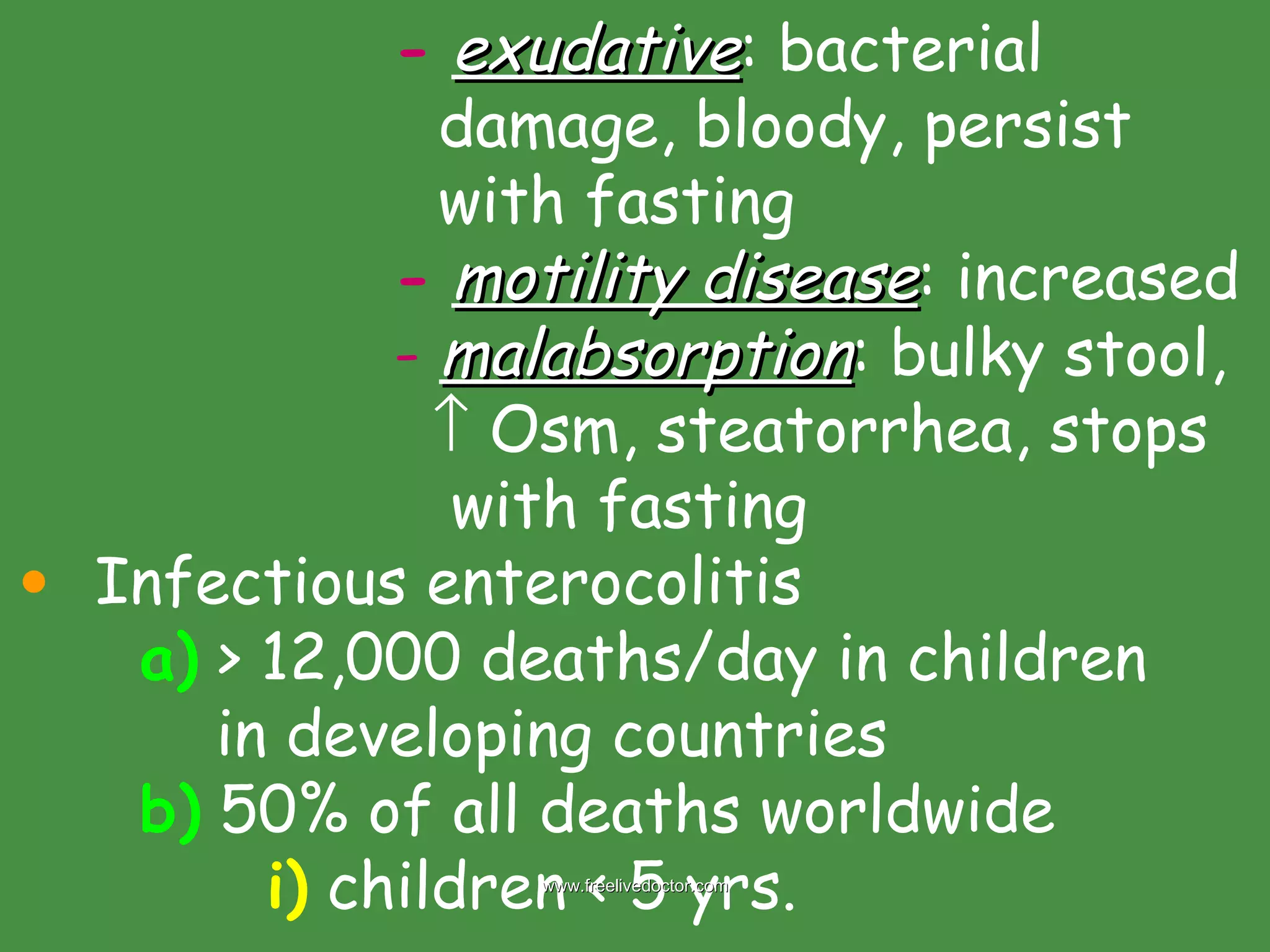
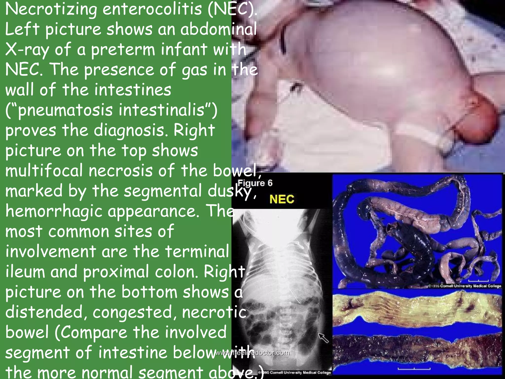
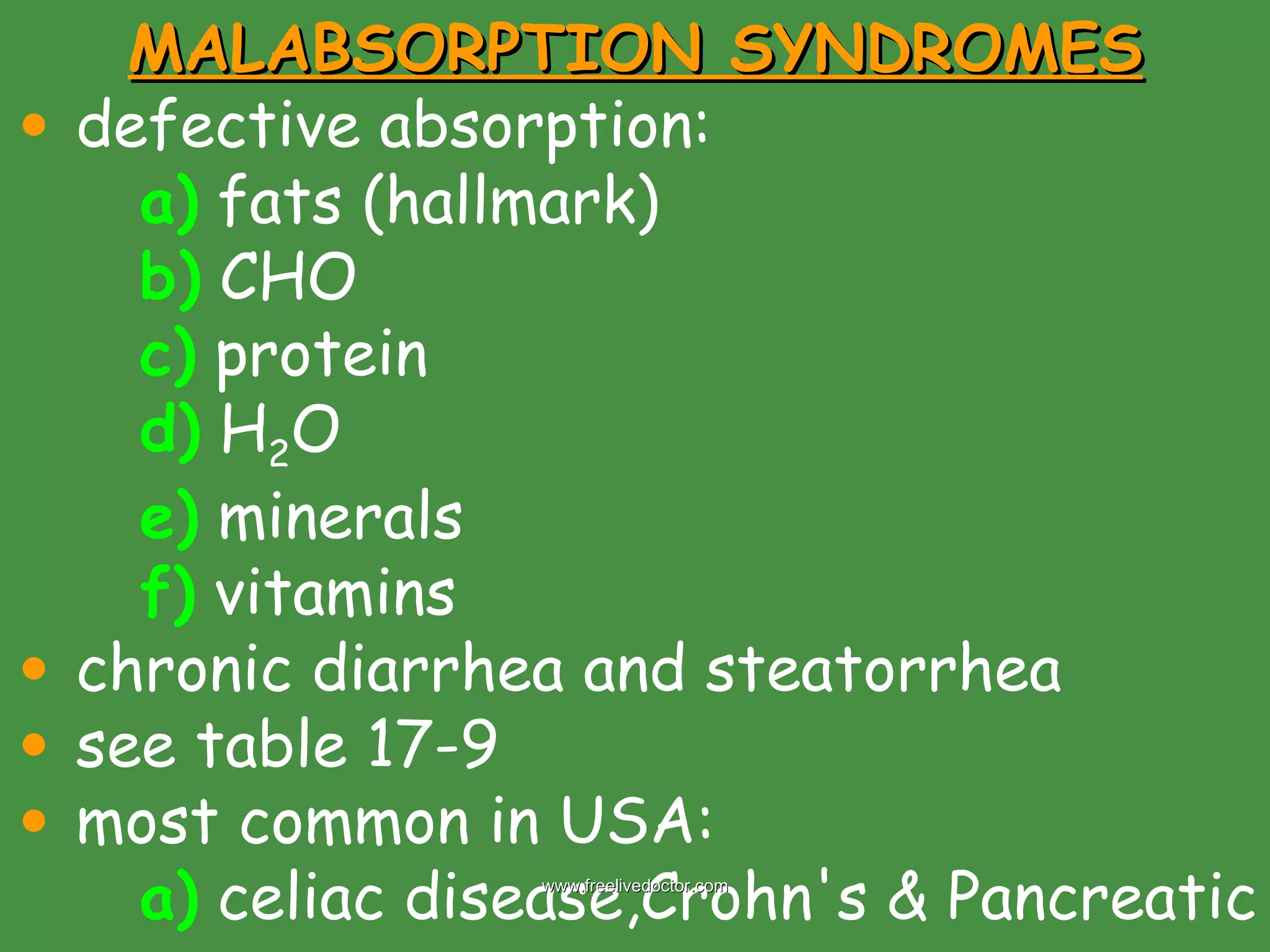
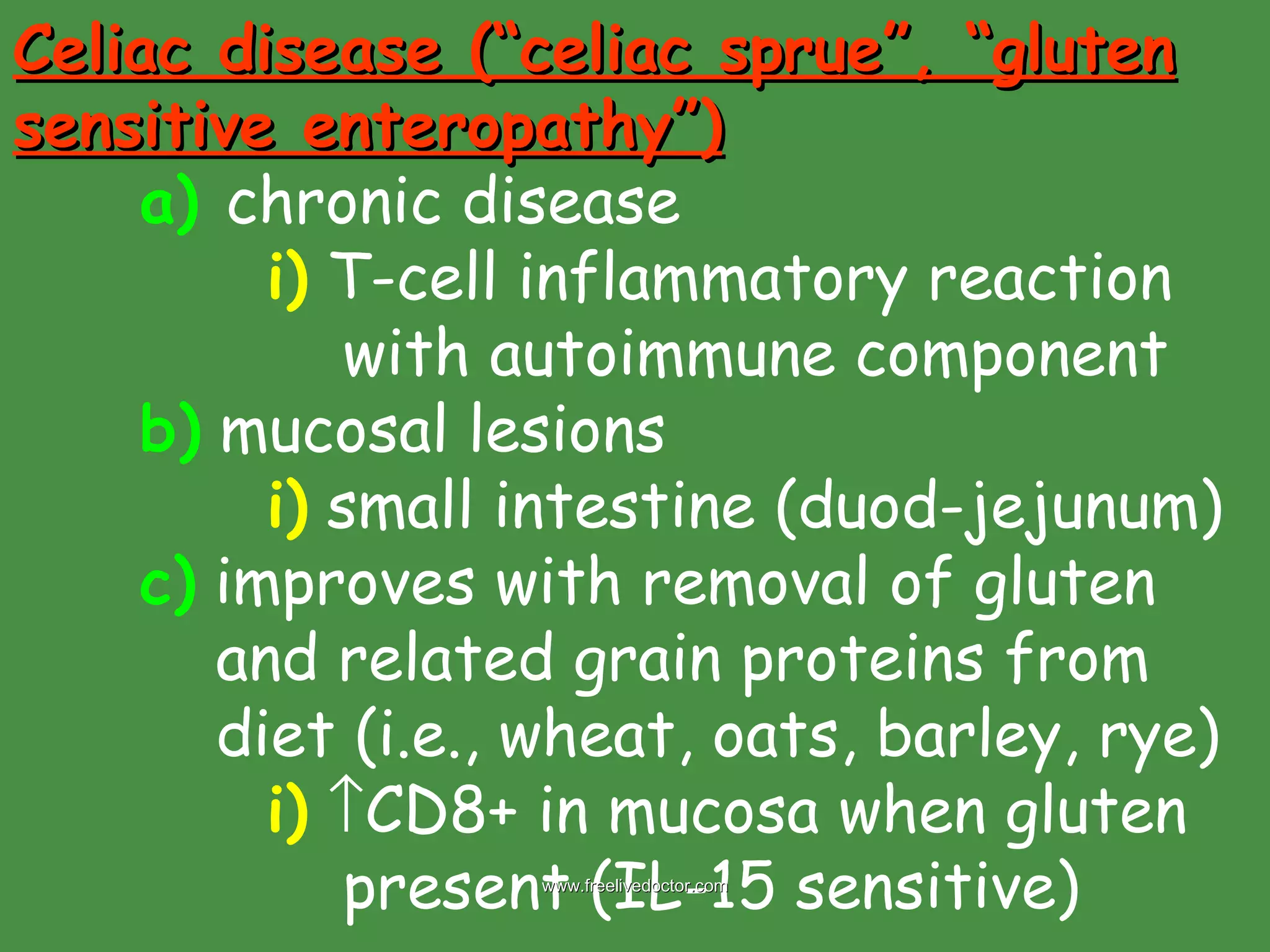
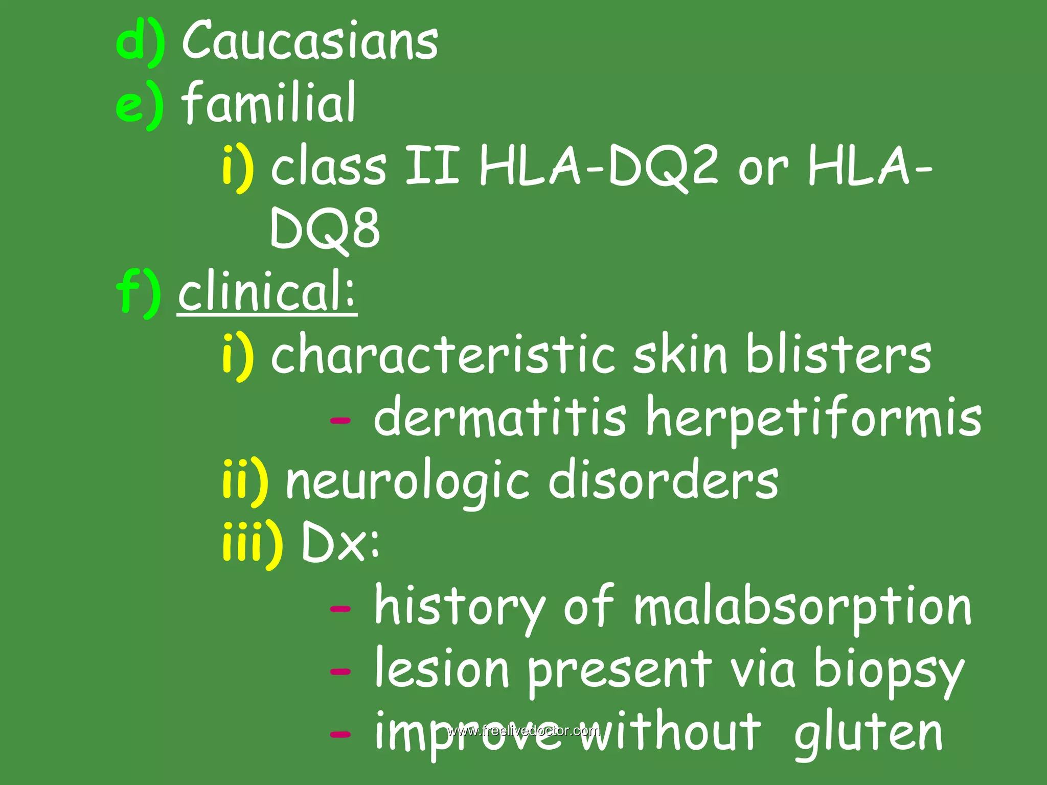
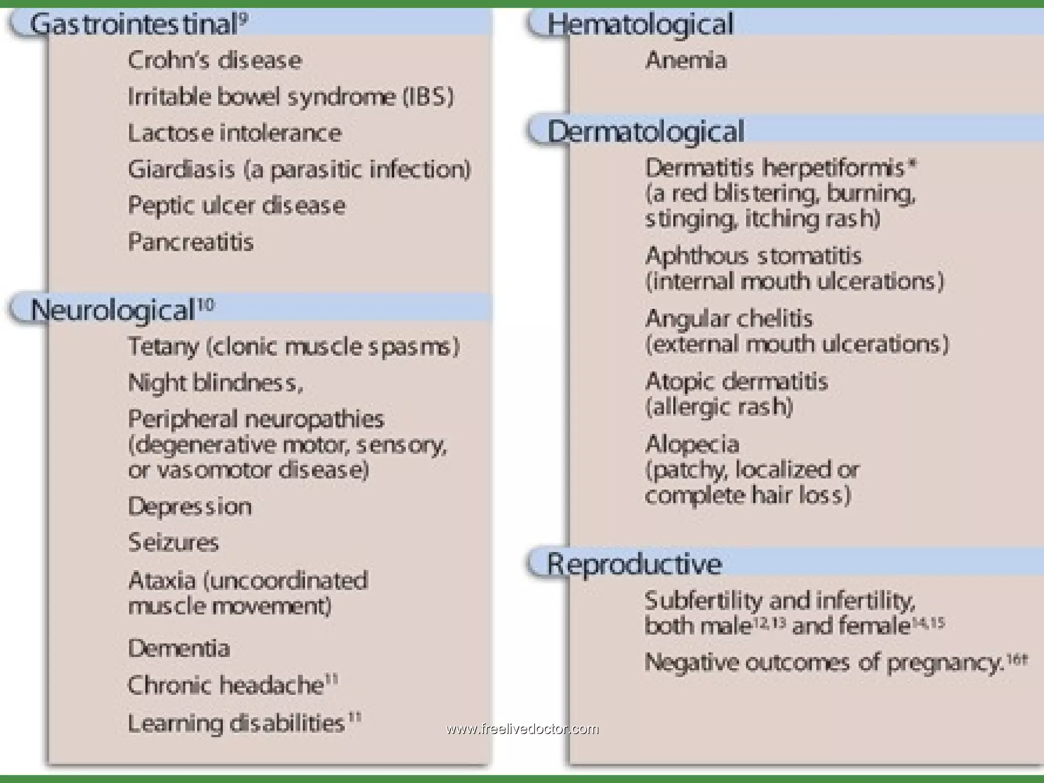
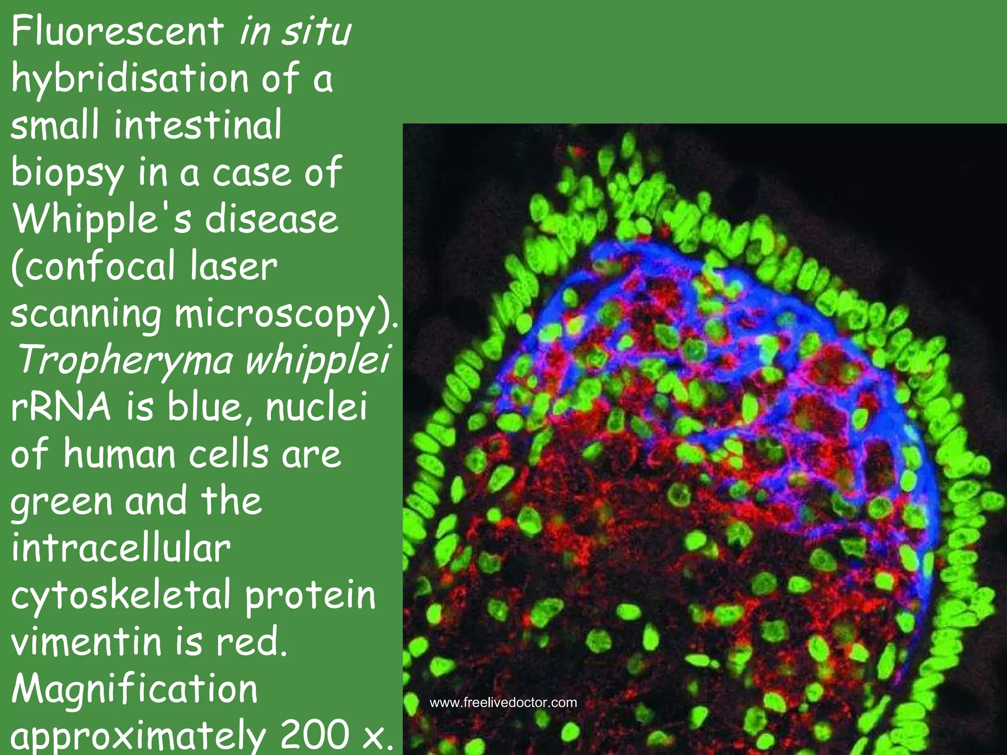
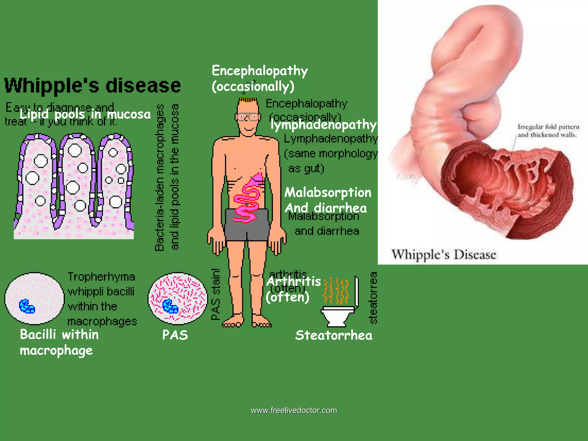
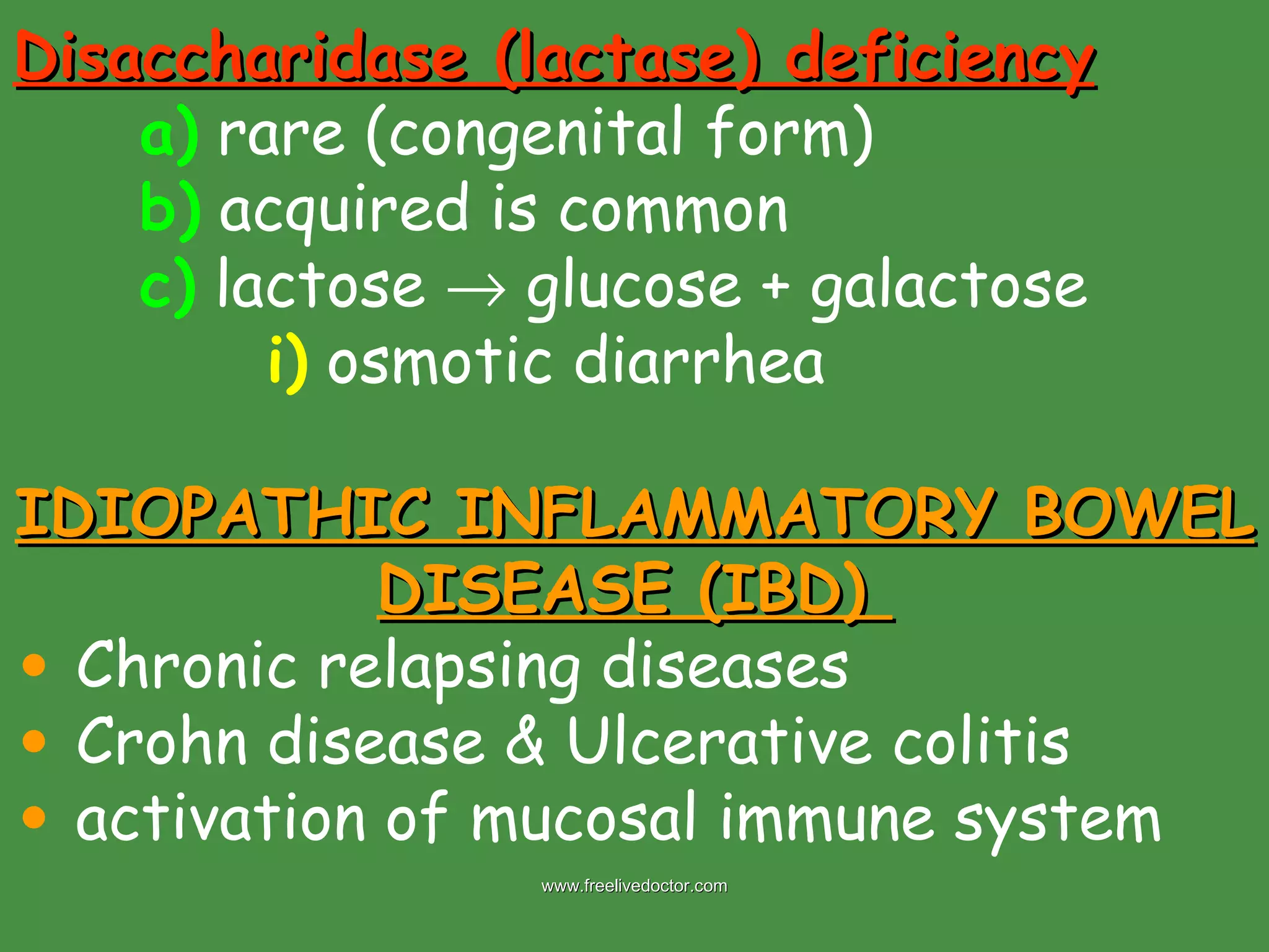
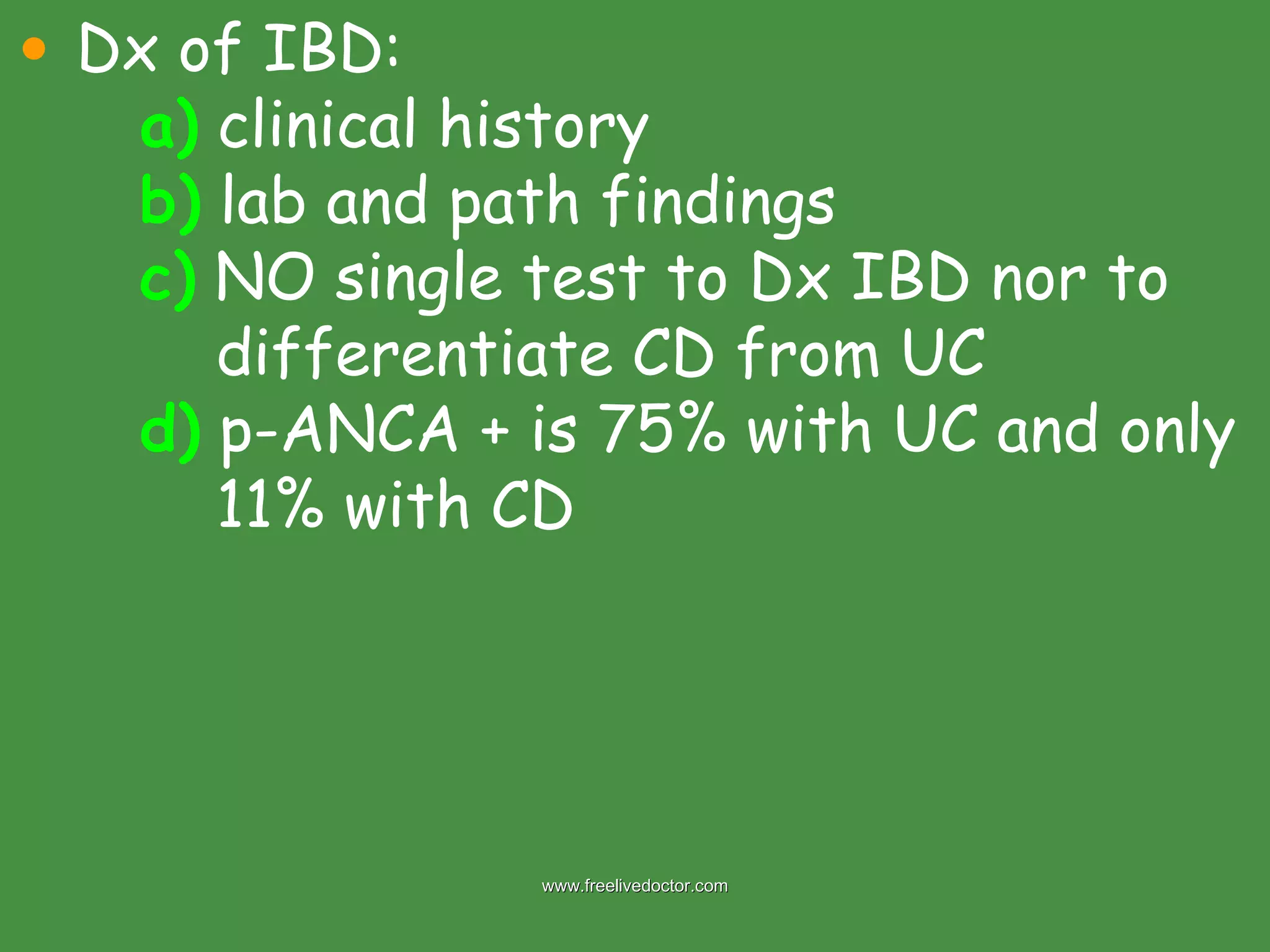
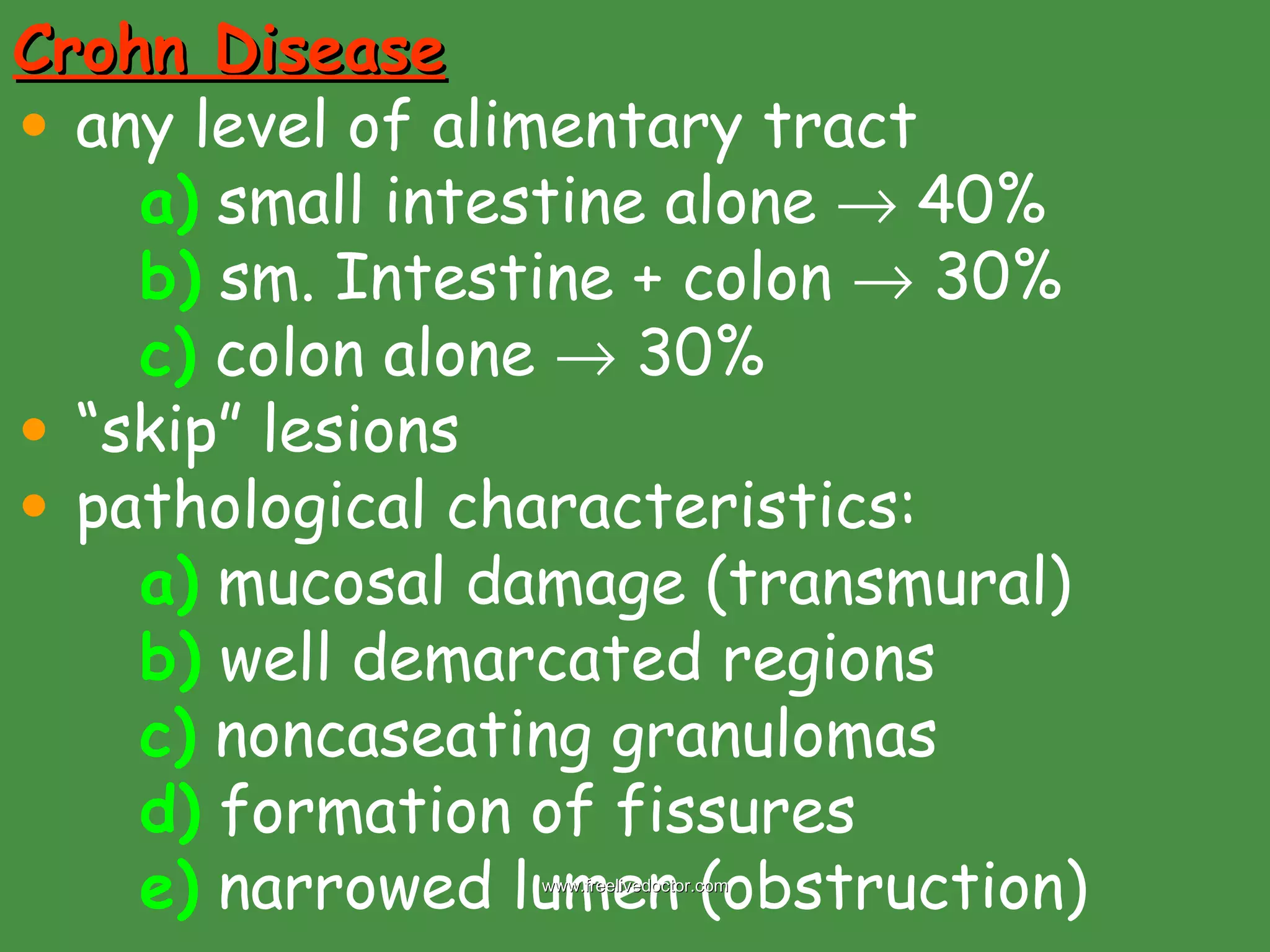
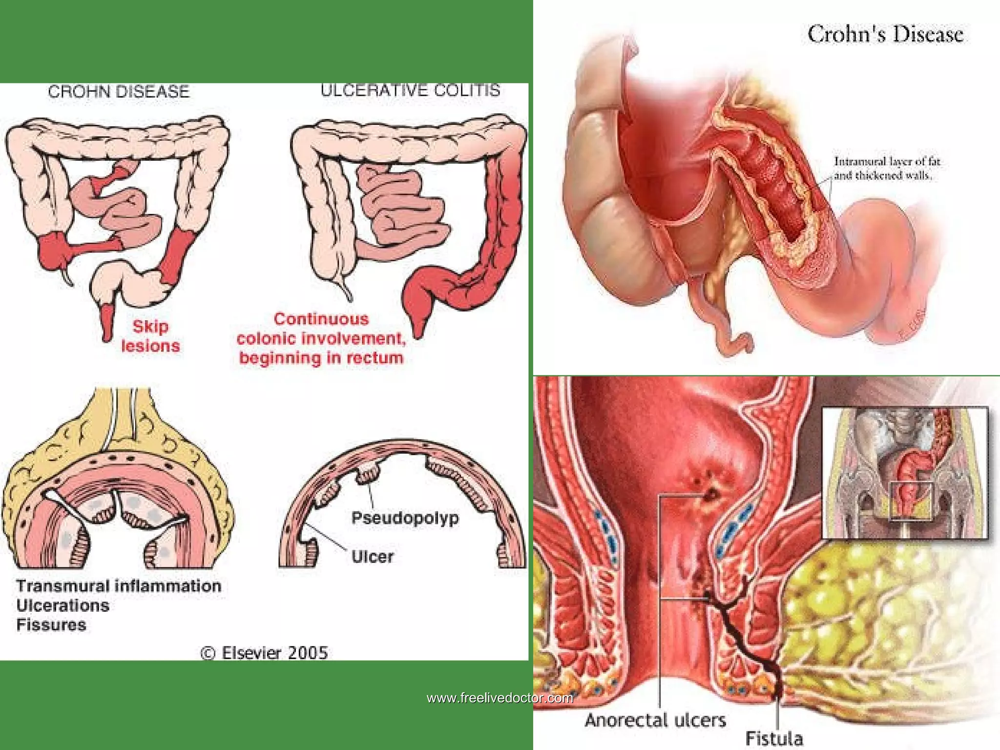
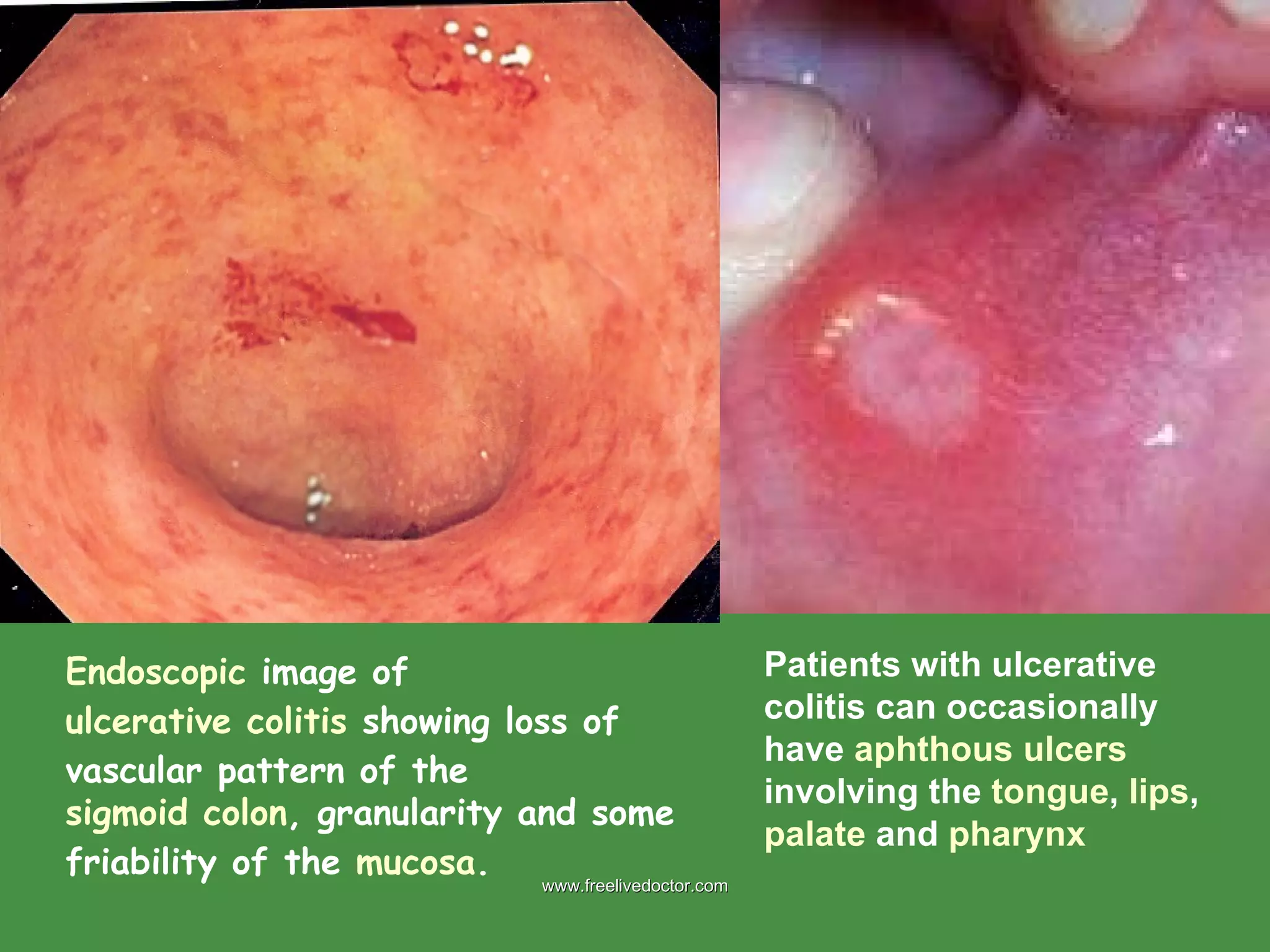
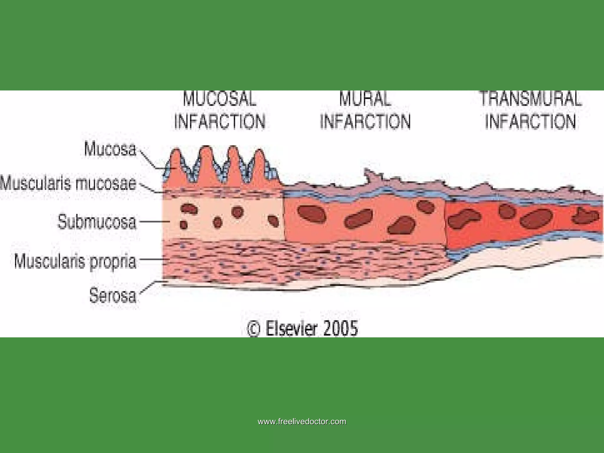
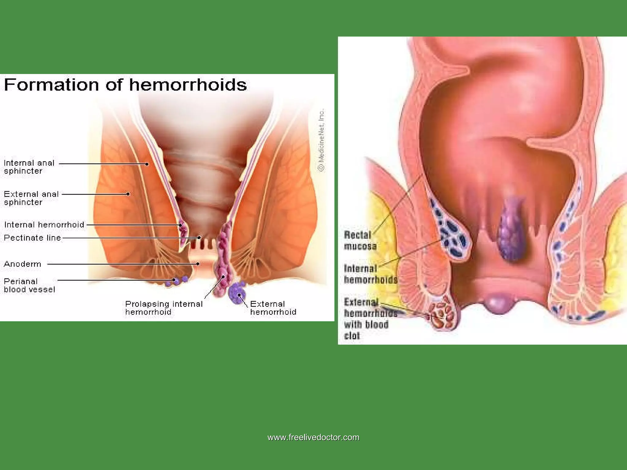
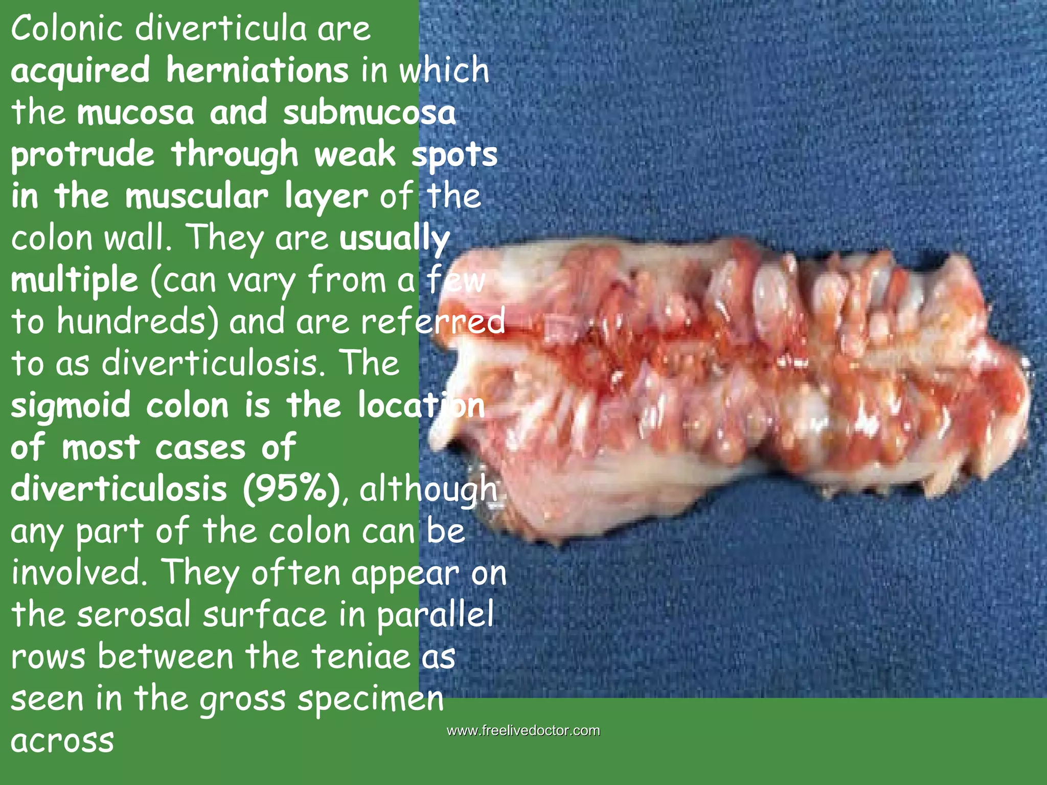
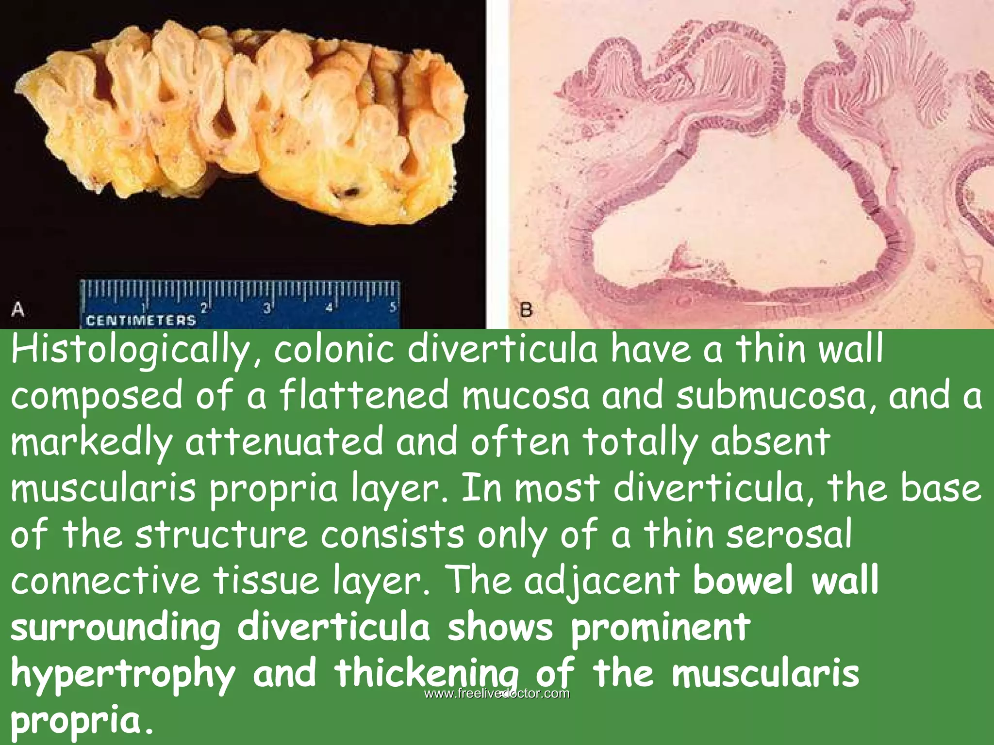
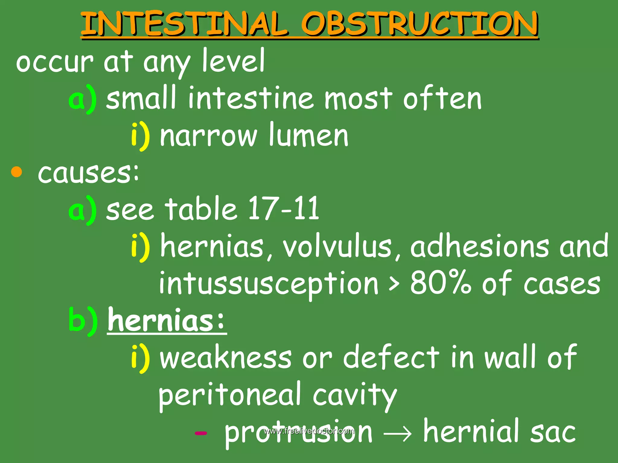
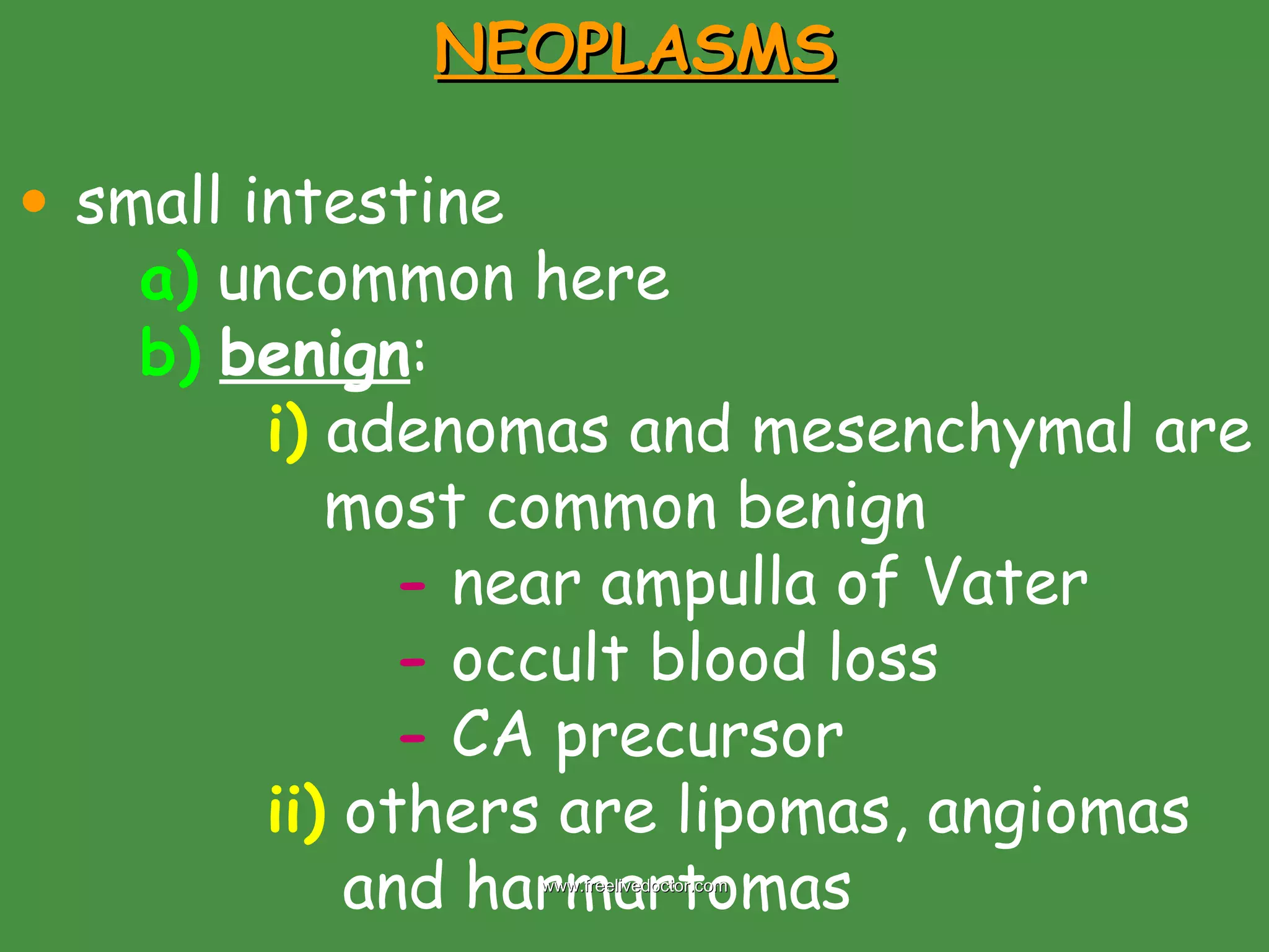
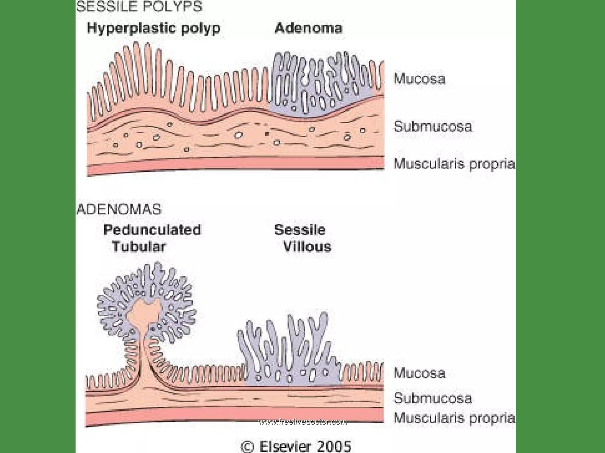
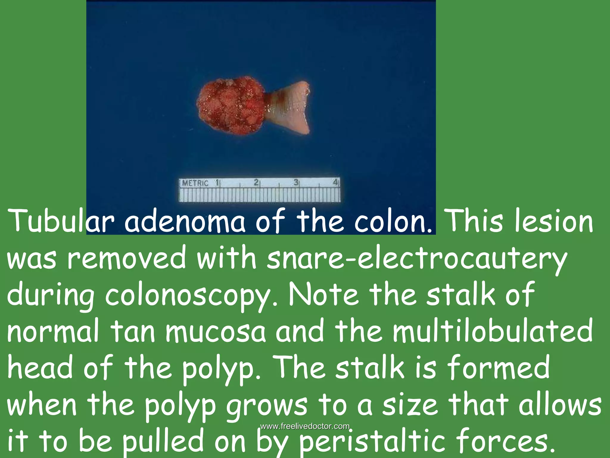
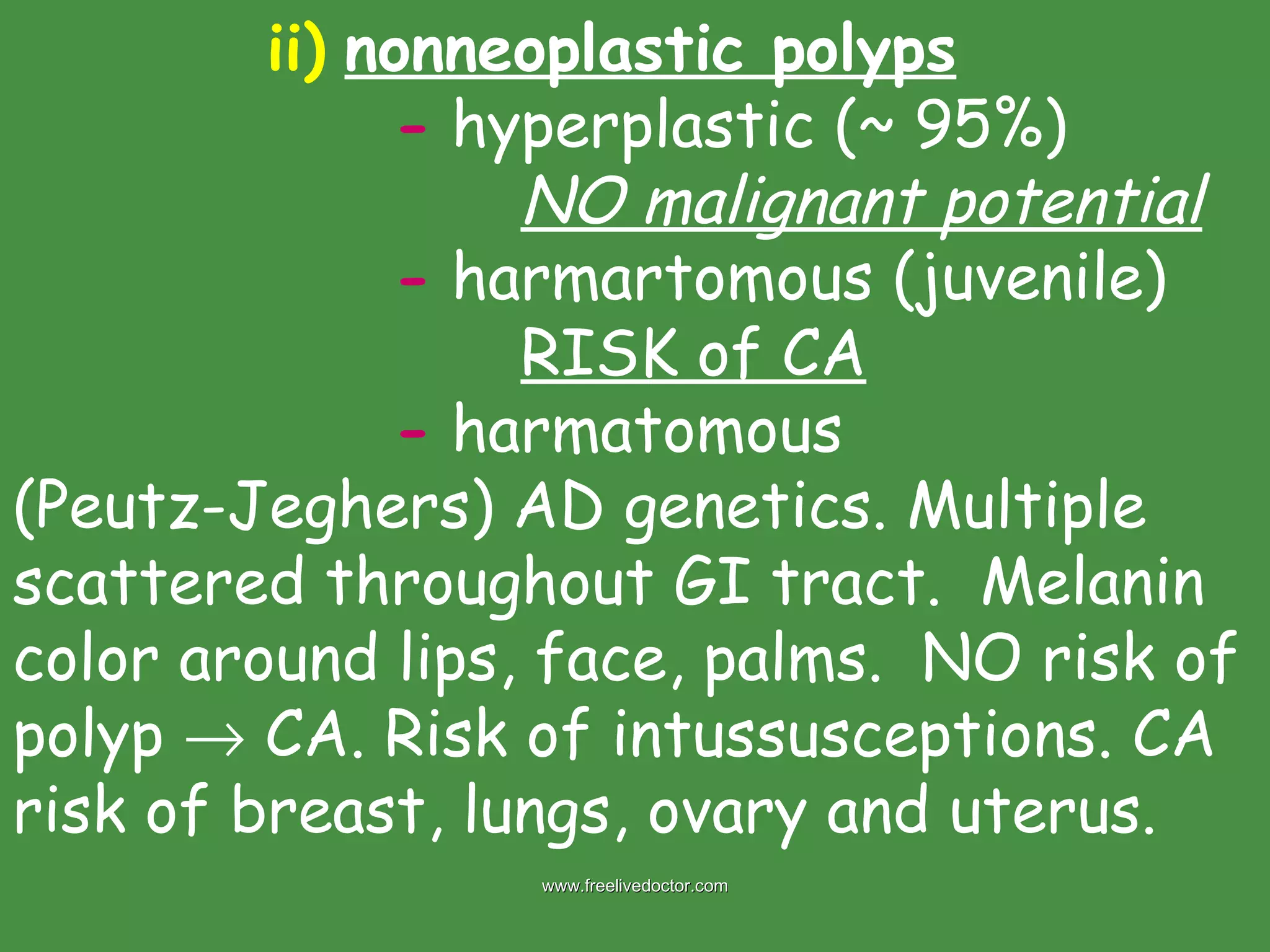
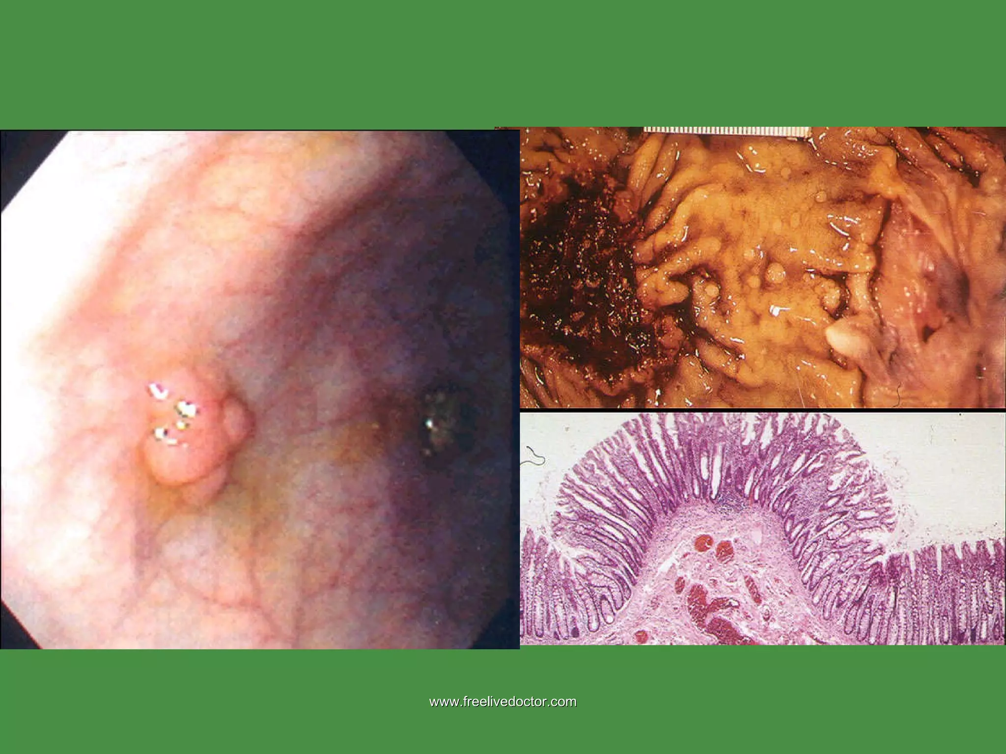
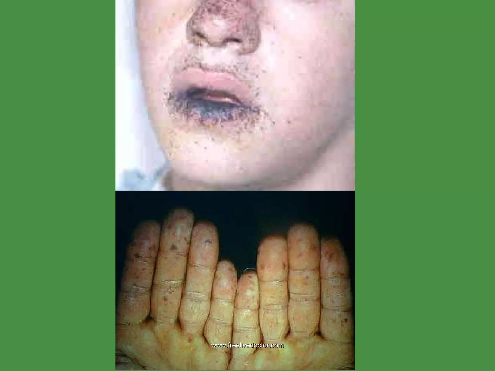
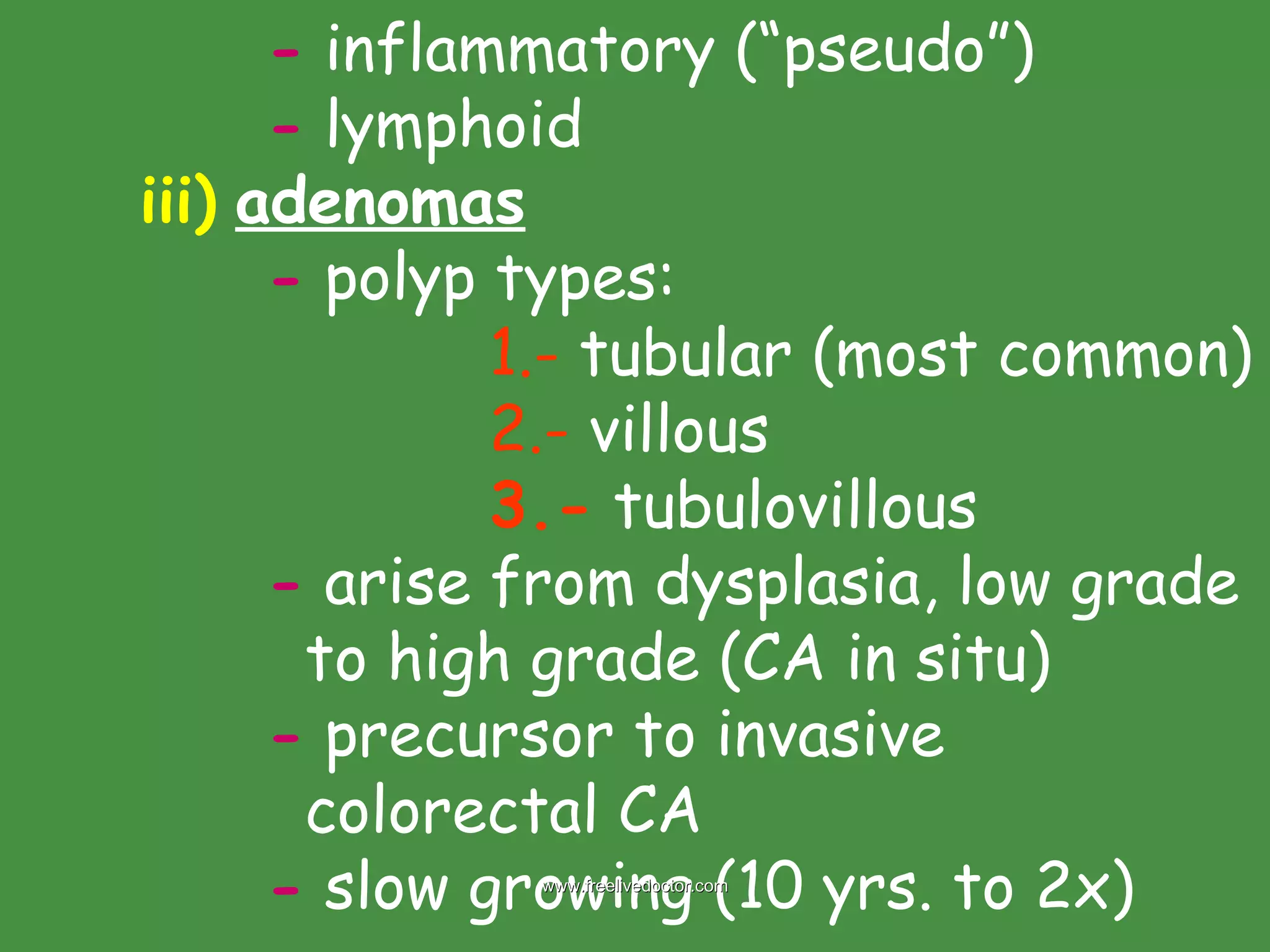
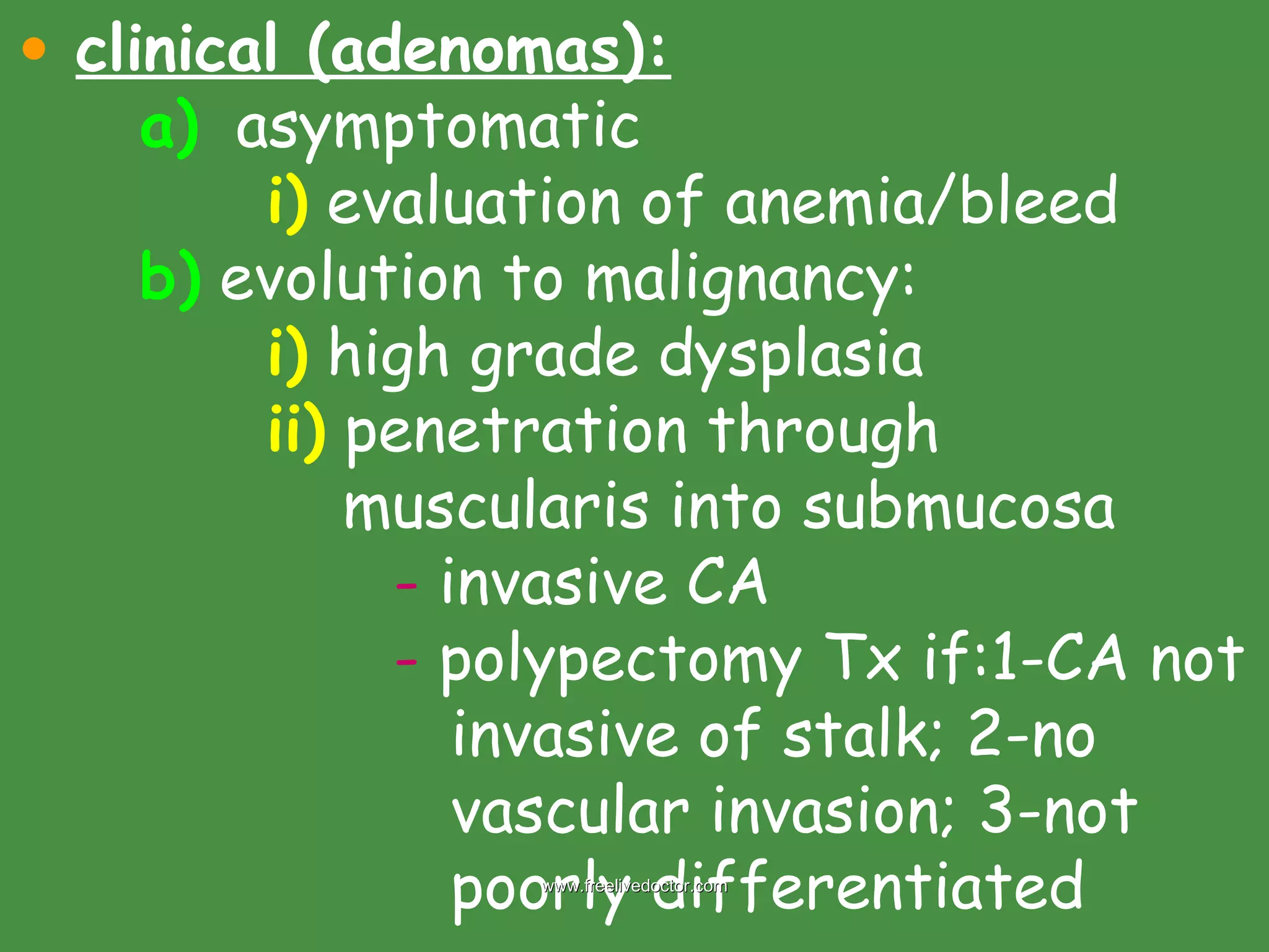
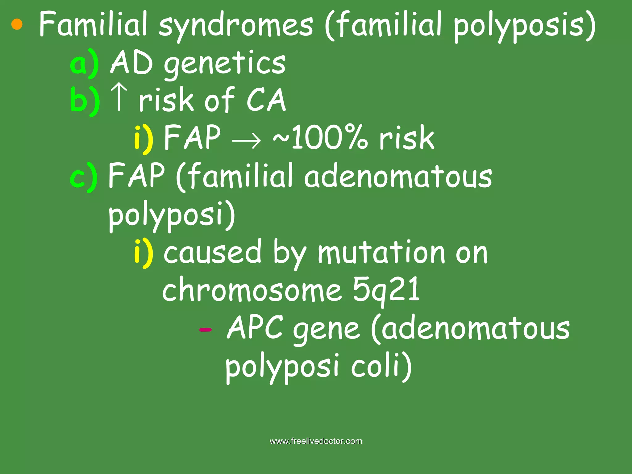
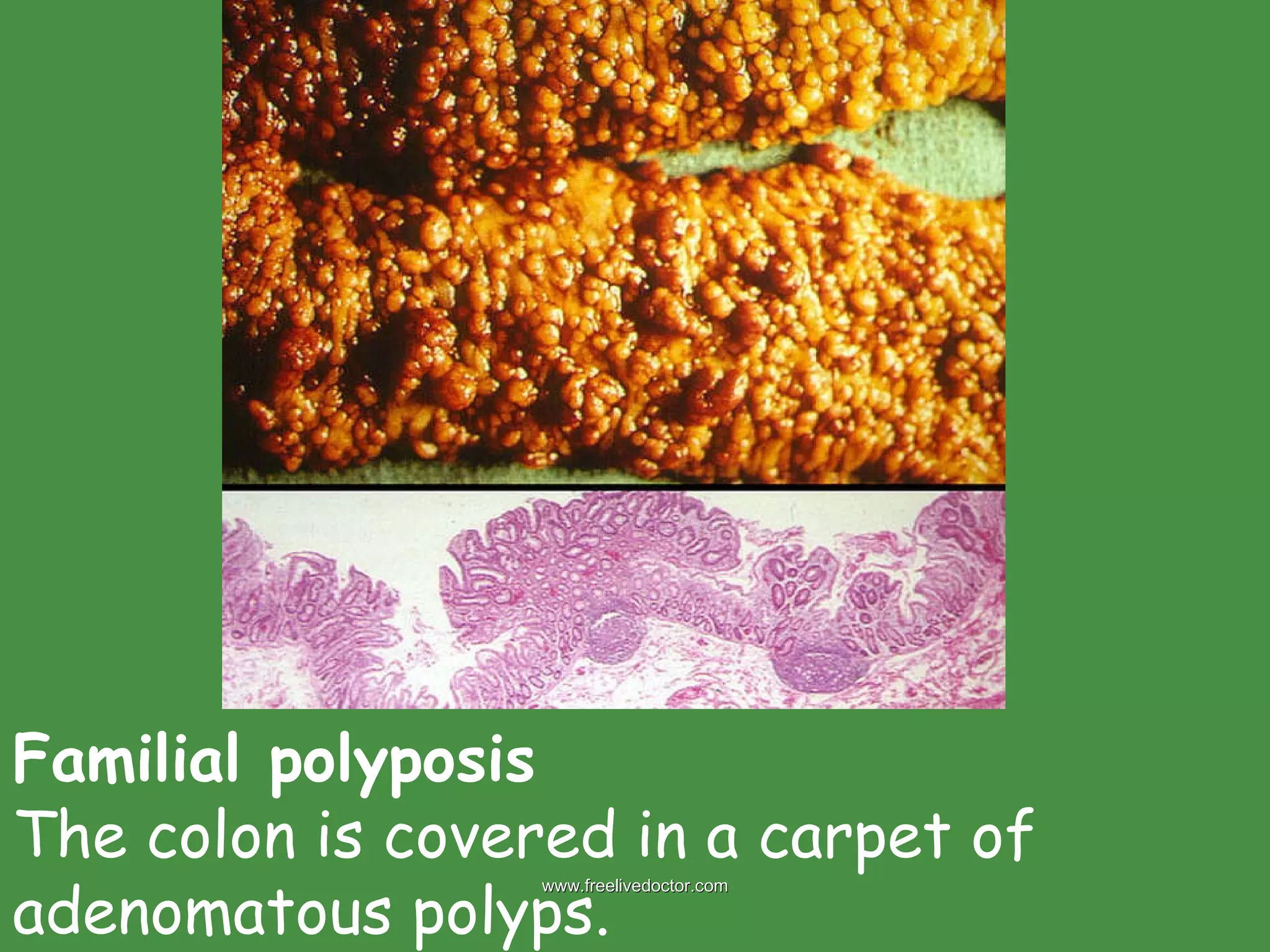
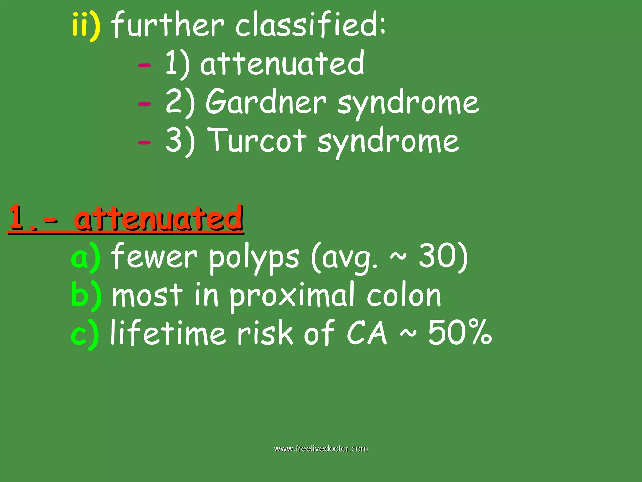
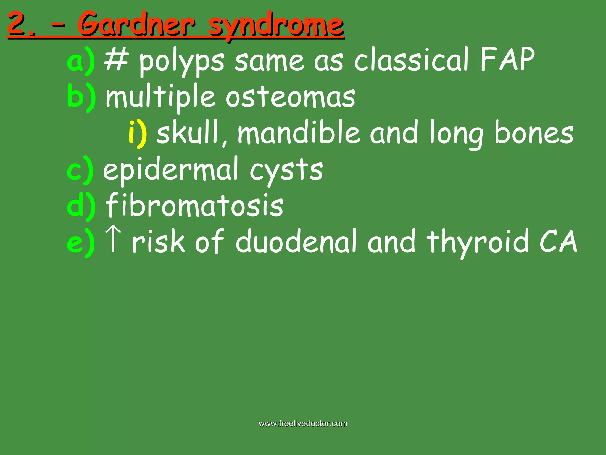
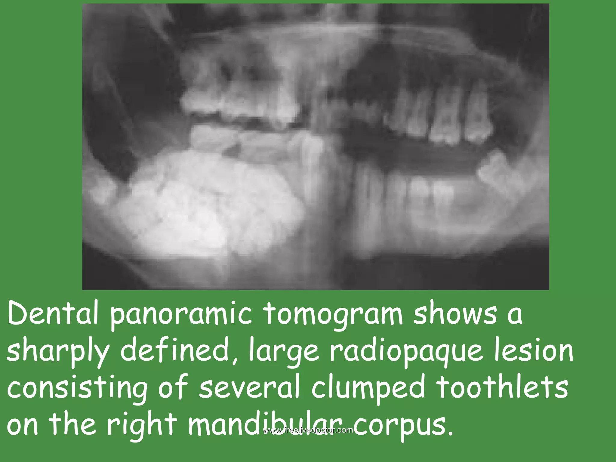
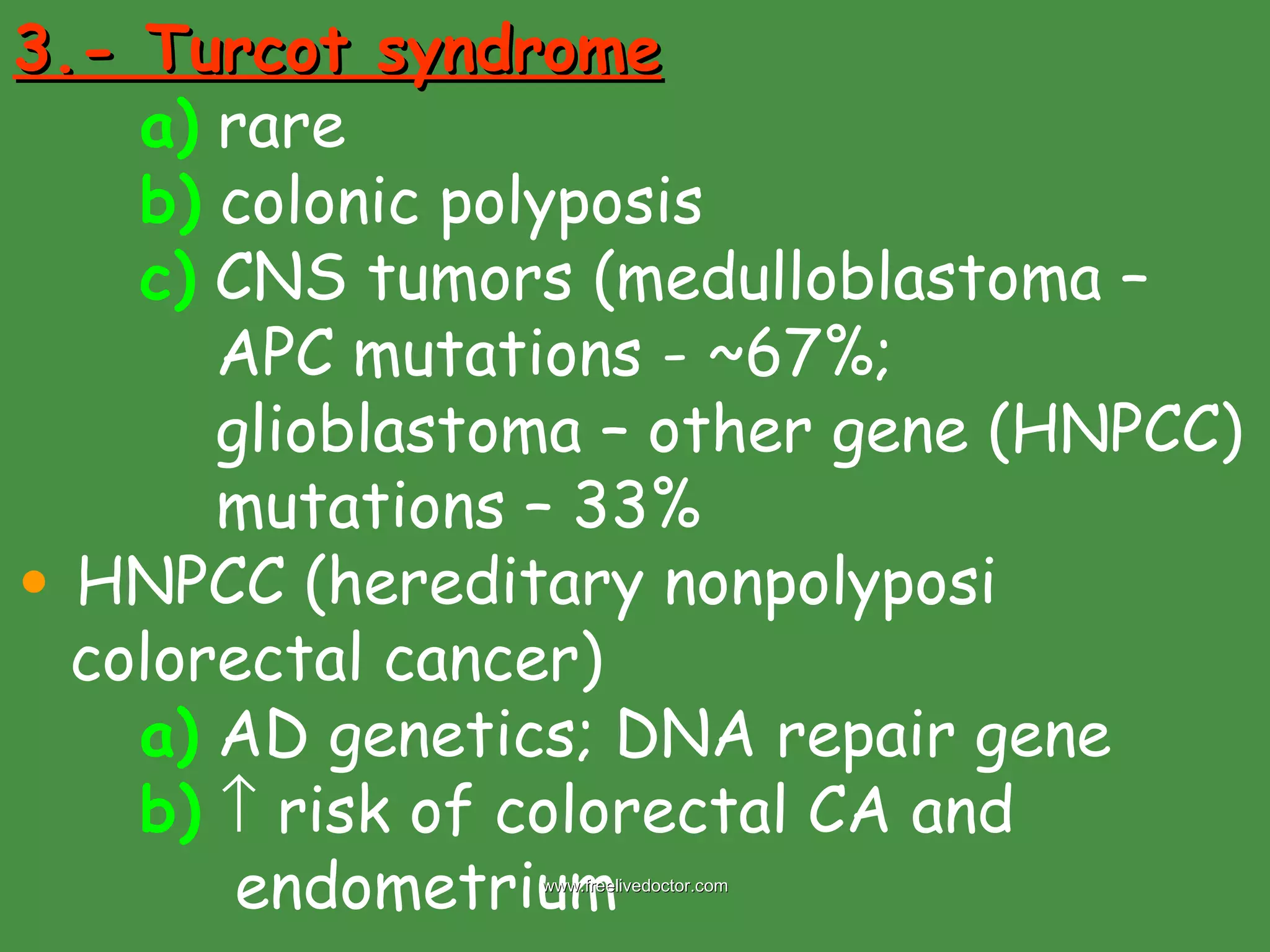
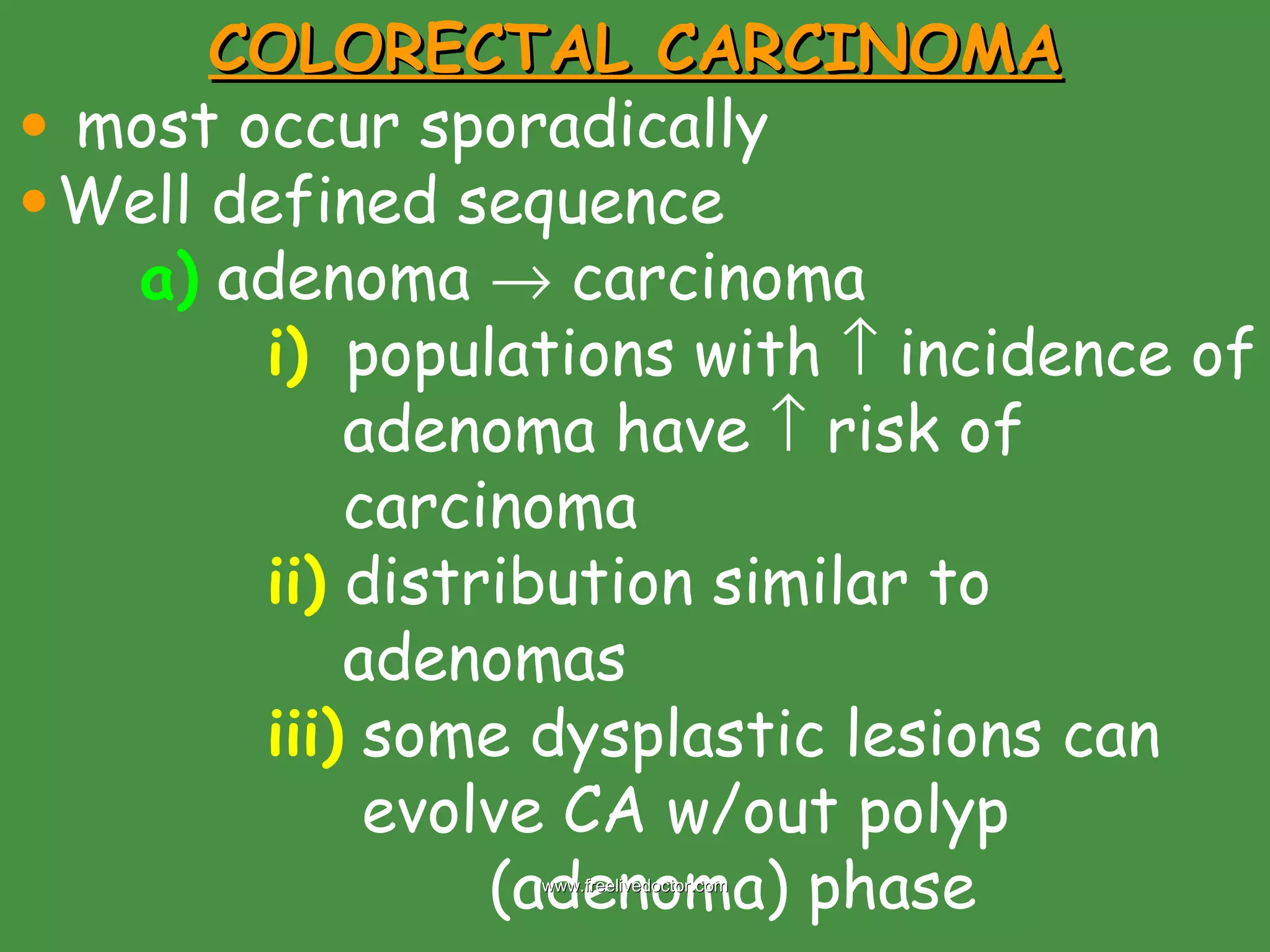
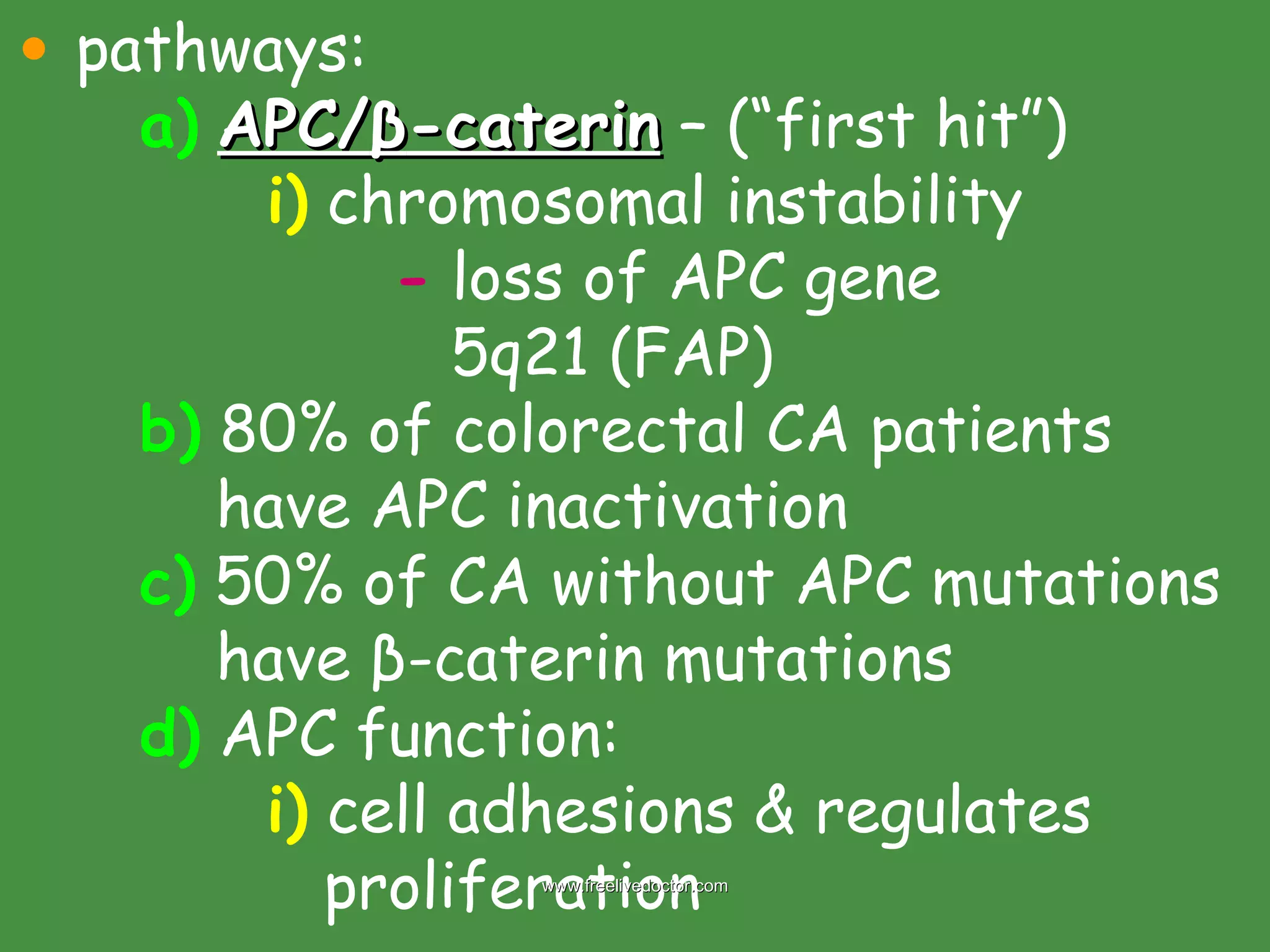
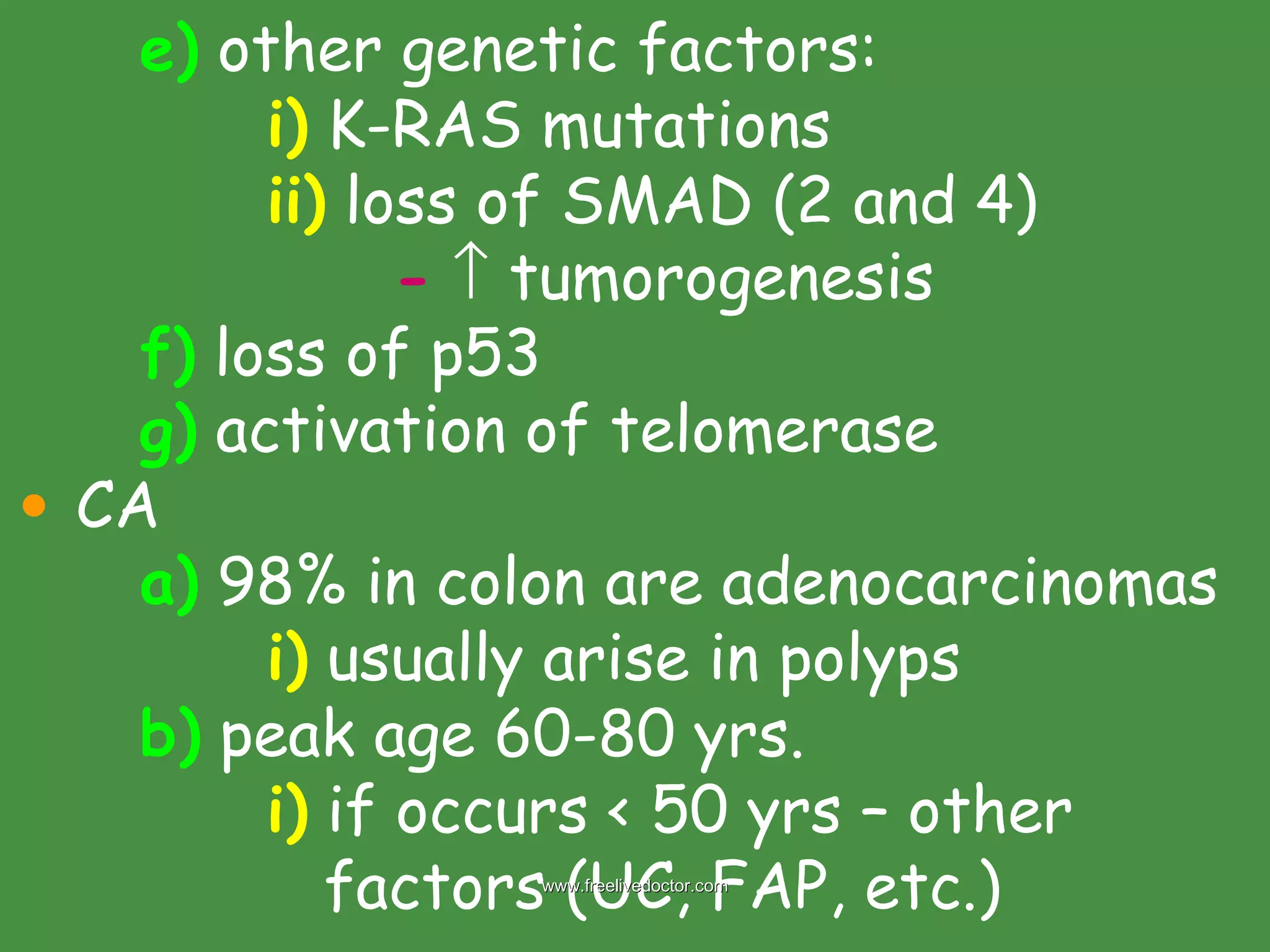
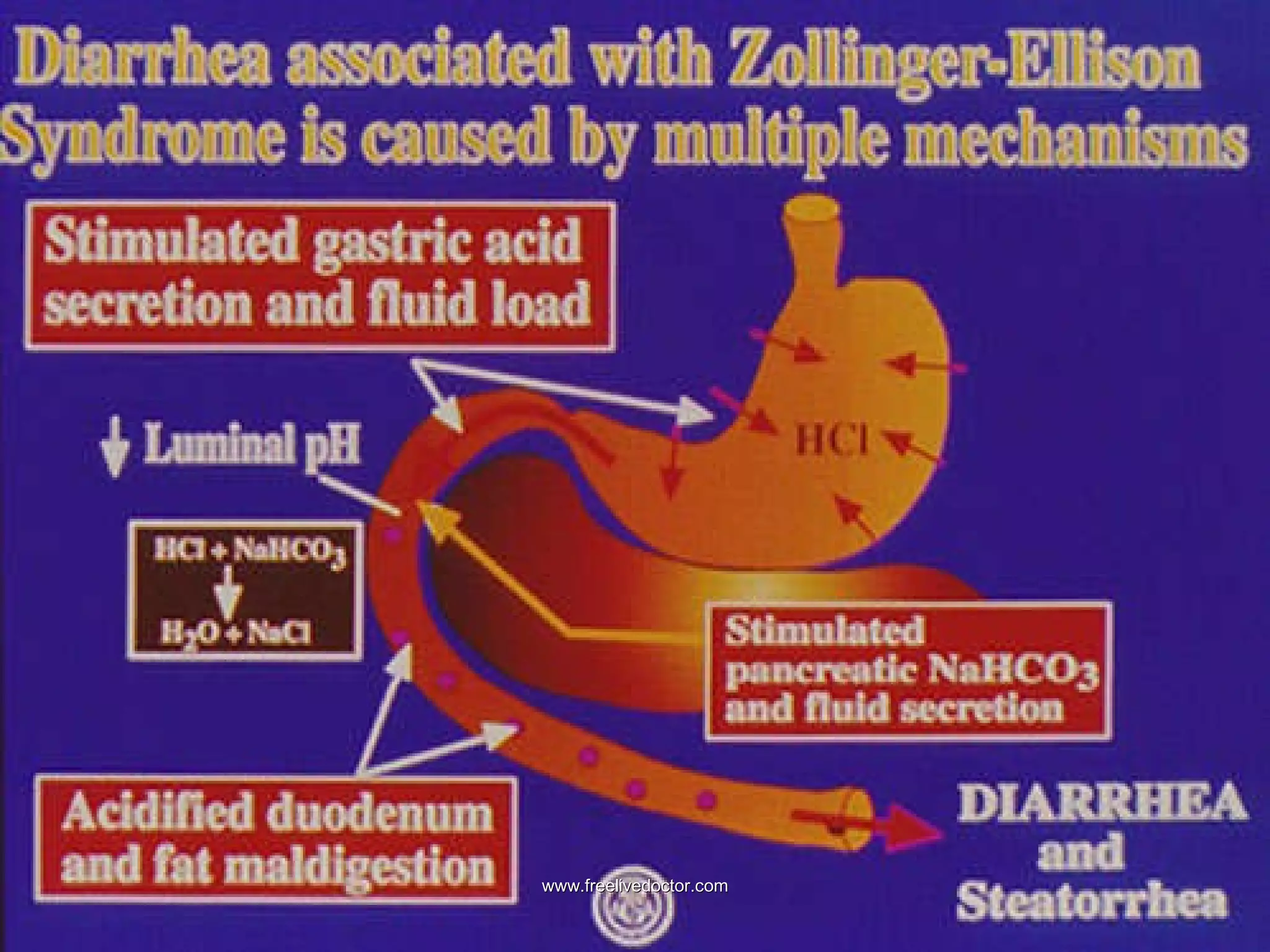
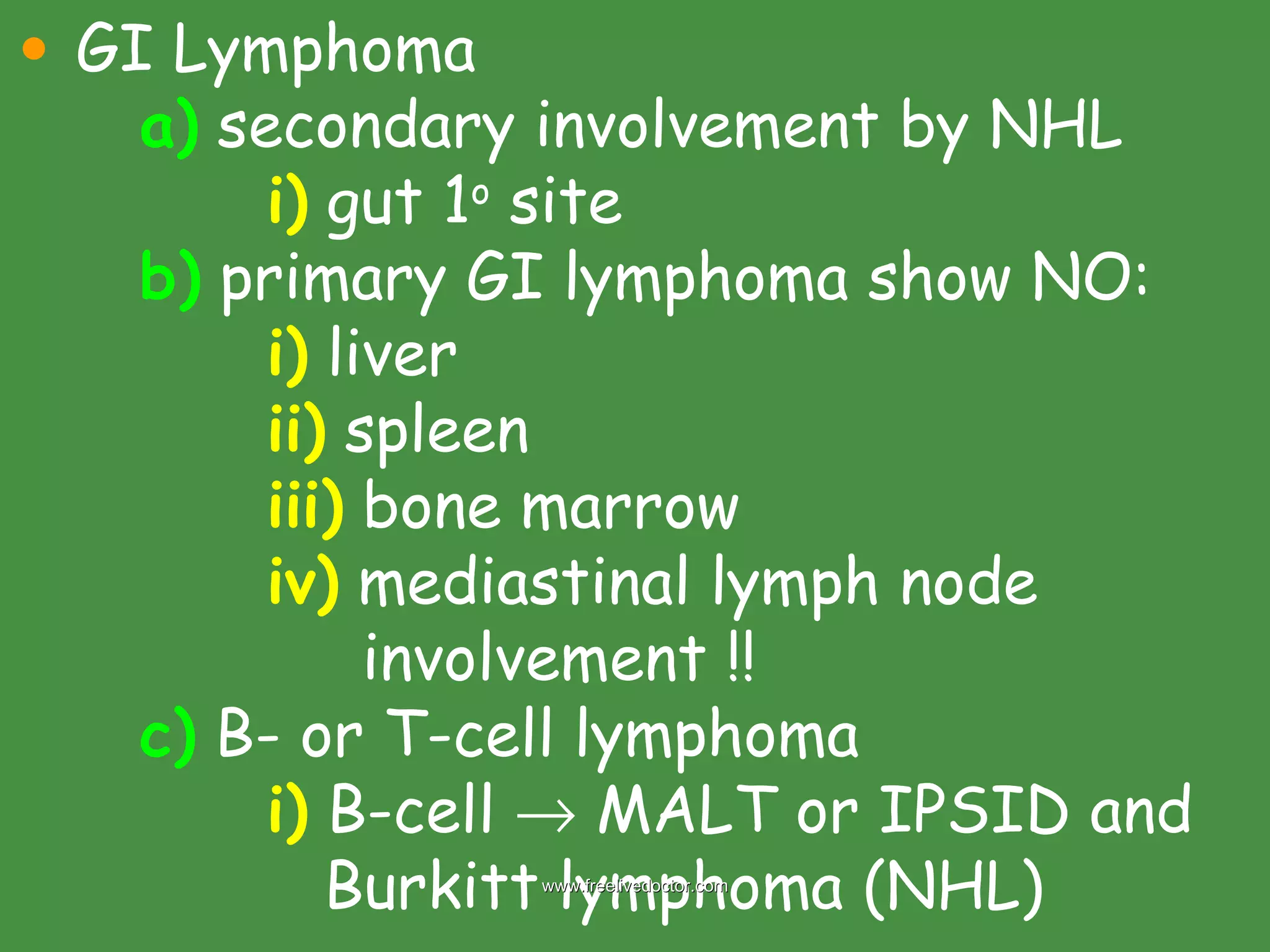
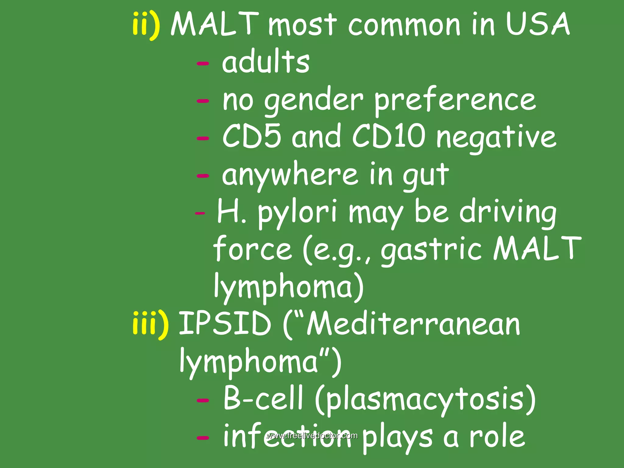
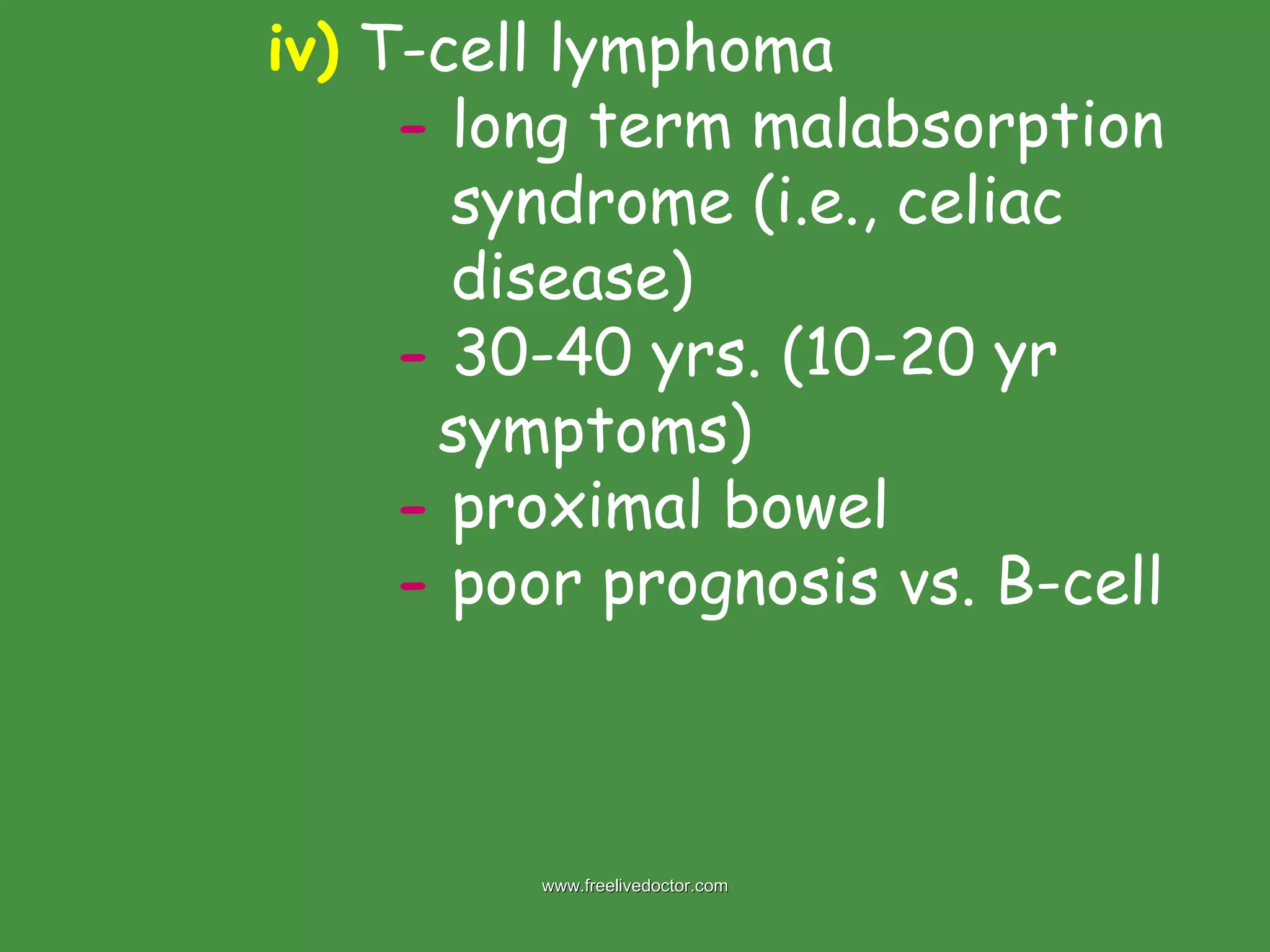
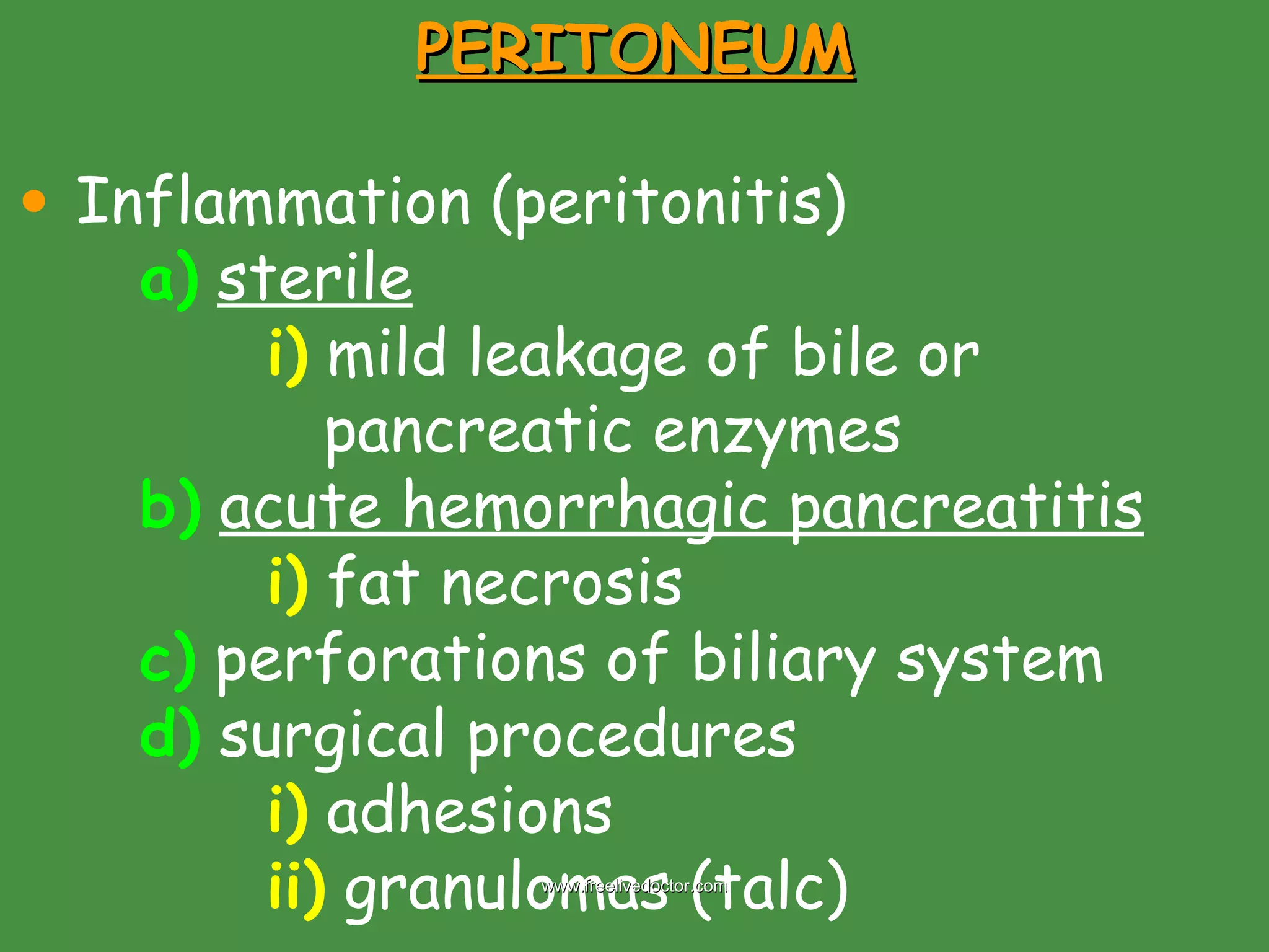
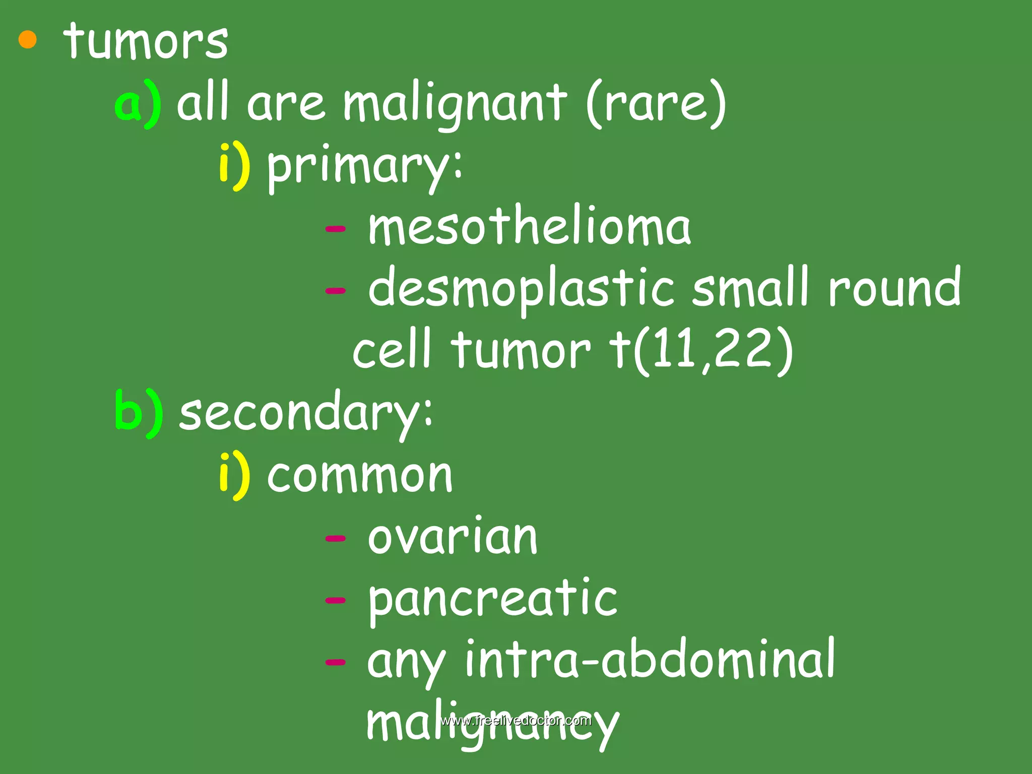
SMALL AND LARGE INTESTINES Congenital anomalies a)  Meckel diverticulum i)  blind pouch located on    antimesenteric side of small    bowel -  within 2 feet on ileocecal    valve ii)  true diverticulum -  contains all 3 layers iii)  usually asymptomatic www.freelivedoctor.com
www.freelivedoctor.com
Hirschsprung disease (aganglionic megacolon) a)  lack of neural connection i)  devoid of Meisnner and    Auerbach myenteric plexus b)  functional obstruction i)  dilation proximal to affected    segment ii)  normal ganglia in dilated    segment iii)  loss of ganglia in contracted    segment www.freelivedoctor.com
www.freelivedoctor.com
iv)  rectum always affected v)  “short disease” involves   rectum and sigmoid vi)  “long disease” involves rectum   and entire colon vii)  male 4:1 -  10% with Down’s syndrome viii)  enterocolitis, perforations    with peritonitis are major    causes of death www.freelivedoctor.com
c)  acquired Hirschsprung i)  Chagas disease ii)  organic obstruction -  neoplasm -  inflammatory stricture -  toxic (UC or Crohn’s) ENTEROCOLITIS Diarrheal diseases a)  infections b)  malabsorption c)  inflammatory bowel disease www.freelivedoctor.com
Diarrhea and dysentery  a)   diarrhea     > 250 grams stool/day i)  70-95% water b)   dysentery     low volume, painful,    bloody diarrhea c)  causes: see table 17-6 i)  5 categories -   secretory : > 500 ml fluid    stool, isotonic, with fasting -  osmotic : > 500 ml fluid    stool, stops with fasting,    Osm > plasma (50 mOsm) www.freelivedoctor.com
-   exudative : bacterial    damage, bloody, persist    with fasting -   motility disease : increased -   malabsorption : bulky stool,       Osm, steatorrhea, stops    with fasting Infectious enterocolitis a)  > 12,000 deaths/day in children   in developing countries b)  50% of all deaths worldwide i)  children < 5 yrs.  www.freelivedoctor.com
ii)  self-limited diarrhea mostly    caused by viruses c)   viral gastroenteritis i)  see table 17-7 ii)   rotavirus   -  children 6-24 months -  young children &    debilitated adults -  selectively destroys    enterocytes in small    intestine    malabsorption,   secretory and Osm diarrhea www.freelivedoctor.com
-  peds. in hospitals and day-   care centers -  Ab in moms milk       infections seen at time of    weaning iii)   adenovirus   -  Ad31, Ad40 & Ad41 most   common diarrhea in children -  malabsorption and    secretory diarrhea www.freelivedoctor.com
iv)   calicivirus   -   Sapporo-like  (rare) -   Norwalk-like  (common);      majority of nonbacterial    food-borne epidemic    gastroenteritis in all age    groups  v)   astrovirus -   1 o  children d)  necrotizing enterocolitis i)  neonates, premature, low    birth weight (sm intest)  www.freelivedoctor.com
Necrotizing enterocolitis (NEC). Left picture shows an abdominal X-ray of a preterm infant with NEC. The presence of gas in the wall of the intestines (“pneumatosis intestinalis”) proves the diagnosis. Right picture on the top shows multifocal necrosis of the bowel, marked by the segmental dusky, hemorrhagic appearance. The most common sites of involvement are the terminal ileum and proximal colon. Right picture on the bottom shows a distended, congested, necrotic bowel (Compare the involved segment of intestine below with the more normal segment above.)  www.freelivedoctor.com
MALABSORPTION SYNDROMES defective absorption: a)  fats (hallmark) b)  CHO c)  protein d)  H 2 O e)  minerals f)  vitamins chronic diarrhea and steatorrhea  see table 17-9 most common in USA: a)  celiac disease,Crohn's & Pancreatic www.freelivedoctor.com
Celiac disease (“celiac sprue”, “gluten sensitive enteropathy”) a)   chronic disease i)  T-cell inflammatory reaction    with autoimmune component b)  mucosal lesions i)  small intestine (duod-jejunum) c)  improves with removal of gluten    and related grain proteins from    diet (i.e., wheat, oats, barley, rye) i)    CD8+ in mucosa when gluten    present (IL-15 sensitive)   www.freelivedoctor.com
d)  Caucasians e)  familial i)  class II HLA-DQ2 or HLA-   DQ8 f)   clinical: i)  characteristic skin blisters -  dermatitis herpetiformis ii)  neurologic disorders iii)  Dx: -  history of malabsorption -  lesion present via biopsy -  improve without  gluten www.freelivedoctor.com
www.freelivedoctor.com
g)  long term risk: i)  NHL ii)  adenocarcinoma iii)  esophageal carcinoma Tropical sprue a)  same characteristics as celiac b)  Caribbean (not Jamaica), India,    Africa, Asia  c)  NO specific causal agent found i)  bacterial overgrowth ? -  E. coli; Hemophilus  www.freelivedoctor.com
d)  injury seen at all levels of small    intestine e)  usually folate/B 12  deficiency f)  broad spectrum antibiotics i)  bacterial origin ? g)  no carcinoma susceptibility  Whipple disease a)  rare i)  bacterium   -  Tropheryma whippelii b)  systemic condition www.freelivedoctor.com
Fluorescent  in situ  hybridisation of a small intestinal biopsy in a case of Whipple's disease (confocal laser scanning microscopy).  Tropheryma whipplei  rRNA is blue, nuclei of human cells are green and the intracellular cytoskeletal protein vimentin is red. Magnification approximately 200 x.  www.freelivedoctor.com
i)  affect any body part ii)  mainly intestines, CNS and    joints (1 o  presentation) iii)  small intestines: -  distended macrophages -  mucosal edema -  lymphatic distension:      lipid deposition in villi       “ lipid dystrophy” iv)  Caucasians; 10:1 male  v)  Dx = PAS+ macrophages -  with rod shaped organisms www.freelivedoctor.com
PAS Bacilli within macrophage Arthritis (often) Steatorrhea  Encephalopathy (occasionally)  Malabsorption And diarrhea lymphadenopathy Lipid pools in mucosa www.freelivedoctor.com
Disaccharidase (lactase) deficiency a)  rare (congenital form) b)  acquired is common c)  lactose    glucose + galactose i)  osmotic diarrhea IDIOPATHIC INFLAMMATORY BOWEL DISEASE (IBD)  Chronic relapsing diseases Crohn disease & Ulcerative colitis activation of mucosal immune system www.freelivedoctor.com
Dx of IBD: a)  clinical history b)  lab and path findings c)  NO single test to Dx IBD nor to    differentiate CD from UC d)  p-ANCA + is 75% with UC and only    11% with CD  www.freelivedoctor.com
Crohn Disease any level of alimentary tract a)  small intestine alone    40% b)  sm. Intestine + colon    30% c)  colon alone    30% “ skip” lesions pathological characteristics: a)  mucosal damage (transmural) b)  well demarcated regions c)  noncaseating granulomas d)  formation of fissures  e)  narrowed lumen (obstruction) www.freelivedoctor.com
clinical: a)  more subtle than UC b)  diarrhea, fever, abdominal pain i)  lower right pain -  mimic appendicitis or -  perforation  c)  colonic involvement    blood i)  anemia over time d)  bimodal age distribution i)  10-30 and 50-70 yrs e)  Caucasians 5:1 vs. noncaucasians www.freelivedoctor.com
f)  chronic course may lead to: i)  fibrosing strictures -  terminal ileum -  fistulas other areas  ii)  protein loss iii)  Vit B 12  loss iv)  bile salt loss -  steatorrhea  v)  linear serpentine ulcers www.freelivedoctor.com
e)  extraintestinal:   (altered immunity) i)  polyarthritis  ii)  erythema nodosum iii)  clubbing of fingers iv)  ankylosing spondylitis  v)     risk of GI carcinoma -  less than UC www.freelivedoctor.com
www.freelivedoctor.com
Ulcerative colitis inflammatory disease limited to colon affecting mucosa and submucosa a)  except in most severe cases i)  transmural extends in continuous fashion a)  from rectum well formed granulomas are absent as with CD, UC is systemic disease    incidence vs. CD age peak 20-30 yrs www.freelivedoctor.com
involves rectum and extends retrograde to involve entire colon a)  “pancolitis” b)  disease of continuity i)  NO skip lesions c)  more common than CD d)  NO mural thickening vs. CD e)  with severe cases    toxic        megacolon i)  muscularis and neural plexus   involvement  -  neuromuscular failure  www.freelivedoctor.com
f)  ulceration of distal colon or   throughout its length  g)  evolution of UC    dysplasia         carcinoma clinical: a)  relapsing bloody mucoid diarrhea i)  relieved by defecation -  1 st  initial signs b)  long term complication    CA i)     with pancolitis (25X normal) c)  see table 17-10  i)  Crohn vs. UC www.freelivedoctor.com
Patients with ulcerative colitis can occasionally have  aphthous ulcers  involving the  tongue ,  lips ,  palate  and  pharynx   Endoscopic  image of  ulcerative colitis  showing loss of vascular pattern of the  sigmoid colon , granularity and some friability of the  mucosa .   www.freelivedoctor.com
VASCULAR DISORDERS Ischemic bowel disease  a)  small and/or large intestine i)  depending on vessel(s) -  superior mesenteric -  celiac -  inferior mesenteric Types: a)   transmural infarcts i)  mechanical compromise  -  major mesenteric vessels www.freelivedoctor.com
b)   mucosal (i.e., mural) infarcts i)  hypoperfusion c)   venous compromise is a less      frequent cause of infarcts Causes: a)   arterial thrombosis i)  atheroma (origin of vessel),    angiography (i.e., Ca ++ ),  BC pills b)   arterial embolism i)  atheroemboli (i.e., plaques),    cardiac vegetations www.freelivedoctor.com
c)   nonocclusive ischemia i)  cardiac failure ii)  shock iii)  vasoconstrictive drugs d)   venous thrombosis i)  BC pills ii)  hypercoagulable states iii)  peritonitis iv)  invasive neoplasms v)  cirrhosis vi)  abdominal trauma e)   other  (radiation, herniation, etc.) www.freelivedoctor.com
www.freelivedoctor.com
clinical: a)  embolic injury mainly affects  i)  SMA b)  injury i)  initial hypoxic insult ii)  reperfusion injury -  most of intestinal injury c)  bowel infarction is uncommon i)  50-75% lethality ii)  older population (disease) d)  abdominal pain e)  bloody diarrhea www.freelivedoctor.com
f)  vasculitides: i)  PAN ii)  WG iii)  Henoch-Schönlein disease Hemorrhoids a)  vertical dilations of anal and      perianal venous plexuses i)  causes: -  straining with constipation -  venous stasis of pregnancy www.freelivedoctor.com
www.freelivedoctor.com
DIVERTICULAR DISEASE Blind pouch a)  congenital i)  involve all 3 layers -  Meckel diverticulum b)  acquired i)  lack or attenuated muscularis  ii)  most common location -  left side of colon -  majority in sigmoid www.freelivedoctor.com
www.freelivedoctor.com
iii)  rare < 30 yrs iv)  > 60 yrs common (~ 50%) v)  occur in multiples -  “diverticulosis” pathogenesis: a)  focal weakness in colonic wall b)     intraluminal pressure i)  lack of fiber in diet ii)  causes “sequestration” clinical: a)  cramping; feeling “not able to      empty”; blood loss www.freelivedoctor.com
Colonic diverticula are  acquired herniations  in which the  mucosa and submucosa protrude through weak spots in the muscular layer  of the colon wall. They are  usually multiple  (can vary from a few to hundreds) and are referred to as diverticulosis. The  sigmoid colon is the location of most cases of diverticulosis (95%) , although any part of the colon can be involved. They often appear on the serosal surface in parallel rows between the teniae as seen in the gross specimen across  www.freelivedoctor.com
Histologically, colonic diverticula have a thin wall composed of a flattened mucosa and submucosa, and a markedly attenuated and often totally absent muscularis propria layer. In most diverticula, the base of the structure consists only of a thin serosal connective tissue layer. The adjacent  bowel wall surrounding diverticula shows prominent hypertrophy and thickening of the muscularis propria. www.freelivedoctor.com
INTESTINAL OBSTRUCTION occur at any level a)  small intestine most often i)  narrow lumen causes: a)  see table 17-11 i)  hernias, volvulus, adhesions and    intussusception > 80% of cases b)   hernias: i)  weakness or defect in wall of    peritoneal cavity -  protrusion    hernial sac www.freelivedoctor.com
ii)  inguinal, umbilical and scar    areas -  most are small bowel www.freelivedoctor.com
NEOPLASMS small intestine a)  uncommon here b)   benign : i)  adenomas and mesenchymal are    most common benign -  near ampulla of Vater -  occult blood loss -  CA precursor  ii)  others are lipomas, angiomas    and harmartomas www.freelivedoctor.com
c)   malignant : i)  adenocarcinoma, carcinoid,    lymphomas and sarcomas ii)  most in the duodenum iii)  near ampulla of Vater may    cause obstructive jaundice iv)  obstruction major complaint -  pain, cramping, nausea,    vomiting, weight loss, tired    (due to blood loss) v)     risk from IBD (e.g., CD) and    celiac disease, etc. www.freelivedoctor.com
large intestine (colon and rectum) a)  colorectal CA very common      malignancies in Western countries b)   benign : i)   polyps -  tumorous mass protruding    into lumen -  sessile (without stalk) or -  pedunculated (stalk) www.freelivedoctor.com
www.freelivedoctor.com
Tubular adenoma of the colon. This lesion was removed with snare-electrocautery during colonoscopy. Note the stalk of normal tan mucosa and the multilobulated head of the polyp. The stalk is formed when the polyp grows to a size that allows it to be pulled on by peristaltic forces.  www.freelivedoctor.com
ii)   nonneoplastic polyps -  hyperplastic (~ 95%) NO malignant potential -  harmartomous (juvenile) RISK of CA -  harmatomous  (Peutz-Jeghers) AD genetics. Multiple scattered throughout GI tract.  Melanin color around lips, face, palms.  NO risk of polyp    CA. Risk of intussusceptions. CA risk of breast, lungs, ovary and uterus. www.freelivedoctor.com
www.freelivedoctor.com
Enterography: Lobulated polyps in the small bowel (arrows) cause intermitting obstruction (arrows).  www.freelivedoctor.com
www.freelivedoctor.com
-  inflammatory (“pseudo”) -  lymphoid iii)   adenomas -  polyp types:   1.-  tubular (most common)   2.-  villous   3.-  tubulovillous  -  arise from dysplasia, low grade    to high grade (CA in situ) -  precursor to invasive    colorectal CA -  slow growing (10 yrs. to 2x) www.freelivedoctor.com
clinical (adenomas): a)   asymptomatic i)  evaluation of anemia/bleed b)  evolution to malignancy: i)  high grade dysplasia ii)  penetration through    muscularis into submucosa -  invasive CA -  polypectomy Tx if:1-CA not    invasive of stalk; 2-no    vascular invasion; 3-not    poorly differentiated www.freelivedoctor.com
Familial syndromes (familial polyposis) a)  AD genetics b)     risk of CA i)  FAP    ~100% risk c)  FAP (familial adenomatous    polyposi) i)  caused by mutation on    chromosome 5q21 -  APC gene (adenomatous    polyposi coli) www.freelivedoctor.com
Familial polyposis The colon is covered in a carpet of adenomatous polyps.  www.freelivedoctor.com
ii)  further classified: -  1) attenuated -  2) Gardner syndrome -  3) Turcot syndrome 1.- attenuated a)  fewer polyps (avg. ~ 30) b)  most in proximal colon c)  lifetime risk of CA ~ 50% www.freelivedoctor.com
2. – Gardner syndrome a)  # polyps same as classical FAP b)  multiple osteomas i)  skull, mandible and long bones c)  epidermal cysts d)  fibromatosis e)     risk of duodenal and thyroid CA www.freelivedoctor.com
Dental panoramic tomogram shows a sharply defined, large radiopaque lesion consisting of several clumped toothlets on the right mandibular corpus.  www.freelivedoctor.com
3.- Turcot syndrome a)  rare b)  colonic polyposis c)  CNS tumors (medulloblastoma –    APC mutations - ~67%;    glioblastoma – other gene (HNPCC)   mutations – 33% HNPCC (hereditary nonpolyposi colorectal cancer) a)  AD genetics; DNA repair gene b)     risk of colorectal CA and      endometrium www.freelivedoctor.com
COLORECTAL CARCINOMA most occur sporadically Well defined sequence a)  adenoma    carcinoma i)   populations with    incidence of    adenoma have    risk of    carcinoma ii)  distribution similar to    adenomas iii)  some dysplastic lesions can    evolve CA w/out polyp    (adenoma) phase www.freelivedoctor.com
pathways: a)   APC/ β -caterin  – (“first hit”) i)  chromosomal instability -  loss of APC gene   5q21 (FAP) b)  80% of colorectal CA patients    have APC inactivation c)  50% of CA without APC mutations    have  β -caterin mutations d)  APC function: i)  cell adhesions & regulates    proliferation www.freelivedoctor.com
e)  other genetic factors: i)  K-RAS mutations ii)  loss of SMAD (2 and 4) -     tumorogenesis f)  loss of p53 g)  activation of telomerase CA a)  98% in colon are adenocarcinomas i)  usually arise in polyps b)  peak age 60-80 yrs. i)  if occurs < 50 yrs – other    factors (UC, FAP, etc.) www.freelivedoctor.com
c)  environmental factors i)  diet !! -  immigrants from low risk    CA countries coming to    USA develop increased risk    of CA -  implicated are: 1.     fiber intake 2.     caloric intake vs.    requirement 3.     unrefined CHO 4.  red meat www.freelivedoctor.com
clinical: a)  in proximal colon i)  polypoid lesion -  obstruction uncommon -  bleeding b)  in distal colon i)  encircling ring -  “napkin ring” constriction -  constipation & reduced    caliber of stool -  dx earlier vs. proximal c)  asymptomatic for years www.freelivedoctor.com
Figure 17-61 Carcinoma of the cecum. The fungating carcinoma projects into the lumen but has not caused obstruction. Figure 17-62 Carcinoma of the descending colon. This circumferential tumor has heaped-up edges and an ulcerated central portion. The arrows identify separate mucosal polyps. www.freelivedoctor.com
d)  iron deficiency anemia in older      male means GI CA  until disproved !   e)  metastasize to regional lymph      nodes, liver, bone etc. f)  Most important prognostic        indicator i)  extent or STAGE of tumor at    time of diagnosis -  TNM classification see Table 17-14 www.freelivedoctor.com
Figure 17-64 Pathologic staging of colorectal cancer. Staging is based on the depth of tumor invasion www.freelivedoctor.com
Carcinoid tumors a)   possess endocrine cells i)  lung also had endocrine cells ii)  most carcinoids are from gut b)  ~ 2% of colorectal CA i)  ~ 50% of small intestine CA c)  most common site is appendix,      followed by small intestine (ileum) i)  appendix and rectal carcinoids    rarely metastasize ! d)  solid, yellow-tan appearance e)  rarely produce local symptoms www.freelivedoctor.com
f)  can release hormones directly   into circulation i)  Zollinger-Ellison syndrome -     gastrin from pancreatic    carcinoid    many peptic    ulcers g)  “carcinoid” syndrome (see table    17-15) i)     serotonin (5-HT) and its    metabolite (5-HIAA)    hydroxyindoleacetic acid www.freelivedoctor.com
www.freelivedoctor.com
GI Lymphoma a)  secondary involvement by NHL i)  gut 1 o  site b)  primary GI lymphoma show NO: i)  liver ii)  spleen iii)  bone marrow iv)  mediastinal lymph node     involvement !! c)  B- or T-cell lymphoma i)  B-cell    MALT or IPSID and    Burkitt lymphoma (NHL) www.freelivedoctor.com
ii)  MALT most common in USA -  adults -  no gender preference -  CD5 and CD10 negative -  anywhere in gut -  H. pylori may be driving    force (e.g., gastric MALT    lymphoma) iii)  IPSID (“Mediterranean    lymphoma”)  -  B-cell (plasmacytosis) -  infection plays a role www.freelivedoctor.com
iv)  T-cell lymphoma -  long term malabsorption    syndrome (i.e., celiac    disease) -  30-40 yrs. (10-20 yr    symptoms) -  proximal bowel -  poor prognosis vs. B-cell www.freelivedoctor.com
PERITONEUM Inflammation (peritonitis) a)   sterile   i)  mild leakage of bile or    pancreatic enzymes b)   acute hemorrhagic pancreatitis i)  fat necrosis c)  perforations of biliary system d)  surgical procedures i)  adhesions ii)  granulomas (talc) www.freelivedoctor.com
tumors a)  all are malignant (rare) i)  primary: -  mesothelioma -  desmoplastic small round    cell tumor t(11,22) b)  secondary: i)  common -  ovarian -  pancreatic -  any intra-abdominal      malignancy www.freelivedoctor.com

Small and large intestine pathology

  • 1.
    SMALL AND LARGEINTESTINES Congenital anomalies a) Meckel diverticulum i) blind pouch located on antimesenteric side of small bowel - within 2 feet on ileocecal valve ii) true diverticulum - contains all 3 layers iii) usually asymptomatic www.freelivedoctor.com
  • 2.
  • 3.
    Hirschsprung disease (aganglionicmegacolon) a) lack of neural connection i) devoid of Meisnner and Auerbach myenteric plexus b) functional obstruction i) dilation proximal to affected segment ii) normal ganglia in dilated segment iii) loss of ganglia in contracted segment www.freelivedoctor.com
  • 4.
  • 5.
    iv) rectumalways affected v) “short disease” involves rectum and sigmoid vi) “long disease” involves rectum and entire colon vii) male 4:1 - 10% with Down’s syndrome viii) enterocolitis, perforations with peritonitis are major causes of death www.freelivedoctor.com
  • 6.
    c) acquiredHirschsprung i) Chagas disease ii) organic obstruction - neoplasm - inflammatory stricture - toxic (UC or Crohn’s) ENTEROCOLITIS Diarrheal diseases a) infections b) malabsorption c) inflammatory bowel disease www.freelivedoctor.com
  • 7.
    Diarrhea and dysentery a) diarrhea  > 250 grams stool/day i) 70-95% water b) dysentery  low volume, painful, bloody diarrhea c) causes: see table 17-6 i) 5 categories - secretory : > 500 ml fluid stool, isotonic, with fasting - osmotic : > 500 ml fluid stool, stops with fasting, Osm > plasma (50 mOsm) www.freelivedoctor.com
  • 8.
    - exudative : bacterial damage, bloody, persist with fasting - motility disease : increased - malabsorption : bulky stool,  Osm, steatorrhea, stops with fasting Infectious enterocolitis a) > 12,000 deaths/day in children in developing countries b) 50% of all deaths worldwide i) children < 5 yrs. www.freelivedoctor.com
  • 9.
    ii) self-limiteddiarrhea mostly caused by viruses c) viral gastroenteritis i) see table 17-7 ii) rotavirus - children 6-24 months - young children & debilitated adults - selectively destroys enterocytes in small intestine  malabsorption, secretory and Osm diarrhea www.freelivedoctor.com
  • 10.
    - peds.in hospitals and day- care centers - Ab in moms milk  infections seen at time of weaning iii) adenovirus - Ad31, Ad40 & Ad41 most common diarrhea in children - malabsorption and secretory diarrhea www.freelivedoctor.com
  • 11.
    iv) calicivirus - Sapporo-like (rare) - Norwalk-like (common); majority of nonbacterial food-borne epidemic gastroenteritis in all age groups v) astrovirus - 1 o children d) necrotizing enterocolitis i) neonates, premature, low birth weight (sm intest) www.freelivedoctor.com
  • 12.
    Necrotizing enterocolitis (NEC).Left picture shows an abdominal X-ray of a preterm infant with NEC. The presence of gas in the wall of the intestines (“pneumatosis intestinalis”) proves the diagnosis. Right picture on the top shows multifocal necrosis of the bowel, marked by the segmental dusky, hemorrhagic appearance. The most common sites of involvement are the terminal ileum and proximal colon. Right picture on the bottom shows a distended, congested, necrotic bowel (Compare the involved segment of intestine below with the more normal segment above.) www.freelivedoctor.com
  • 13.
    MALABSORPTION SYNDROMES defectiveabsorption: a) fats (hallmark) b) CHO c) protein d) H 2 O e) minerals f) vitamins chronic diarrhea and steatorrhea see table 17-9 most common in USA: a) celiac disease,Crohn's & Pancreatic www.freelivedoctor.com
  • 14.
    Celiac disease (“celiacsprue”, “gluten sensitive enteropathy”) a) chronic disease i) T-cell inflammatory reaction with autoimmune component b) mucosal lesions i) small intestine (duod-jejunum) c) improves with removal of gluten and related grain proteins from diet (i.e., wheat, oats, barley, rye) i)  CD8+ in mucosa when gluten present (IL-15 sensitive) www.freelivedoctor.com
  • 15.
    d) Caucasianse) familial i) class II HLA-DQ2 or HLA- DQ8 f) clinical: i) characteristic skin blisters - dermatitis herpetiformis ii) neurologic disorders iii) Dx: - history of malabsorption - lesion present via biopsy - improve without gluten www.freelivedoctor.com
  • 16.
  • 17.
  • 18.
    g) longterm risk: i) NHL ii) adenocarcinoma iii) esophageal carcinoma Tropical sprue a) same characteristics as celiac b) Caribbean (not Jamaica), India, Africa, Asia c) NO specific causal agent found i) bacterial overgrowth ? - E. coli; Hemophilus www.freelivedoctor.com
  • 19.
    d) injuryseen at all levels of small intestine e) usually folate/B 12 deficiency f) broad spectrum antibiotics i) bacterial origin ? g) no carcinoma susceptibility Whipple disease a) rare i) bacterium  - Tropheryma whippelii b) systemic condition www.freelivedoctor.com
  • 20.
    Fluorescent insitu hybridisation of a small intestinal biopsy in a case of Whipple's disease (confocal laser scanning microscopy). Tropheryma whipplei rRNA is blue, nuclei of human cells are green and the intracellular cytoskeletal protein vimentin is red. Magnification approximately 200 x. www.freelivedoctor.com
  • 21.
    i) affectany body part ii) mainly intestines, CNS and joints (1 o presentation) iii) small intestines: - distended macrophages - mucosal edema - lymphatic distension:  lipid deposition in villi  “ lipid dystrophy” iv) Caucasians; 10:1 male v) Dx = PAS+ macrophages - with rod shaped organisms www.freelivedoctor.com
  • 22.
    PAS Bacilli withinmacrophage Arthritis (often) Steatorrhea Encephalopathy (occasionally) Malabsorption And diarrhea lymphadenopathy Lipid pools in mucosa www.freelivedoctor.com
  • 23.
    Disaccharidase (lactase) deficiencya) rare (congenital form) b) acquired is common c) lactose  glucose + galactose i) osmotic diarrhea IDIOPATHIC INFLAMMATORY BOWEL DISEASE (IBD) Chronic relapsing diseases Crohn disease & Ulcerative colitis activation of mucosal immune system www.freelivedoctor.com
  • 24.
    Dx of IBD:a) clinical history b) lab and path findings c) NO single test to Dx IBD nor to differentiate CD from UC d) p-ANCA + is 75% with UC and only 11% with CD www.freelivedoctor.com
  • 25.
    Crohn Disease anylevel of alimentary tract a) small intestine alone  40% b) sm. Intestine + colon  30% c) colon alone  30% “ skip” lesions pathological characteristics: a) mucosal damage (transmural) b) well demarcated regions c) noncaseating granulomas d) formation of fissures e) narrowed lumen (obstruction) www.freelivedoctor.com
  • 26.
    clinical: a) more subtle than UC b) diarrhea, fever, abdominal pain i) lower right pain - mimic appendicitis or - perforation c) colonic involvement  blood i) anemia over time d) bimodal age distribution i) 10-30 and 50-70 yrs e) Caucasians 5:1 vs. noncaucasians www.freelivedoctor.com
  • 27.
    f) chroniccourse may lead to: i) fibrosing strictures - terminal ileum - fistulas other areas ii) protein loss iii) Vit B 12 loss iv) bile salt loss - steatorrhea v) linear serpentine ulcers www.freelivedoctor.com
  • 28.
    e) extraintestinal: (altered immunity) i) polyarthritis ii) erythema nodosum iii) clubbing of fingers iv) ankylosing spondylitis v)  risk of GI carcinoma - less than UC www.freelivedoctor.com
  • 29.
  • 30.
    Ulcerative colitis inflammatorydisease limited to colon affecting mucosa and submucosa a) except in most severe cases i) transmural extends in continuous fashion a) from rectum well formed granulomas are absent as with CD, UC is systemic disease  incidence vs. CD age peak 20-30 yrs www.freelivedoctor.com
  • 31.
    involves rectum andextends retrograde to involve entire colon a) “pancolitis” b) disease of continuity i) NO skip lesions c) more common than CD d) NO mural thickening vs. CD e) with severe cases  toxic megacolon i) muscularis and neural plexus involvement - neuromuscular failure www.freelivedoctor.com
  • 32.
    f) ulcerationof distal colon or throughout its length g) evolution of UC  dysplasia  carcinoma clinical: a) relapsing bloody mucoid diarrhea i) relieved by defecation - 1 st initial signs b) long term complication  CA i)  with pancolitis (25X normal) c) see table 17-10 i) Crohn vs. UC www.freelivedoctor.com
  • 33.
    Patients with ulcerativecolitis can occasionally have aphthous ulcers involving the tongue , lips , palate and pharynx   Endoscopic image of ulcerative colitis showing loss of vascular pattern of the sigmoid colon , granularity and some friability of the mucosa . www.freelivedoctor.com
  • 34.
    VASCULAR DISORDERS Ischemicbowel disease a) small and/or large intestine i) depending on vessel(s) - superior mesenteric - celiac - inferior mesenteric Types: a) transmural infarcts i) mechanical compromise - major mesenteric vessels www.freelivedoctor.com
  • 35.
    b) mucosal (i.e., mural) infarcts i) hypoperfusion c) venous compromise is a less frequent cause of infarcts Causes: a) arterial thrombosis i) atheroma (origin of vessel), angiography (i.e., Ca ++ ), BC pills b) arterial embolism i) atheroemboli (i.e., plaques), cardiac vegetations www.freelivedoctor.com
  • 36.
    c) nonocclusive ischemia i) cardiac failure ii) shock iii) vasoconstrictive drugs d) venous thrombosis i) BC pills ii) hypercoagulable states iii) peritonitis iv) invasive neoplasms v) cirrhosis vi) abdominal trauma e) other (radiation, herniation, etc.) www.freelivedoctor.com
  • 37.
  • 38.
    clinical: a) embolic injury mainly affects i) SMA b) injury i) initial hypoxic insult ii) reperfusion injury - most of intestinal injury c) bowel infarction is uncommon i) 50-75% lethality ii) older population (disease) d) abdominal pain e) bloody diarrhea www.freelivedoctor.com
  • 39.
    f) vasculitides:i) PAN ii) WG iii) Henoch-Schönlein disease Hemorrhoids a) vertical dilations of anal and perianal venous plexuses i) causes: - straining with constipation - venous stasis of pregnancy www.freelivedoctor.com
  • 40.
  • 41.
    DIVERTICULAR DISEASE Blindpouch a) congenital i) involve all 3 layers - Meckel diverticulum b) acquired i) lack or attenuated muscularis ii) most common location - left side of colon - majority in sigmoid www.freelivedoctor.com
  • 42.
  • 43.
    iii) rare< 30 yrs iv) > 60 yrs common (~ 50%) v) occur in multiples - “diverticulosis” pathogenesis: a) focal weakness in colonic wall b)  intraluminal pressure i) lack of fiber in diet ii) causes “sequestration” clinical: a) cramping; feeling “not able to empty”; blood loss www.freelivedoctor.com
  • 44.
    Colonic diverticula are acquired herniations in which the mucosa and submucosa protrude through weak spots in the muscular layer of the colon wall. They are usually multiple (can vary from a few to hundreds) and are referred to as diverticulosis. The sigmoid colon is the location of most cases of diverticulosis (95%) , although any part of the colon can be involved. They often appear on the serosal surface in parallel rows between the teniae as seen in the gross specimen across www.freelivedoctor.com
  • 45.
    Histologically, colonic diverticulahave a thin wall composed of a flattened mucosa and submucosa, and a markedly attenuated and often totally absent muscularis propria layer. In most diverticula, the base of the structure consists only of a thin serosal connective tissue layer. The adjacent bowel wall surrounding diverticula shows prominent hypertrophy and thickening of the muscularis propria. www.freelivedoctor.com
  • 46.
    INTESTINAL OBSTRUCTION occurat any level a) small intestine most often i) narrow lumen causes: a) see table 17-11 i) hernias, volvulus, adhesions and intussusception > 80% of cases b) hernias: i) weakness or defect in wall of peritoneal cavity - protrusion  hernial sac www.freelivedoctor.com
  • 47.
    ii) inguinal,umbilical and scar areas - most are small bowel www.freelivedoctor.com
  • 48.
    NEOPLASMS small intestinea) uncommon here b) benign : i) adenomas and mesenchymal are most common benign - near ampulla of Vater - occult blood loss - CA precursor ii) others are lipomas, angiomas and harmartomas www.freelivedoctor.com
  • 49.
    c) malignant : i) adenocarcinoma, carcinoid, lymphomas and sarcomas ii) most in the duodenum iii) near ampulla of Vater may cause obstructive jaundice iv) obstruction major complaint - pain, cramping, nausea, vomiting, weight loss, tired (due to blood loss) v)  risk from IBD (e.g., CD) and celiac disease, etc. www.freelivedoctor.com
  • 50.
    large intestine (colonand rectum) a) colorectal CA very common malignancies in Western countries b) benign : i) polyps - tumorous mass protruding into lumen - sessile (without stalk) or - pedunculated (stalk) www.freelivedoctor.com
  • 51.
  • 52.
    Tubular adenoma ofthe colon. This lesion was removed with snare-electrocautery during colonoscopy. Note the stalk of normal tan mucosa and the multilobulated head of the polyp. The stalk is formed when the polyp grows to a size that allows it to be pulled on by peristaltic forces. www.freelivedoctor.com
  • 53.
    ii) nonneoplastic polyps - hyperplastic (~ 95%) NO malignant potential - harmartomous (juvenile) RISK of CA - harmatomous (Peutz-Jeghers) AD genetics. Multiple scattered throughout GI tract. Melanin color around lips, face, palms. NO risk of polyp  CA. Risk of intussusceptions. CA risk of breast, lungs, ovary and uterus. www.freelivedoctor.com
  • 54.
  • 55.
  • 56.
    Enterography: Lobulated polypsin the small bowel (arrows) cause intermitting obstruction (arrows). www.freelivedoctor.com
  • 57.
  • 58.
    - inflammatory(“pseudo”) - lymphoid iii) adenomas - polyp types: 1.- tubular (most common) 2.- villous 3.- tubulovillous - arise from dysplasia, low grade to high grade (CA in situ) - precursor to invasive colorectal CA - slow growing (10 yrs. to 2x) www.freelivedoctor.com
  • 59.
    clinical (adenomas): a) asymptomatic i) evaluation of anemia/bleed b) evolution to malignancy: i) high grade dysplasia ii) penetration through muscularis into submucosa - invasive CA - polypectomy Tx if:1-CA not invasive of stalk; 2-no vascular invasion; 3-not poorly differentiated www.freelivedoctor.com
  • 60.
    Familial syndromes (familialpolyposis) a) AD genetics b)  risk of CA i) FAP  ~100% risk c) FAP (familial adenomatous polyposi) i) caused by mutation on chromosome 5q21 - APC gene (adenomatous polyposi coli) www.freelivedoctor.com
  • 61.
    Familial polyposis Thecolon is covered in a carpet of adenomatous polyps. www.freelivedoctor.com
  • 62.
    ii) furtherclassified: - 1) attenuated - 2) Gardner syndrome - 3) Turcot syndrome 1.- attenuated a) fewer polyps (avg. ~ 30) b) most in proximal colon c) lifetime risk of CA ~ 50% www.freelivedoctor.com
  • 63.
    2. – Gardnersyndrome a) # polyps same as classical FAP b) multiple osteomas i) skull, mandible and long bones c) epidermal cysts d) fibromatosis e)  risk of duodenal and thyroid CA www.freelivedoctor.com
  • 64.
    Dental panoramic tomogramshows a sharply defined, large radiopaque lesion consisting of several clumped toothlets on the right mandibular corpus. www.freelivedoctor.com
  • 65.
    3.- Turcot syndromea) rare b) colonic polyposis c) CNS tumors (medulloblastoma – APC mutations - ~67%; glioblastoma – other gene (HNPCC) mutations – 33% HNPCC (hereditary nonpolyposi colorectal cancer) a) AD genetics; DNA repair gene b)  risk of colorectal CA and endometrium www.freelivedoctor.com
  • 66.
    COLORECTAL CARCINOMA mostoccur sporadically Well defined sequence a) adenoma  carcinoma i) populations with  incidence of adenoma have  risk of carcinoma ii) distribution similar to adenomas iii) some dysplastic lesions can evolve CA w/out polyp (adenoma) phase www.freelivedoctor.com
  • 67.
    pathways: a) APC/ β -caterin – (“first hit”) i) chromosomal instability - loss of APC gene 5q21 (FAP) b) 80% of colorectal CA patients have APC inactivation c) 50% of CA without APC mutations have β -caterin mutations d) APC function: i) cell adhesions & regulates proliferation www.freelivedoctor.com
  • 68.
    e) othergenetic factors: i) K-RAS mutations ii) loss of SMAD (2 and 4) -  tumorogenesis f) loss of p53 g) activation of telomerase CA a) 98% in colon are adenocarcinomas i) usually arise in polyps b) peak age 60-80 yrs. i) if occurs < 50 yrs – other factors (UC, FAP, etc.) www.freelivedoctor.com
  • 69.
    c) environmentalfactors i) diet !! - immigrants from low risk CA countries coming to USA develop increased risk of CA - implicated are: 1.  fiber intake 2.  caloric intake vs. requirement 3.  unrefined CHO 4. red meat www.freelivedoctor.com
  • 70.
    clinical: a) in proximal colon i) polypoid lesion - obstruction uncommon - bleeding b) in distal colon i) encircling ring - “napkin ring” constriction - constipation & reduced caliber of stool - dx earlier vs. proximal c) asymptomatic for years www.freelivedoctor.com
  • 71.
    Figure 17-61 Carcinomaof the cecum. The fungating carcinoma projects into the lumen but has not caused obstruction. Figure 17-62 Carcinoma of the descending colon. This circumferential tumor has heaped-up edges and an ulcerated central portion. The arrows identify separate mucosal polyps. www.freelivedoctor.com
  • 72.
    d) irondeficiency anemia in older male means GI CA until disproved ! e) metastasize to regional lymph nodes, liver, bone etc. f) Most important prognostic indicator i) extent or STAGE of tumor at time of diagnosis - TNM classification see Table 17-14 www.freelivedoctor.com
  • 73.
    Figure 17-64 Pathologicstaging of colorectal cancer. Staging is based on the depth of tumor invasion www.freelivedoctor.com
  • 74.
    Carcinoid tumors a) possess endocrine cells i) lung also had endocrine cells ii) most carcinoids are from gut b) ~ 2% of colorectal CA i) ~ 50% of small intestine CA c) most common site is appendix, followed by small intestine (ileum) i) appendix and rectal carcinoids rarely metastasize ! d) solid, yellow-tan appearance e) rarely produce local symptoms www.freelivedoctor.com
  • 75.
    f) canrelease hormones directly into circulation i) Zollinger-Ellison syndrome -  gastrin from pancreatic carcinoid  many peptic ulcers g) “carcinoid” syndrome (see table 17-15) i)  serotonin (5-HT) and its metabolite (5-HIAA) hydroxyindoleacetic acid www.freelivedoctor.com
  • 76.
  • 77.
  • 78.
    GI Lymphoma a) secondary involvement by NHL i) gut 1 o site b) primary GI lymphoma show NO: i) liver ii) spleen iii) bone marrow iv) mediastinal lymph node involvement !! c) B- or T-cell lymphoma i) B-cell  MALT or IPSID and Burkitt lymphoma (NHL) www.freelivedoctor.com
  • 79.
    ii) MALTmost common in USA - adults - no gender preference - CD5 and CD10 negative - anywhere in gut - H. pylori may be driving force (e.g., gastric MALT lymphoma) iii) IPSID (“Mediterranean lymphoma”) - B-cell (plasmacytosis) - infection plays a role www.freelivedoctor.com
  • 80.
    iv) T-celllymphoma - long term malabsorption syndrome (i.e., celiac disease) - 30-40 yrs. (10-20 yr symptoms) - proximal bowel - poor prognosis vs. B-cell www.freelivedoctor.com
  • 81.
    PERITONEUM Inflammation (peritonitis)a) sterile i) mild leakage of bile or pancreatic enzymes b) acute hemorrhagic pancreatitis i) fat necrosis c) perforations of biliary system d) surgical procedures i) adhesions ii) granulomas (talc) www.freelivedoctor.com
  • 82.
    tumors a) all are malignant (rare) i) primary: - mesothelioma - desmoplastic small round cell tumor t(11,22) b) secondary: i) common - ovarian - pancreatic - any intra-abdominal malignancy www.freelivedoctor.com