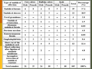
The umbilicus is the remnant of the fetal maternal connection . In the developing fetus , the various component structures of the umbilical cord pass through the ventral abdominal wall . These comprise the umbilical vein which leads to the liver , paired umbilical arteries which arise from the iliac arteries as well as the urachus which passes to the bladder.
This document discusses fetal mummification and maceration in animals. Fetal mummification is the shriveling of the fetus caused by the absorption of fluids from the fetus and uterus, leading to the death of the fetus. It can be caused by genetic abnormalities, hormonal issues, or problems with the placenta or umbilical cord. Maceration occurs when the fetus dies but is not aborted, causing it to become putrefied inside the uterus. Both conditions require veterinary intervention such as medications to induce abortion or surgical removal of the remains if abortion does not occur naturally.
1. Hernias are protrusions of abdominal contents through openings or weaknesses in the abdominal wall. They can be classified by location (e.g. umbilical, inguinal), contents (e.g. bowel, omentum, bladder), and reducibility.
2. Umbilical hernias occur through the umbilicus, and may be congenital or develop from straining. Ventral hernias develop through other parts of the abdominal wall, often from trauma.
3. Treatment involves reducing the hernia contents and repairing the abdominal wall defect surgically, usually with sutures or mesh. Postoperative management focuses on rest and reduced feeding to aid healing.
Uterine torsion occurs when the uterus rotates around its longitudinal axis, causing dystocia. It is most common in cattle and buffaloes. Risk factors include multiparity, oversized fetus, and lack of uterine tone. Clinical signs include difficulty calving, abdominal pain, and anorexia. Diagnosis involves vaginal and rectal examination to determine the degree and direction of torsion. Treatment options include rolling the dam, rotating the fetus through the birth canal, laparotomy, or caesarean section if correction fails or the cervix remains closed. Torsion can cause damage to the uterus and complications like rupture, hemorrhage, or infection if not addressed.
Uterine torsion in cattle occurs when the pregnant uterus rotates around its longitudinal axis, obstructing the birth canal and preventing the calf from entering the cervix. It is more common in multiparous cows and can result from sudden movements that cause the fetus to move violently within the uterus. Left untreated, it can lead to fetal death from reduced blood flow and oxygen. Veterinarians diagnose uterine torsion based on symptoms like pain and straining, as well as physical exams that reveal a twisted vagina and evidence of broad ligament twisting seen rectally. Treatment methods include manually rotating the fetus and uterus if slight, rolling the cow, using a plank to roll the cow and fix the uterus, or a cesare
This document discusses tail amputation procedures in various animal species. It describes the anatomy of the tail and indications for amputation such as trauma, infection, or cosmetic purposes. The procedure typically involves clipping and preparing the tail, retracting the skin, identifying the desired transection site, ligating vessels, disarticulating the tail between vertebrae, and suturing skin edges. Considerations for puppies, adults, and different animal species are provided. Complications can include tetanus or vertebral necrosis.
Caesarean section, also known as a C-section, is a surgical procedure where incisions are made through a woman's abdomen and uterus to deliver one or more babies. Indications for a C-section include uterine inertia, obstructive dystocia, rupture of the uterus, animal in compromised condition, twin pregnancy in mares, and incomplete cervical dilation. The ideal conditions for surgery include a clean area with minimal airborne contamination to prevent post-operative complications. The incision site varies by species, with dogs receiving a ventral midline incision and cattle receiving a left lower flank incision. During the procedure, the fetus is removed, the umbilical cord is ligated and
Chemical destruction of the teat involves injecting solutions such as diluted formalin, silver nitrate, acriflavine, copper sulfate, or chlorhexidine into the teat to destroy the teat tissue. This is done as a last resort when the teat cannot otherwise be salvaged due to extensive damage or infection. The solutions work to kill the teat tissue over the course of a few days through their disinfectant and caustic properties. Pretreating with a local anesthetic like megludyne can help reduce pain from the chemical destruction process.
This document discusses the anatomy, diagnosis, and treatment of guttural pouch diseases in horses. It describes the guttural pouch as a mucous sac located in the cranial cavity that connects the pharynx to the eustachian tube. Common guttural pouch diseases include empyema (accumulation of exudate), emphysema (abnormal filling with air), and impactions. Diagnosis involves endoscopy, radiography, or ultrasound imaging of the pouch. Treatment involves systemic antibiotics, flushing the pouch, or surgical drainage and removal of debris through approaches like Viborg's triangle or hyovertebrotomy. Post-operative care includes antibiotics, analgesics
This document discusses surgical anatomy and diseases of the oesophagus. It describes the oesophagus' location and surrounding structures. Common surgical diseases include obstruction, diverticula, stenosis, and wounds/fistulas. Obstruction is often caused by foreign bodies and treated conservatively via catheterization or surgically via oesophagotomy. Stenosis is corrected by longitudinal incision and transverse suturing to enlarge the lumen. Care must be taken during surgery to control hemorrhage and avoid nerve damage.
This document summarizes a study that compared three techniques for treating auricular hematoma in dogs: conventional suturing, skin staples, and surgical glue. Fifteen dogs were divided into three groups and treated with one of the three techniques. The techniques were compared based on closure time, cosmetic appearance, and healing time. Surgical glue had the fastest closure time, the best cosmetic appearance results, and the shortest healing time, making it the most effective technique according to this study.
Gutteral pouches, By Dr. Rekha Pathak, senior scientist IVRIRekha Pathak
- Empyema of the guttural pouches is a condition where pus collects in the guttural pouches of horses, usually due to infection spreading from the pharynx through the eustachian tube.
- Symptoms include intermittent nasal discharge, difficulty swallowing and breathing, and swelling of lymph nodes.
- Treatment involves early antibiotic therapy if possible, but once pus has formed, surgical drainage of the affected pouch is needed, which can be done through the incising along the atlas bone or through the viborg's triangle approach in the neck.
Canine pyometra is a uterine infection in intact female dogs that occurs during diestrus. It is caused by bacterial infection, usually E. coli, within the uterus under the influence of progesterone. Clinical signs include vaginal discharge, fever, lethargy, vomiting, and polyuria/polydipsia. Diagnosis involves abdominal palpation, ultrasonography, and clinical pathology. Treatment of choice is ovariohysterectomy, while antibiotics and prostaglandins may be used for medical management in some cases. Prognosis is good with early diagnosis and intervention to prevent systemic complications.
Ear new affection of ear and its treatmentBikas Puri
Otitis, or ear infections, can affect the outer, middle, or inner ear in dogs and cats and are commonly caused by parasites, bacteria, yeast, or skin issues. Symptoms include ear scratching, redness, discharge, and in severe cases neurological signs. Treatment involves cleaning and flushing the ears under anesthesia followed by topical and oral antibiotics, antifungals, or other medications based on diagnostic tests and addressing any underlying issues.
1) Displaced abomasum, commonly known as twisted stomach, occurs when a dairy cow's fourth stomach, the abomasum, moves from its normal position on the right side of the abdomen. This is most common in high producing dairy cows after calving.
2) The main causes of displaced abomasum are abomasal atony due to hypocalcemia and metabolic issues as well as increased gas production in the abomasum from diets high in grains. Clinical signs include reduced appetite, milk production and a ping sound heard on the left side.
3) Treatment involves surgical correction through approaches like the left flank or percutaneous toggle techniques to return the abomasum to its normal position
This document discusses causes of abortion in large animals. It describes infectious causes such as bacterial (e.g. Brucella abortus, Campylobacter fetus, Listeria monocytogenes), fungal (e.g. Aspergillus sp.), and protozoal (e.g. Neospora caninum, Tritrichomonas foetus) agents. It also discusses non-infectious causes including genetic factors, nutritional deficiencies, heat stress, and various toxins. The document provides details on abortion rates, timing, lesion presentation, and samples needed for diagnosis for many of the common infectious causes. It emphasizes the importance of rapid and proper sample collection and handling for determining the etiology of abortion
Hernia is defined as the protrusion of body cavity contents through an opening in the cavity wall. A hernia consists of a ring, sac, and contents. Diaphragmatic hernia is the passage of abdominal viscera into the thoracic cavity through a congenital or acquired opening in the diaphragm. Clinical signs include reduced appetite, bloating, and breathing difficulties. Diagnosis involves physical exam, x-rays, and exploratory surgery. Surgical repair involves replacing herniated organs, closing diaphragmatic openings, and suturing muscle layers. Postoperative care focuses on respiratory support, antibiotics, and pain management.
Uterine prolapse occurs when the uterus slips from its normal position into the vaginal canal. It has four stages based on how far the cervix protrudes. Causes include difficult births, weak pelvic muscles, surgery, and excessive calf size. Treatment involves manually pushing the uterus back into place, applying disinfectants, sutures to prevent reoccurrence, and antibiotics. The condition has a better prognosis if treated early, and survival rates depend on the cow's posture and presence of other complications like milk fever. Fertility can be impacted after uterine prolapse.
Rajeev mishra ,castration of small animal(dog and cat).Raaz Eve Mishra
This document provides information about neutering male dogs and cats. It discusses the surgical procedure for neutering, which involves making an incision to remove the testicles. The benefits of neutering include preventing unwanted litters and reducing behavioral issues. Potential risks include weight gain and delayed growth. Neutering can be done between 5 to 16 months old in dogs and at around 6 months in cats. Post-operative care involves monitoring wounds, limiting activity, and providing pain relief if needed.
The document describes a study of 65 cases of benign and malignant neoplasms in ruminants over a 3 year period. The neoplasms included 2 fibromas, 1 fibromatous epulis, 1 fibroadenoma, 4 fibropapillomas, 25 cutaneous papillomas, 8 keratoacanthomas, 2 fibrosarcomas, 21 squamous cell carcinomas, and 1 lymphosarcoma. The neoplasms were surgically treated and followed up for 4-6 months. The study aimed to describe the gross and histopathological features of various ruminant neoplasms and determine which cases were suitable for surgical treatment.
This document summarizes a study on the surgical management of certain ano-rectal conditions in farm animals. A total of 65 cases were examined between 2003-2006, including congenital defects like atresia ani (24 cases), acquired conditions like rectal prolapse (15 cases), and rare cases of anus vaginalis. Surgical techniques are described for reconstructing the anal opening in atresia ani and repairing rectal prolapse. Early surgical correction of atresia ani is important for animal survival. Histopathology was used to diagnose neoplastic masses. The most common condition was atresia ani, especially in lambs and calves.
This document contains images and descriptions of various fetal abnormalities and congenital defects in cattle, including:
- Six-legged calves resulting from duplication of the hind legs.
- Three-legged calves due to complete absence of a front or hind leg.
- Conjoined twins sharing body parts like chests or hindquarters.
- Other defects include parasitic extra limbs, cyclopia, dropsy/anasarca, vertebral defects like schistosomus reflexus, and achondroplasia.
The document provides photos and notes on various rare congenital defects observed in cattle fetuses and calves.
This document discusses several metabolic diseases that can affect large ruminants, with a focus on milk fever. Milk fever is a hypocalcemia condition seen around the time of calving, characterized by muscle weakness and depression. It is caused by a severe drop in blood calcium levels due to increased calcium demands of lactation. The document outlines the risk factors, stages of clinical signs, diagnosis through calcium testing, and treatment using calcium supplements. Prevention focuses on dietary management of calcium and phosphorus levels before and after calving.
Intussusception is a condition where a segment of intestine invaginates into the adjacent bowel, obstructing the gastrointestinal tract. It is most commonly seen in the small intestine of cattle. Clinical signs include abdominal pain, decreased appetite, and signs of obstruction. Ultrasound can be used to diagnose intussusception based on the "target-like" or "bullseye" appearance. Treatment involves surgical resection of the affected segment through an abdominal incision. The surgery is ideally performed with the animal under general anesthesia to allow for thorough exploration and resection. Untreated intussusception is fatal as the blocked blood supply causes necrosis of the invaginated bowel.
This document discusses nalid deg disease, a condition affecting cattle and buffalo that feed on rice straw. It is more common in winter months in rice growing areas. Clinically, it causes gangrenous lesions on the hoof, ear, and tail. It may be caused by excessive ingestion of selenium from contaminated rice straw grown in selenium-rich soil or from mycotoxins produced when rice straw is stored in moist conditions, allowing fungal growth. Treatment involves washing lesions, applying ointment, administering pentasulfate orally or antibiotics if needed, and potentially surgery to dock the tail. Prevention includes treating rice straw with sodium hydroxide or supplementing animals' diets with copper,
A 4-day-old male infant was admitted to the hospital for vomiting. He was diagnosed with sepsis neonatorum and omphalitis (umbilical infection). He received intravenous antibiotics and wound care. His condition improved and he was discharged after 6 days with oral antibiotics to complete the course of treatment.
This document discusses disorders of the umbilicus that can occur in pediatric patients. During development, the umbilicus functions as a channel for blood flow between the placenta and fetus and plays a role in intestinal and urinary development. After birth, the umbilicus should show no evidence of these connections. However, umbilical disorders are common in pediatric surgery and range from umbilical hernias to potentially life-threatening infections. Patients typically present with drainage or a mass at the umbilicus. Understanding the umbilicus' anatomy and embryology is important for properly diagnosing and treating these conditions.
This document describes a case of omphalitis, an infection of the umbilical stump, in a 7-day-old male infant. The infant presented with fever, yellowish umbilical discharge, and hypoactivity. Laboratory tests showed elevated white blood cell count and C-reactive protein. The infant was diagnosed with omphalitis and sepsis and started on intravenous antibiotics and supportive care. Omphalitis is a potentially serious infection in neonates that requires prompt treatment with antibiotics and sometimes surgery.
Calves 1-2 weeks old commonly experience umbilical swelling, fever, loss of appetite, and depression due to umbilical abscesses. Treatment includes intramuscular injections of broad-spectrum antibiotics for 5-7 days, symptomatic relief with analgesics, fluids, and corticosteroids, and surgical opening and cleaning of abscesses with disinfectants and local antibiotics.
This document summarizes newborn calf management from before birth through weaning. It discusses providing proper nutrition to dams during pregnancy, assisting with calving, disinfecting and tagging calves after birth, ensuring calves receive colostrum within 1/2 hour, and providing vaccinations and antibiotics. It also discusses housing calves individually for the first week, dehorning at 5 days, moving calves to group housing at 2 weeks, and weaning after 4 months. The most common health issues of omphalitis (navel ill), enteritis (diarrhea), and pneumonia are described along with their treatments. Special emphasis is placed on colostrum management and gradual weaning once calves can consistently consume 1kg
This document provides an overview of intestinal obstruction, including its classification, common causes, clinical features, investigations, and treatment options. Intestinal obstruction can result from mechanical blockage from structural abnormalities (dynamic obstruction) or paralysis without physical obstruction (ileus). Common causes of dynamic obstruction include adhesions, hernias, volvulus, and intussusception. Clinical features include colicky pain, vomiting, abdominal distention, and constipation. Investigations may include blood tests, abdominal x-rays, and CT scans. Treatment involves resuscitation, nasogastric decompression, IV fluids, and potentially surgery to resolve the obstruction. Complications can include bleeding, infection, leakage, and reoccurrence of obstruction
Spigelian Hernia: A Rare Hernia With Peculiar Anatomy. (Case Report And Revie...KETAN VAGHOLKAR
Background: Spigelian hernia best described as
spontaneous lateral ventral hernia is an extremely rare type of
hernia. The anatomical peculiarities and diagnostic challenges
need to be understood in order to surgically mange this hernia.
Introduction: Spigelian hernia occurs through a defect in the
spigelian fascia typically lying in the spigelian zone.
Case report: A case of a large incarcerated spigelian hernia
is presented to highlight the diagnostic and anatomical
peculiarities of this hernia.
Discussion: The anatomical basis of this hernia along with
clinical presentation, diagnostic modalities and treatment
options is discussed.
Conclusion: Clinical suspicion confirmed by imaging is
necessary for diagnosis. Surgery is the mainstay of treatment.
Third-degree Rectovestibular Lacerations in mares and mare donkeysProf.Dr. Gamal Karrouf
Six mares and a she donkey suffering from third-degree rectovestibular lacerations during difficult births were admitted to a veterinary clinic. Surgical correction was performed 5-7 weeks later through a single-stage repair using interrupted vertical mattress sutures of polydioxanone. Primary healing of the reconstructed rectovestibular shelves and perineal bodies occurred in six animals, though one mare developed anal sphincter incompetence and another a rectovestibular fistula due to post-operative straining.
Colonic incarceration in an adult umbilical hernia: case report and review of...KETAN VAGHOLKAR
Umbilical hernia is one of the commonest ventral hernias constituting ten percent of all hernias. It affects obese individuals and has a high recurrence rate if repaired by suture techniques. Incarceration of the colon in an umbilical hernia is quite rare. A case of colonic incarceration in an umbilical hernia is presented to highlight the diagnostic and technical challenges in managing such a hernia. Contrast enhanced computerized tomography is essential to ascertain the contents. Open surgery is the main stay of treatment especially in such rare cases. A combined tissue and mesh repair provides excellent results.
Successful Repeated CT-Guided Drainage Of Rectal Mucocele After LAleksandr Reznichenko
This document describes a case study of a rectal mucocele that was successfully treated with repeated CT-guided drainage after a patient underwent low anterior resection for rectal prolapse. A rectal mucocele developed as a fluid-filled cyst near the rectal stump that caused symptoms. It was drained multiple times under CT guidance, with catheters inserted each time. Analysis of the fluid indicated it was a rectal mucocele rather than an abscess. This case demonstrates that repeated CT-guided drainage can successfully treat a rectal mucocele in a patient who was not a candidate for surgical resection.
This document summarizes information about hypospadias, a congenital malformation where the opening of the urethra is on the underside of the penis instead of at the tip. It discusses the presentation, causes, classification, associated anomalies, surgical repair techniques, and complications of hypospadias. The goal of surgery is to completely repair the penis by placing the urethral opening at the tip and achieving a symmetrical, conical glans with uniform caliber urethra and satisfactory cosmetic coverage.
Laparoscopic sterilization was the first popular minimal access surgical procedure ever performed. Laparoscopic sterilization is very straightforward procedure. Worldwide laparoscopic sterilization is now the most commonly applied method for family planning
This case report describes a successful case of managing placenta percreta with invasion into the bladder. A 33-year-old woman at 35 weeks of gestation was found to have placenta previa and suspected placenta percreta. During a cesarean section and hysterectomy, it was discovered that newly formed vessels from the placenta had invaded the bladder wall. Prophylactic balloon occlusion of the lower abdominal aorta was performed to control hemorrhaging. The placenta, uterus, and part of the invaded bladder wall were removed. The massive intraoperative hemorrhage was successfully controlled and the patient recovered well. The management of newly formed vessels is crucial for effective treatment of placent
The document discusses the anatomy and histology of the cervix. It describes the squamocolumnar junction (SCJ) as the boundary between squamous and columnar epithelium. The location of the SCJ changes throughout a woman's life due to hormonal influences and metaplastic changes. The transformation zone is the region between the original and new SCJ where columnar epithelium is replaced by squamous epithelium. Precancerous lesions and cancers of the cervix primarily originate from cells within the transformation zone, making it an important area.
Incarcerated infraumbilical incisional hernia: a surgical challengeDrKetanVagholkar
Incisional hernia continues to be the most challenging type of hernia. Variability in the anatomy and supervening
complications add to its complexity. Infraumbilical incisional hernias are usually due to gynecological operations.
This may range from a scar of tubal ligation procedure to a Pfannenstiel incision or an infraumbilical scar of caesarian
section. The sparse volume of strong anatomical structures in this region poses the biggest challenge during repair. A
54-year-old lady presented with a hernia arising from a scar of previous tubal ligation surgery. The hernia was
irreducible with a large mass of omentum in the hernial sac. Laparoscopy was difficult to perform in view of the
current state. Hence open surgery was performed. The technique used was creation of a preperitoneal space followed
by creation of space between external oblique aponeurosis and underlying muscle. A mesh as placed between the
muscular and aponeurotic layer. The post-operative course was uneventful with no recurrence. The anatomical basis
of placing the mesh between the muscular and aponeurotic layer or intermediate placement technique is discussed.
Creation of space below the aponeurotic level is pivotal in managing infraumbilical incisional hernia. Placing a mesh
at this layer below the aponeurosis ensures least complications with excellent result.
New microsoft office power point presentationRiyadhWaheed
This document discusses the role of 3D ultrasound in evaluating uterine diseases and anomalies. It begins by reviewing female reproductive tract embryology. It then describes the American Fertility Society's 7-class system for classifying Müllerian duct anomalies, including uterine agenesis, unicornuate uterus, bicornuate uterus, septate uterus, and DES exposure anomalies. It discusses how 3D ultrasound and MRI can be used to distinguish between septate and bicornuate anomalies. The document also discusses how 3D ultrasound can be used to measure endometrial volume and vascularity, which are important indicators of endometrial receptivity and pregnancy potential.
This document provides information about various veterinary surgical procedures including caesarean section, rumenotomy, and castration. It discusses the indications, techniques, and post-operative care for each procedure. Specifically, it describes how caesarean section involves opening the abdomen and uterus to deliver calves when natural birth is difficult. It also explains that rumenotomy requires an incision into the rumen to remove contents and relieve impaction. Finally, it notes that castration removes the testicles in animals like dogs and cattle.
Conservative surgeries for genital prolapseNikhil Bansal
The document discusses various conservative surgical procedures for genital prolapse that preserve menstrual and childbearing functions. It describes procedures like anterior and posterior colporrhaphy, Fothergill's repair, Shirodkar's procedure, and abdominal sling operations. Each procedure is explained detailing its principles, indications, steps, and potential complications. Conservative surgeries presented aim to correct prolapse of the uterus, bladder, rectum or vagina without removing organs or abolishing functions.
This document discusses degrees of lacerations of the birth canal, from first to fourth degree. It also discusses risk factors for more severe lacerations, morbidity risks, episiotomy procedures including types, timing and repair techniques. Layered repair techniques are described for third and fourth degree lacerations extending to the anal sphincter. Postoperative pain management options are also summarized.
Vesicouterine Fistula Following Cesarean Delivery – Ultrasound Diagnosis and ...Michelle Fynes
Vesicouterine fistulae are uncommon, with most units reporting 1–5 cases over 5–15 year periods. To date there has been a paucity of case reports regarding this problem and only a few case series. In this report we outline the presentation and management of a vesicouterine fistula complicating a repeat Cesarean delivery, specifically describing the role of transvaginal ultrasound.
This case report describes a 4-day old male neonate admitted with severe birth asphyxia, anemia, and sepsis who developed massive pneumoperitoneum. Abdominal x-rays showed a large amount of free gas under the diaphragm compressing the abdominal viscera. Emergency laparotomy revealed a 9cm longitudinal tear in the descending colon and perforation in the sigmoid colon with fecal soilage. The affected colon segments were resected and anastomosis performed. Post-operatively, the patient recovered well and was discharged at 6 weeks old. The report discusses pneumoperitoneum causes in neonates, signs on imaging, and importance of immediate surgical intervention for perforated viscus with per
Hirschsprung disease is a congenital disorder where the enteric nervous system fails to develop in parts of the colon, most commonly in the rectum. This leads to obstruction and complications like enterocolitis. The diagnosis is confirmed with biopsy showing lack of ganglion cells. Treatment involves surgical removal of the aganglionic segment and reconnection of the bowel, such as with a Swenson or Duhamel procedure. Early diagnosis and treatment are important to prevent complications.
This document discusses genital prolapse, which is the descent of one or more genital organs through the pelvic floor. It describes the different types of prolapse including vaginal, uterine, and vault prolapse. Factors that can influence pelvic floor support like childbirth injury, age, and endocrine factors are examined. Symptoms and examination findings are outlined. Treatment options discussed include conservative measures like pessaries as well as various surgical repair techniques for the anterior vaginal wall, posterior vaginal wall, and uterus.
This document discusses laparoscopic assisted vaginal hysterectomy (LAVH). It begins with a brief history of laparoscopic hysterectomy and incidence rates of different hysterectomy methods from various studies. It then describes the classification, indications, procedure, complications, and comparison of LAVH, vaginal hysterectomy, and abdominal hysterectomy. While laparoscopic hysterectomy may be preferred for its minimal invasiveness, the document emphasizes that vaginal hysterectomy should be performed when possible to avoid unnecessary procedures. In conclusion, it states that the main goal of LAVH is to reduce abdominal hysterectomy rates by utilizing it where vaginal hysterectomy is contraindicated.
This document discusses cervical insufficiency, including its definition, causes, diagnosis, and treatment. Cervical insufficiency is the inability of the cervix to retain a pregnancy due to structural or functional defects in the absence of labor contractions. It can be congenital or acquired due to conditions like cervical trauma. Diagnosis involves history, examination finding an open cervix, and transvaginal ultrasound to measure cervical length. Treatment options include cervical cerclage surgery to provide structural support to the cervix, progesterone to help maintain the cervix, or a pessary. Cerclage is indicated for women with a history suggestive of cervical insufficiency or a short cervix on ultrasound.
This document provides rationales for questions from the 2007 ACR Diagnostic Radiology In-Training Exam related to ultrasound imaging. It discusses the correct answers and rationales for questions regarding various pathologies seen on ultrasound of the uterus, testes, kidneys, abdomen and fetal anatomy. Key details include identifying a cornual pregnancy based on its location, tubular ectasia of the rete testis in an older male, medullary nephrocalcinosis seen as echogenic renal pyramids, the left renal vein in the transverse abdominal image, and measuring fetal head circumference at the level of the thalami and cavum septi pellucidi.
At Techbox Square, in Singapore, we're not just creative web designers and developers, we're the driving force behind your brand identity. Contact us today.
How are Lilac French Bulldogs Beauty Charming the World and Capturing Hearts....Lacey Max
“After being the most listed dog breed in the United States for 31
years in a row, the Labrador Retriever has dropped to second place
in the American Kennel Club's annual survey of the country's most
popular canines. The French Bulldog is the new top dog in the
United States as of 2022. The stylish puppy has ascended the
rankings in rapid time despite having health concerns and limited
color choices.”
Company Valuation webinar series - Tuesday, 4 June 2024FelixPerez547899
This session provided an update as to the latest valuation data in the UK and then delved into a discussion on the upcoming election and the impacts on valuation. We finished, as always with a Q&A
The Genesis of BriansClub.cm Famous Dark WEb PlatformSabaaSudozai
BriansClub.cm, a famous platform on the dark web, has become one of the most infamous carding marketplaces, specializing in the sale of stolen credit card data.
How MJ Global Leads the Packaging Industry.pdfMJ Global
MJ Global's success in staying ahead of the curve in the packaging industry is a testament to its dedication to innovation, sustainability, and customer-centricity. By embracing technological advancements, leading in eco-friendly solutions, collaborating with industry leaders, and adapting to evolving consumer preferences, MJ Global continues to set new standards in the packaging sector.
Unveiling the Dynamic Personalities, Key Dates, and Horoscope Insights: Gemin...my Pandit
Explore the fascinating world of the Gemini Zodiac Sign. Discover the unique personality traits, key dates, and horoscope insights of Gemini individuals. Learn how their sociable, communicative nature and boundless curiosity make them the dynamic explorers of the zodiac. Dive into the duality of the Gemini sign and understand their intellectual and adventurous spirit.
3 Simple Steps To Buy Verified Payoneer Account In 2024SEOSMMEARTH
Buy Verified Payoneer Account: Quick and Secure Way to Receive Payments
Buy Verified Payoneer Account With 100% secure documents, [ USA, UK, CA ]. Are you looking for a reliable and safe way to receive payments online? Then you need buy verified Payoneer account ! Payoneer is a global payment platform that allows businesses and individuals to send and receive money in over 200 countries.
If You Want To More Information just Contact Now:
Skype: SEOSMMEARTH
Telegram: @seosmmearth
Gmail: seosmmearth@gmail.com
SATTA MATKA SATTA FAST RESULT KALYAN TOP MATKA RESULT KALYAN SATTA MATKA FAST RESULT MILAN RATAN RAJDHANI MAIN BAZAR MATKA FAST TIPS RESULT MATKA CHART JODI CHART PANEL CHART FREE FIX GAME SATTAMATKA ! MATKA MOBI SATTA 143 spboss.in TOP NO1 RESULT FULL RATE MATKA ONLINE GAME PLAY BY APP SPBOSS
How to Implement a Real Estate CRM SoftwareSalesTown
To implement a CRM for real estate, set clear goals, choose a CRM with key real estate features, and customize it to your needs. Migrate your data, train your team, and use automation to save time. Monitor performance, ensure data security, and use the CRM to enhance marketing. Regularly check its effectiveness to improve your business.
Easily Verify Compliance and Security with Binance KYCAny kyc Account
Use our simple KYC verification guide to make sure your Binance account is safe and compliant. Discover the fundamentals, appreciate the significance of KYC, and trade on one of the biggest cryptocurrency exchanges with confidence.
HOW TO START UP A COMPANY A STEP-BY-STEP GUIDE.pdf46adnanshahzad
How to Start Up a Company: A Step-by-Step Guide Starting a company is an exciting adventure that combines creativity, strategy, and hard work. It can seem overwhelming at first, but with the right guidance, anyone can transform a great idea into a successful business. Let's dive into how to start up a company, from the initial spark of an idea to securing funding and launching your startup.
Introduction
Have you ever dreamed of turning your innovative idea into a thriving business? Starting a company involves numerous steps and decisions, but don't worry—we're here to help. Whether you're exploring how to start a startup company or wondering how to start up a small business, this guide will walk you through the process, step by step.
Understanding User Needs and Satisfying ThemAggregage
https://www.productmanagementtoday.com/frs/26903918/understanding-user-needs-and-satisfying-them
We know we want to create products which our customers find to be valuable. Whether we label it as customer-centric or product-led depends on how long we've been doing product management. There are three challenges we face when doing this. The obvious challenge is figuring out what our users need; the non-obvious challenges are in creating a shared understanding of those needs and in sensing if what we're doing is meeting those needs.
In this webinar, we won't focus on the research methods for discovering user-needs. We will focus on synthesis of the needs we discover, communication and alignment tools, and how we operationalize addressing those needs.
Industry expert Scott Sehlhorst will:
• Introduce a taxonomy for user goals with real world examples
• Present the Onion Diagram, a tool for contextualizing task-level goals
• Illustrate how customer journey maps capture activity-level and task-level goals
• Demonstrate the best approach to selection and prioritization of user-goals to address
• Highlight the crucial benchmarks, observable changes, in ensuring fulfillment of customer needs
❼❷⓿❺❻❷❽❷❼❽ Dpboss Matka Result Satta Matka Guessing Satta Fix jodi Kalyan Final ank Satta Matka Dpbos Final ank Satta Matta Matka 143 Kalyan Matka Guessing Final Matka Final ank Today Matka 420 Satta Batta Satta 143 Kalyan Chart Main Bazar Chart vip Matka Guessing Dpboss 143 Guessing Kalyan night
Building Your Employer Brand with Social MediaLuanWise
Presented at The Global HR Summit, 6th June 2024
In this keynote, Luan Wise will provide invaluable insights to elevate your employer brand on social media platforms including LinkedIn, Facebook, Instagram, X (formerly Twitter) and TikTok. You'll learn how compelling content can authentically showcase your company culture, values, and employee experiences to support your talent acquisition and retention objectives. Additionally, you'll understand the power of employee advocacy to amplify reach and engagement – helping to position your organization as an employer of choice in today's competitive talent landscape.
[To download this presentation, visit:
https://www.oeconsulting.com.sg/training-presentations]
This PowerPoint compilation offers a comprehensive overview of 20 leading innovation management frameworks and methodologies, selected for their broad applicability across various industries and organizational contexts. These frameworks are valuable resources for a wide range of users, including business professionals, educators, and consultants.
Each framework is presented with visually engaging diagrams and templates, ensuring the content is both informative and appealing. While this compilation is thorough, please note that the slides are intended as supplementary resources and may not be sufficient for standalone instructional purposes.
This compilation is ideal for anyone looking to enhance their understanding of innovation management and drive meaningful change within their organization. Whether you aim to improve product development processes, enhance customer experiences, or drive digital transformation, these frameworks offer valuable insights and tools to help you achieve your goals.
INCLUDED FRAMEWORKS/MODELS:
1. Stanford’s Design Thinking
2. IDEO’s Human-Centered Design
3. Strategyzer’s Business Model Innovation
4. Lean Startup Methodology
5. Agile Innovation Framework
6. Doblin’s Ten Types of Innovation
7. McKinsey’s Three Horizons of Growth
8. Customer Journey Map
9. Christensen’s Disruptive Innovation Theory
10. Blue Ocean Strategy
11. Strategyn’s Jobs-To-Be-Done (JTBD) Framework with Job Map
12. Design Sprint Framework
13. The Double Diamond
14. Lean Six Sigma DMAIC
15. TRIZ Problem-Solving Framework
16. Edward de Bono’s Six Thinking Hats
17. Stage-Gate Model
18. Toyota’s Six Steps of Kaizen
19. Microsoft’s Digital Transformation Framework
20. Design for Six Sigma (DFSS)
To download this presentation, visit:
https://www.oeconsulting.com.sg/training-presentations
Innovation Management Frameworks: Your Guide to Creativity & Innovation
Umbilical affections
1. بسم ا الرحمن الرحيم
”قالوا سبحانك لعلم
لنا ال ما علمتنا انك أنت
العليم الحكيم“
صدق ا العظيم
2. Surgical Management Of Certain
Umbilical Affections In
Calves And Foals
By
Karrouf, G.I.A. and Mosbah, E
Dept. of Surgery, Anesthesiology & Radiology
.Fac. of Vet.Med., Mansoura University
3. Introduction
The umbilicus is the remnant of the fetal
maternal connection . In the developing
fetus , the various component structures
of the umbilical cord pass through the
ventral abdominal wall . These comprise
the umbilical vein which leads to the liver ,
paired umbilical arteries which arise from
the iliac arteries as well as the urachus
which passes to the bladder.
4. Separation of the umbilical cord
allows the umbilical arteries and
urachus to retract into the abdomen,
where they close by smooth muscle
contraction while the umbilical vein
and remnants of amniotic membrane
remain outside the body wall
6. Aim of the work
The present work aimed to declare the
commonly and rarely occurred surgical
affections observed at the umbilical
region in calves and foals as well as
their surgical management .
7. Materials and Methods
The present investigation was performed
on 88 calves ( 46 cow calves and 42 buffalo
calves ) and 17 foals suffered different
umbilical affections (Table 1). These cases
were admitted to the Surgery Clinic of the
Fac. of Vet. Med ., Mansoura University
and collected also from different localities
and farms in Dakahlia Province .
8. The age of the affected animals ranged
from birth to 6 months old . The type and
number of cases, the species of the
affected animals and the percentage ( % )
of each umbilical affection as well as
history , physical examination and clinical
signs were recorded . Histopatholgical
examination of navel granulomas as well
as surgical treatment of these affections
were described .
9. Our results revealed 11 surgical conditions
affecting the umbilicus . Umbilical hernia was
the most common one ( 37.1% ) followed by
umbilical abscess ( 20.9% ) , short cut of the
umbilical cord ( 11.4% ) , omphalophlebities
( 8.7% ) , blood collection after ligation of
amniotic membrane ( 4.8% ), incomplete
retraction of umbilical contents ( 4.8% ) , navel
granuloma ( 3.8% ) and pervious urachus
( 4.8% ) . Uncommon umbilical affections as
abomaso-umbilical fistula, umbilical
eventration, and enterocutaneous fistula were
recorded in this study
10.
11. In the present study , umbilical
hernia was the most common
umbilical affection recorded in
calves and foals (37.1 %) . These
cases could be corrected surgically
without any clear postoperative
complications
12. Spontaneous healing was obtained in cases of
reducible umbilical hernias with small rings ( 12 fingers ) when the animals get older (6–8
months) . The explanations for spontaneous
recovery could only presumably the abdomen
becomes deeper while the mesentery becomes
relatively shorter as the animal grows the viscera
is thus withdrawn within the abdominal cavity
while the umbilical ring cicatrize behind it.
13. The present study agreed that most
umbilical hernia repairs were
elective procedures and were
performed in normal animals for
cosmetic reasons. Slight thickening
at the operation site noticed in large
umbilical hernias may be due to
excessive fibrous tissue formation
around the used hernial tape
14. Fig.2: Umbilical hernias in
buffalo calf (A) and cow
calf (B) and closure of the
hernial ring by a series of
interrupted
horizontal
mattress sutures using of
Polyglycolic acid (C) .
15. Fig.3: Umbilical hernia in
foal (A), after dissection
and freeing of the hernial
sac 1cm peripheral to the
hernial
ring
(B).
Herniorrhaphy
was
obtained by a series of
interrupted
horizontal
mattress sutures using of
silk (C) .
16. Fig.9: Umbilical hernias in buffalo calf (A) and cow
calves (B&C) and closure of the hernial ring by a
series of interrupted horizontal mattress sutures
using of silk (D) .
18. Abomaso-umbilical fistula
Abomaso-umbilical fistula had been
reported in association with umbilical
infection. Diagnosis was relatively simple
and was based on the observation of
luminal contents from an umbilical fistula.
Attempts to over sew the fistula in situ
without resection had not been successful
.Treatment involves resection of the
fistula with contaminated umbilicus and
involved abomasal wall
19. Fig.14: A fistula discharging fluidy ingesta at the umbilicus in
3 months female calf (A, arrows). Undigested piece of food
protruded from a narrow necrotic orifice (B), undigested food
( C ), necrotic tissue and fluidy ingesta (D) were seen after an
elliptical skin incision and freeing of the abomasums.
20. Fig.15: The abomasal wound closure (A) and the skin (B) ,
slight oedema around the healed wound 10 days postopreation
(C,arrow) which was disappeared with complete healing at 35
days postopreation (D,arrow) .
21. Enterocutaneous fistula
Enterocutaneous fistula was recorded
in a male cow calf with a history of
discharging fluid ingesta from a small
narrow orifice 3 weeks after umbilical
herniorrhaphy .It was treated by a
simple technique by allowing second
intention healing with a resultant
granulation tissue formation 3 weeks
after closure .
22. Fig.16: A narrow orifice
discharging ingesta near the
umbilicus (Enterocutaneous
fistula A, arrow), a pursestring suture was applied after
thorough debridement (B)
and healthy granulation was
formed and fill the orifice 3
weeks
postopreation
(C,arrow).
23. In the present study abdominal fissure at
the umbilicus associated with eventration
of the intestine were recorded in 2 calves
.
presented following a natural delivery
24. The aetiology of abdominal fissures at the
umbilicus is still obscure while its
frequency is said to be uncommon
(Leipold and dennis 1980). During fetal
development, abdominal viscera normally
pass through the umbilicus to lie outside
the abdominal cavity as term approaches
the viscera was normally drawn within the
abdominal cavity and the umbilical
opening closes around the cord
25. The condition was considered as a congenital
defect where the yolk stalk may be persist from
jejunum to the umbilicus as fibrous cord which
allows direct communication to the surface of
the body resulting in variable degree of
eventration of the abdominal contents
particularly the intestines. Umbilical
eventration should be differentiated from cases
of Schistosoma reflexus which characterized by
severe closure defects of the thoracic and
abdominal cavities together with curvature and
twisted deformities of the spinal column
26. Clinical examination revealed contaminated
and congested fully exposed jejunum.
Satisfactory recovery was obtained 14 days
postoperation after reduction of the
prolapsed part and closure of the abdominal
defect by a series of interrupted horizontal
mattress sutures
27. Fig.5: Eventration of small
intenstine through the
umbilicus in a newly born
buffalo calf (A), after
reduction and closure of
the abdominal defect (B) .
28. Fig.12: Eventration of
small intenstine through
the umbilicus in a newly
born calf (A), after
reduction and application
of a retention sutures (B) .
29. Fig.13: The previous
case after skin closure
(A) and 14 days
postoperation with
(. complete healing (B
30. Persistent or patent urachus is a condition of
young animal in which the urachus fails to close
spontaneously at or shortly after parturition
In the present study pervious urachus was more
frequent in foals (4 cases) than calves (1case).
I referred this condition to the relatively larger
umbilical cord of the foal than of the calf and it
is more commonly the site of the navel infection,
persistent urachus and umbilical hernia
31. Patent
urachus was manifested as periodic
stream of urine issuing from the umbilicus.
The navel remains wet. Conservative
treatment was performed by cotton swab
dipped in 5 % tincture of iodine introduced 45 cm inside the urachus towards the urinary
bladder . This was repeated once daily for 3-5
successive days. Surgical resection of the
urachus was performed in 2 foals when
spontaneous closures does not follow by this
time the urachus was resected along with the
cutaneous navel.
32. Fig.6: Drippling of urine
from the umbilicus due to
patent urachus in 9 days old
filly foal (A, arrow) and
resection of the urachus
after mildline celiotomy
(B) .
33. Fig.7: The previous
case after closure of
abdominal muscles (A)
and the subcutaneous
tissues and the skin
(B) .
34. Navel granulomas
Navel granuloma was diagnosed in 2-4
weeks old 4 buffalo calves (3 males and a
female) as a small ulcerated piece of bright
red fleshy mass that protruded from the
navel ( Fig.11A,B&C ) . Good healing was
obtained after surgical excision in all cases .
Microscopical examination revealed that the
granuloma consisted of focal aggregation of
chronic inflammatory cells mainly fibroblasts
(Fig.11D ) .
35. Fig.11: Different shapes of navel granulomas in
buffalo calves (A,B&C) and microcpic examination
(D) revealed focal aggregation of chronic
inflammatory cells mainly fibroblasts (H&E . X : 300)
.
36. Umbilical abscesses
Twenty two calves were presented with an
umbilical abscesses . Examination of the
affected calves revealed a hot , painful ,
circumscribed swelling at the umbilicus
( Fig.10A ) . Omphalophlepitis was recorded
in 9 cases, suppurative discharge from an
inflamed navel was seen (Fig.10B) the affected
calves often suffered from poor performance
and recurring fevers.
38. Surgical interference : Omphalophlebitis
In cases of omphalophlebitis , all
suppurative materials were completely
drained and the wound was saturated with
povidone iodine 3 times daily . Surgical
excision of the chronicaly infected umbilical
remnant was performed in one case (Fig. 8(
which did not respond to the previous
treatment .
39. Fig.8: Surgical excision of chronic inflamed
umbilical remnants in a buffalo calf (A,B&C)
and the excised mass (D).
40. : Surgical interference
Short cut of the umbilical cord
In cases of short cut of the umbilical cord,
the wound edges were debrided and the
skin was sutured by simple interrupted
suture using silk. Cases of blood
collection after ligation of aminiotic
membrane were treated by lancing of and
discharging of the infected blood . The
umbilicus was touched 3 times daily with
Betadine antispetic.
41. Fig.17: Short cut of the
umbilical cord immediately
after birth (A, arrow ) and
after complete healing ( B,
arrow ).
42. Surgical interference :
Incomplete retraction of umbilical contents
In cases of incomplete retraction of umbilical
contents , the amniotic membrane was
opened and the blood vessels were double
ligated at the most highest point near the
abdominal wall , then resected . The skin at
the umbilicus was debrided and sutured .
43. Fig.18: Blood collection after ligation of
amniotic membrane in a buffalo calf ( A ) and
cow calves ( C&D ). The swelling was incised
and soaked with Betadine ( B, arrow ) .
45. Conclusion
Our results revealed 11 surgical conditions affecting the
umbilicus . Umbilical hernia was the most common one
( 37.1% ) followed by umbilical abscess ( 20.9% ) , short cut
of the umbilical cord ( 11.4% ) , omphalophlebities ( 8.7% ) ,
blood collection after ligation of amniotic membrane ( 4.8% ),
incomplete retraction of umbilical contents ( 4.8% ) , navel
granuloma ( 3.8% ). Uncommon umbilical affections as
abomaso-umbilical fistula, umbilical eventration ,
enterocutaneous fistula and pervious urachus were recorded
in this study . Good healing with satisfactory recovery was
obtained after surgical interference . Navel infection could be
minimized through good sanitary conditions during and after
parturition .
46. Conclusion
In conclusion, the umbilicus is subjected to many
surgical conditions which could be corrected
successfully after early diagnosis. It is recommended
that the veterinarians as well as the farmers should pay
regular attention to the umbilicus of calves and foals as
a routine husbandry. Directly after birth the umbilicus
should be cleaned and touched with povidone iodine
without umbilical cord ligation as blood is usually
collected and in the presence of contamination,
omphalitis, omphalophlebitis, omphaloarteritis and
urachal infection may be occurred with subsequent
development of various umbilical affections.
48. Results
Short cut of the umbilical cord
Good healing was obtained in cases of short
cut of the umbilical cord (Fig.17) , blood
collection within the amniotic membrane
(Fig.18)as well as cases of incomplete
retraction of umbilical contents ( Fig.19).
49. Meanwhile Goligher ( 1971 ) and Roback and
Nicoloff ( 1972 ) recorded that simple closure of
an enterocutaneous fistula was generally
unsuccessful . They advised en bloc resection of
the body wall and intestine, as the most
satisfactory treatment. Nonsurgical management of
two horses with presumptive large colon fistulae
by Bristol (1994) has resulted in resolution of the
fistulae without complication
50. Abomaso-umbilical fistula had been reported in
association with umbilical infection. Diagnosis was
relatively simple and was based on the observation
of luminal contents from an umbilical fistula.
Attempts to oversew the fistula in situ without
resection had not been successful
Treatment involves resection of the fistula with
contaminated umbilicus and involved abomasal
wall.
51. Results
Pervious urachus
Four foals ( a male and three females ) and a
female calf aged 2 – 10 days has a history of
intermittent dribbling of urine at the umbilical
region, the navel remain wet and the navel
infections were seen in four cases . The associated
concurrent problem includes meconium impaction in
a foal and a mild degree of contracted tendons of
the forelimbs in the calf . Conservative treatment
for the pervious urachus cases gave satisfactory
results in 2 foals and calf within 4-6 days . In the
other 2 cases radical surgery was performed and
good results were obtained.
52. Surgical interference : Umbilical eventration
In cases of umbilical eventration : A mixed breed
male cow calf and a buffalo calf were presented
following natural delivery and suffering from fissure
at the umbilicus and eventration of the intestine
(Fig 5A . The owner was carefully wrapped the
umbilical region and the prolapsed intestine with a
clean cloth and restrict the movement the calves in
an attempted to protect the viscera. The prolapsed
intestine was flushed with warm isotonic saline
solution . The umbilicus was carefully widened using
blunt scissors for intestinal reduction . The wound
edges were refreshed , the abdominal cavity was
lavaged with 2 L of warm saline solution and the
wound was closed using a series of horizontal
interrupted mattress sutures. The s/c tissues and
the skin were routinely closed (Fig . 5B).
54. Surgical repair of the umbilical hernia was
accomplished with either a closed or an opened
reduction. Although the closed reduction avoided
exposing the abdominal viscera but it involved
much dissection of soft tissue with high
recurrence rate and thickening observed at the
repaired rings while an opened approach showed
no recurrence for hernial repair because it permits
removal of umbilical remnants and closure of
freshly debrided abdominal wall
55. Surgical interference : Pervious urachus
A liquid parafin enema was used in a foal which had
meconium impaction . Surgical resection of the urachus was
performed in 2 foals where conservative treatment failed . An
elliptical skin incision was made around the umbilicus . The
linea alba was incised anteriorly and posteriorly to allow
manipulations . After thorough dissection , the urachus and
umbilical vessels were doubly ligated using chromic catgut
No.1 and then severed and the infected mass was dissected
out (Fig. 6). The abdominal wound was closed by modified
Mayo suture pattern (vest over – pants overlapping suture
56. Results
Umbilical eventeration
Clinical examination of the calves with umbilical
eventeration 1 hour after labour , revealed
contaminated and fully exposed congested
jejunum ( Fig.12A ). Satisfactory recovery was
obtained 14 days postoperation after reduction
of the prolapsed part and closure of the
abdominal defect by a series of interrupted
horizontal mattress sutures ( Figs.12B&13) .
57. Abomaso-umbilcal fistula
Abomaso-umbilcal fistula was recorded in 3
months old mixed breed female calf, fluid
ingesta was discharged from the umbilicus
( Fig . 14A ) . An elliptical skin incision was
performed and the skin was dissected ( Fig .
14 B ) . The abomasal fistula was widened
with removal of the ingesta and the necrotic
tissues were excised (Fig.14C&D) .
58. The abomasum was dissected carefully from the
abdominal muscles and closed with 2 raws of
Lembert's sutures using chromic catgut No.2
( Fig.15A ) . The abdominal wound was closed as
usual ( Fig.15B ). Slight inflammatory swelling was
noticed 10 days postoperation ( Fig.15C ). This
swelling was subsided gradually and completely
disappeared 35 days postoperation ( Fig.15D ) The
animal retained its normal health condition with
satisfactory wound healing 3 months postoperation .
60. Results
Umbilical hernia
Umbilical hernia was recorded in 26 calves and 13 foals . The
hernial contents could be easily reduced except in 3 calves .
Two of them had adhesions from previous surgical
interference and the third case had skin ulceration at the
umbilicus. The hernial rings were easily to be palpated , it was
oval( 24cases ) or rounded ( 15cases ) in shape and varied
in admission of fingers from one to four fingers (Fig. 9).
Reducible umbilical hernias were non painful to the affected
animal. Complete healing was obtained after herniorrhaphy or
spontaneously without complications except in 3 calves, the
hernias were recurred . The operations were performed for
the second time and the hernial rings were closed with hernial
tape . Satisfactory recovery was obtained , however, slight
thickening was noticed at the umbilicus .
61. Introduction
Umbilical hernias are the most frequently
encountered equine hernia that are mostly seen in
female and occur in 0.5 to 2 % of all horses (Peyton
1981 and Fretz et al., 1983). Congenital umbilical
hernias are common defect in calves and foals while
acquired hernia has been attributed to traction of
the umbilicus during parturition , umbilical infection
or external trauma (Labik et al.,1977 and Holt
1986). Many small ( 5 cm ) reducible umbilical
hernias closed spontaneously as the animal mature
while surgical repair is indicated in unresolved cases
by the time the foal or calf is 6 months of age
(Hayes 1974 and Fretz et al 1983).
62. Introduction
Leipold and Dennis (1980) described
abdominal fissure as a variable degree of
opening in the ventral abdominal wall with
herniation of the organs . Ramadan and
Abdin-Bey (1998) reported abdominal
fissures associated with eventration of the
intestine at the umbilicus in a foal, 2 lambs
and a goat –kid .
65. The present study aimed to detect the commonly and rarely
occurred surgical affections observed at the umbilical region
in both calves and foals as well as their surgical
management . 105 animals ( 46 cow calves , 42 buffalo
calves and 17 foals ) suffering from different varieties of
umbilical affections were admitted either to the Surgery
Clinic of the Fac. of Vet. Med., Mansoura University or
collected also from different localities and farms in Dakahlia
Province. The age, type, number, species of the affected
animals and the percentage ( % ) of each umbilical affection
were recorded . The history , physical examination and
clinical signs as well as histopathological examination of
navel granuloma and surgical interference of these
affections were described
66. Introduction
Other umbilical abnormalities include haematoma
within the umbilical cord , omphalocele , persistent
patent omphalomesentric duct ( the communication
between the embryonic gut and the yolk sac ) with
bowel mucosa everted were also recorded by Bauer
and Retik (1978) , Barid ( 1993 ) and Textor et al
( 2001) .
67. Introduction
The urachus is an elongated extension of the bladder
during fetal development. Normally spontaneous closure
occurs at birth or soon afterward and the urachus
persists into adult hood as a cicatrical vestige located at
the apex of the bladder. An anomalous urachus remains
patent to the umbilicus constituting a urinary fistula
( Richardson, 1985 ).
Pervious urachus is frequently seen in both calves
and foals . If the condition persists for sometimes
, retrograde infection generally results in infection
of the umbilical area , abscess formation ,
peritonitis and cystitis ( Lundvall , 1988 and
Robertson&Embertson 1988 ) .
68. :Anesthesia
The calves were operated under the influence of
xylazine Hcl 2% ( Xylaject , ADWIA) at a dose of 0.05
mg/kg B.W. and circular infiltration anaesthesia using
lidocaine Hcl 2% ( Xylocaine , Astra Sodertalje ,
Sweden ) while the foals were premedicated with
Acepromazine ( Vetranquil 1% , Libourne Cedex ,
France ) in a dose of 0.05 mg/kg B.W. and chloral
hydrate narcosis at a dose of 5 gm/50 kg B.W., 10%
concentration . The animals were approached either
in lateral or dorsal recumbency .
69. Surgical interference : Navel granulomas
In cases of navel granulomas , an elliptical skin
incision was made around the base of the mass .
The mass was dissected free and the skin incision
was closed. Specimens(5×5×5mm) were cut and
preserved in 10% formalin. Histopathological
processing of the specimens was performed
according to Lillie et al ( 1984 ) .
70. Introduction
Abomaso-umbilical fistula is a fistula
through which ingesta could be
squeezed with severe adhesions.
( Newcomb & Morton, 1970 and
Fubini & Smith, 1984).
71. Surgical interference : Umbilical hernias
Umbilical hernias were treated surgically through either a
closed or opened reduction where an elliptical skin incision
was made on either side of the umbilical scar. The skin
within the two incisions was dissected free to expose the
external sheath of the rectus abdominus muscle .The
dissection was continues 1-2cm peripheral to the hernial
ring where the hernial sac is inverted into the abdominal
cavity. If adhesions were present within the sac or during
the uses of an open reduction ,the sac was opened,
resected and discarded (12 calves) The ring was closed by
a series of horizontal interrupted mattress suture through
its edges using polyglycolic acid (Dexon)(Fig. 2) or silk
(Fig. 3 . Hernial tape was used for large hernial rings ( 4-5
fingers) (Fig. 4) . Five cases ( 3 calves and 2 foals ) were
left without surgical interference where the hernial ring was
admitted one or two fingers and reducible.