Hinduja hospital conducts regular webinars and tweetinars for online users where they can seek advice from expert doctors of hinduja hospital for free. Above is the webinar conducted by hinduja hospital on Osteoporosis where issues like osteoporosis symptoms, osteoporosis prevention, osteoporosis treatment were discussed successfully by Spine Consultant, Dr. Uday Pawar.
To know more about such upcoming webinars and tweetinars from hinduja hospital, visit http://www.hindujahospital.com/communityportal/
This presentation includes four major topics:
1- reviews the essentials of osteoporosis including definition, pathophysiology, etiology, epidemiology, and prognosis
2- talks about the presentation of osteoporosis, including risk factors, symptoms and signs, radiologic manifestations, and complications
3- reviews the workup process to diagnose and define the severity of osteoporosis, including the lab. and radiologic procedures
4- reviews management tools of osteoporosis, including pharmacologic and non pharmacologic methods, with brief description for each pharmacologic or non pharmacologic tool.
Finally, some statements about the education and prevention of osteoporosis.
Osteoporosis is a condition characterized by a decrease in the density of bone, decreasing its strength and resulting in fragile bones. Know the Risk Factors for Osteoporotic Fracture, Preventive Measures and exercise for osteoporosis. For more health Tips, Visit at http://gisurgery.info
Osteoporosis is a disease in which bones become fragile and can easily break. It has no symptoms in its early stages and is a public health threat to more than 44 million Americans. In this community lecture given live on our Berkeley Heights, NJ campus, Dr. Toscano-Zukor, explains how to identify your risk factors for osteoporosis as well as prevent and treat this disease.
Osteoporosis is a progressive systemic skeletal disease characterized by low bone mass and microarchitecture deterioration of bone tissue, leading to enhanced bone fragility and a consequent increase in fracture risk.
Lumbar spondylosis- Diagnosis | management | a brief medical study martinshaji
Lumbar spondylosis is a degenerative condition which affects the lower spine. In a patient with lumbar spondylosis, the spine is compressed by a narrowing of the space between the vertebrae, causing a variety of health problems ranging from back pain tone urological problems.
please comment
thank you
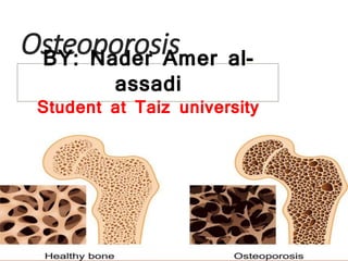
Osteoporosis
BY: Nader Amer al-assadi
Taiz university
1- Definition
2- Epidemiology
3- Risk factor
4-Pathophysiology
5-Classification
6- Osteoporosis Clinical Presentation.
7-Diagnosis
8-Treatment
9-Comblication
10-Prevention
what is Osteoporosis?
is a chronic, progressive disease of multifactorial etiology and it is the most common bone Metabolic disease in humans.
Characterized by:
Low bone mass
Microarchitectural deterioration
Compromised bone strength
Increased risk for fracture
normal minarlization
“Silent disease” until complicated by fractures.
incidence
Globally, osteoporosis is by far the most common metabolic bone disease, estimated to affect over 200 million people worldwide.
1.5 million osteoporotic fractures occur each year:
700,000 are vertebral fractures
300,000 are hip fractures
200,000 are wrist fracture
demographics
-The risk for osteoporosis increases with age as BMD declines. Senile osteoporosis is most common in persons aged 70 years or older.
-Secondary osteoporosis, however, can occur in persons of any age.
-male: female ratio is 1:4 postmenopausal woman
-Men have a higher prevalence of secondary osteoporosis, with an estimated 45-60% of cases being a consequence of hypogonadism, alcoholism, or glucocorticoid excess.
-Osteoporosis can occur in persons of all races and ethnicities. In general, however, whites (especially of northern European descent) and Asians are at increased risk .
The National Osteoporosis Foundation (NOF) has categorized the risk factors into two categories: nonmodifiable and modifiable:
Nonmodifiable risk factors include the following: - Personal history of fracture as an adult - History of fracture in a first-degree relative - White race - Advanced age? - Female sex - Poor health or fragility
This presentation includes four major topics:
1- reviews the essentials of osteoporosis including definition, pathophysiology, etiology, epidemiology, and prognosis
2- talks about the presentation of osteoporosis, including risk factors, symptoms and signs, radiologic manifestations, and complications
3- reviews the workup process to diagnose and define the severity of osteoporosis, including the lab. and radiologic procedures
4- reviews management tools of osteoporosis, including pharmacologic and non pharmacologic methods, with brief description for each pharmacologic or non pharmacologic tool.
Finally, some statements about the education and prevention of osteoporosis.
Osteoporosis is a condition characterized by a decrease in the density of bone, decreasing its strength and resulting in fragile bones. Know the Risk Factors for Osteoporotic Fracture, Preventive Measures and exercise for osteoporosis. For more health Tips, Visit at http://gisurgery.info
Osteoporosis is a disease in which bones become fragile and can easily break. It has no symptoms in its early stages and is a public health threat to more than 44 million Americans. In this community lecture given live on our Berkeley Heights, NJ campus, Dr. Toscano-Zukor, explains how to identify your risk factors for osteoporosis as well as prevent and treat this disease.
Osteoporosis is a progressive systemic skeletal disease characterized by low bone mass and microarchitecture deterioration of bone tissue, leading to enhanced bone fragility and a consequent increase in fracture risk.
Lumbar spondylosis- Diagnosis | management | a brief medical study martinshaji
Lumbar spondylosis is a degenerative condition which affects the lower spine. In a patient with lumbar spondylosis, the spine is compressed by a narrowing of the space between the vertebrae, causing a variety of health problems ranging from back pain tone urological problems.
please comment
thank you
Osteoporosis
BY: Nader Amer al-assadi
Taiz university
1- Definition
2- Epidemiology
3- Risk factor
4-Pathophysiology
5-Classification
6- Osteoporosis Clinical Presentation.
7-Diagnosis
8-Treatment
9-Comblication
10-Prevention
what is Osteoporosis?
is a chronic, progressive disease of multifactorial etiology and it is the most common bone Metabolic disease in humans.
Characterized by:
Low bone mass
Microarchitectural deterioration
Compromised bone strength
Increased risk for fracture
normal minarlization
“Silent disease” until complicated by fractures.
incidence
Globally, osteoporosis is by far the most common metabolic bone disease, estimated to affect over 200 million people worldwide.
1.5 million osteoporotic fractures occur each year:
700,000 are vertebral fractures
300,000 are hip fractures
200,000 are wrist fracture
demographics
-The risk for osteoporosis increases with age as BMD declines. Senile osteoporosis is most common in persons aged 70 years or older.
-Secondary osteoporosis, however, can occur in persons of any age.
-male: female ratio is 1:4 postmenopausal woman
-Men have a higher prevalence of secondary osteoporosis, with an estimated 45-60% of cases being a consequence of hypogonadism, alcoholism, or glucocorticoid excess.
-Osteoporosis can occur in persons of all races and ethnicities. In general, however, whites (especially of northern European descent) and Asians are at increased risk .
The National Osteoporosis Foundation (NOF) has categorized the risk factors into two categories: nonmodifiable and modifiable:
Nonmodifiable risk factors include the following: - Personal history of fracture as an adult - History of fracture in a first-degree relative - White race - Advanced age? - Female sex - Poor health or fragility
Osteoporosis is a chronic, progressive skeletal disease characterized by low bone mass, microarchitecture deterioration of bone tissue, bone fragility, and a consequent increase in fracture risk.
Osteopenia refers to decreased bone mass.
Osteoporosis refers to osteopenia (reduced bone strength/mass) that is severe enough to increase the risk of fracture.
According to WHO, osteoporosis is defined as bone mineral density that falls 2.5 standard deviation below mean for young healthy adult of same sex and race.
Osteoporosis associated fractures :
These are adulthood fractures of any bones (chiefly hip and vertebral fractures) in the setting of trauma less than or equal to fall from standing height with exception of fingers, toes, face and skull.
Drugs associated with osteoporosis
Alcohol
Glucocorticoids
Anticoagulants
Anticonvulsants
Chemotherapy
Excess thyroxine
Endocrine disorders
Cushing syndrome
Hyperparathyroidism
Thyrotoxicosis
Diabetes mellitus (both type I and II)
Acromegaly
CATEGORIZATION OF OSTEOPOROSIS
A.Primary
Idiopathic
Postmenopausal
Senile/age related
B. Secondary (Diseases)
Hypogonadal state, endocrine disorders, nutritional and gastrointestinal disorders, rheumatologic disorders, hematological disorders/malignancy, inherited disorders and others.
Usually asymptomatic until fracture occurs
Vertebral and hip fracture common by simple fall
Loss of height due to multiple vertebral fracture and other deformities like lordoisis, kyphoscoliosis.
Fracture of femur neck, pelvis or spine causes deep vein thrombosis and pulmonary embolism, pneumonia.
INVESTIGATIONS FOR OSTEOPOROSIS
DXA (Dual energy X-ray absorptiometry)
Quantitative CT
Ultrasound
Urea, creatinine and electrolytes
Liver function test and albumin
Renal function test
Full blood count, ESR
Serum calcium and phosphate
Serum vitamin D and alkaline phosphate
Serum PTH
Thyroid function test
Testosterone, estrogen and gonadotropins
Serum cortisol
Bone biopsy
Plain radiography not diagnostic
Following non pharmacological approaches are taken:
Exercise
Appropriate calcium and vitamin D intake (Calcium 1000mg/day and vitamin D 800 IU/daily)
Cessation of smoking
Limit/ Quit alcohol intake
Get up and go exercise
Hip protectors to reduce the risk of fracture.
Pharmacological agents
Bisphosphonates ( decrease osteoclast activity)
Postmenopausal hormone replacement therapy
Denusumab (anti- RANKL antibody)
Anti- sclerostin antibodies
Cathepsin k antibodies
Know everything about Osteoporosis- prevention and management.
Did You Know?
The incidence of hip fracture is 1 woman to 1 man in India
Know more such facts and useful information on prevention of Osteoporosis.
- Video recording of this lecture in English language: https://youtu.be/lK81BzxMqdo
- Video recording of this lecture in Arabic language: https://youtu.be/Ve4P0COk9OI
- Link to download the book free: https://nephrotube.blogspot.com/p/nephrotube-nephrology-books.html
- Link to NephroTube website: www.NephroTube.com
- Link to NephroTube social media accounts: https://nephrotube.blogspot.com/p/join-nephrotube-on-social-media.html
NVBDCP.pptx Nation vector borne disease control programSapna Thakur
NVBDCP was launched in 2003-2004 . Vector-Borne Disease: Disease that results from an infection transmitted to humans and other animals by blood-feeding arthropods, such as mosquitoes, ticks, and fleas. Examples of vector-borne diseases include Dengue fever, West Nile Virus, Lyme disease, and malaria.
New Drug Discovery and Development .....NEHA GUPTA
The "New Drug Discovery and Development" process involves the identification, design, testing, and manufacturing of novel pharmaceutical compounds with the aim of introducing new and improved treatments for various medical conditions. This comprehensive endeavor encompasses various stages, including target identification, preclinical studies, clinical trials, regulatory approval, and post-market surveillance. It involves multidisciplinary collaboration among scientists, researchers, clinicians, regulatory experts, and pharmaceutical companies to bring innovative therapies to market and address unmet medical needs.
Couples presenting to the infertility clinic- Do they really have infertility...Sujoy Dasgupta
Dr Sujoy Dasgupta presented the study on "Couples presenting to the infertility clinic- Do they really have infertility? – The unexplored stories of non-consummation" in the 13th Congress of the Asia Pacific Initiative on Reproduction (ASPIRE 2024) at Manila on 24 May, 2024.
These lecture slides, by Dr Sidra Arshad, offer a quick overview of physiological basis of a normal electrocardiogram.
Learning objectives:
1. Define an electrocardiogram (ECG) and electrocardiography
2. Describe how dipoles generated by the heart produce the waveforms of the ECG
3. Describe the components of a normal electrocardiogram of a typical bipolar leads (limb II)
4. Differentiate between intervals and segments
5. Enlist some common indications for obtaining an ECG
Study Resources:
1. Chapter 11, Guyton and Hall Textbook of Medical Physiology, 14th edition
2. Chapter 9, Human Physiology - From Cells to Systems, Lauralee Sherwood, 9th edition
3. Chapter 29, Ganong’s Review of Medical Physiology, 26th edition
4. Electrocardiogram, StatPearls - https://www.ncbi.nlm.nih.gov/books/NBK549803/
5. ECG in Medical Practice by ABM Abdullah, 4th edition
6. ECG Basics, http://www.nataliescasebook.com/tag/e-c-g-basics
Report Back from SGO 2024: What’s the Latest in Cervical Cancer?bkling
Are you curious about what’s new in cervical cancer research or unsure what the findings mean? Join Dr. Emily Ko, a gynecologic oncologist at Penn Medicine, to learn about the latest updates from the Society of Gynecologic Oncology (SGO) 2024 Annual Meeting on Women’s Cancer. Dr. Ko will discuss what the research presented at the conference means for you and answer your questions about the new developments.
Pulmonary Thromboembolism - etilogy, types, medical- Surgical and nursing man...VarunMahajani
Disruption of blood supply to lung alveoli due to blockage of one or more pulmonary blood vessels is called as Pulmonary thromboembolism. In this presentation we will discuss its causes, types and its management in depth.
Knee anatomy and clinical tests 2024.pdfvimalpl1234
This includes all relevant anatomy and clinical tests compiled from standard textbooks, Campbell,netter etc..It is comprehensive and best suited for orthopaedicians and orthopaedic residents.
HOT NEW PRODUCT! BIG SALES FAST SHIPPING NOW FROM CHINA!! EU KU DB BK substit...GL Anaacs
Contact us if you are interested:
Email / Skype : kefaya1771@gmail.com
Threema: PXHY5PDH
New BATCH Ku !!! MUCH IN DEMAND FAST SALE EVERY BATCH HAPPY GOOD EFFECT BIG BATCH !
Contact me on Threema or skype to start big business!!
Hot-sale products:
NEW HOT EUTYLONE WHITE CRYSTAL!!
5cl-adba precursor (semi finished )
5cl-adba raw materials
ADBB precursor (semi finished )
ADBB raw materials
APVP powder
5fadb/4f-adb
Jwh018 / Jwh210
Eutylone crystal
Protonitazene (hydrochloride) CAS: 119276-01-6
Flubrotizolam CAS: 57801-95-3
Metonitazene CAS: 14680-51-4
Payment terms: Western Union,MoneyGram,Bitcoin or USDT.
Deliver Time: Usually 7-15days
Shipping method: FedEx, TNT, DHL,UPS etc.Our deliveries are 100% safe, fast, reliable and discreet.
Samples will be sent for your evaluation!If you are interested in, please contact me, let's talk details.
We specializes in exporting high quality Research chemical, medical intermediate, Pharmaceutical chemicals and so on. Products are exported to USA, Canada, France, Korea, Japan,Russia, Southeast Asia and other countries.
Lung Cancer: Artificial Intelligence, Synergetics, Complex System Analysis, S...Oleg Kshivets
RESULTS: Overall life span (LS) was 2252.1±1742.5 days and cumulative 5-year survival (5YS) reached 73.2%, 10 years – 64.8%, 20 years – 42.5%. 513 LCP lived more than 5 years (LS=3124.6±1525.6 days), 148 LCP – more than 10 years (LS=5054.4±1504.1 days).199 LCP died because of LC (LS=562.7±374.5 days). 5YS of LCP after bi/lobectomies was significantly superior in comparison with LCP after pneumonectomies (78.1% vs.63.7%, P=0.00001 by log-rank test). AT significantly improved 5YS (66.3% vs. 34.8%) (P=0.00000 by log-rank test) only for LCP with N1-2. Cox modeling displayed that 5YS of LCP significantly depended on: phase transition (PT) early-invasive LC in terms of synergetics, PT N0—N12, cell ratio factors (ratio between cancer cells- CC and blood cells subpopulations), G1-3, histology, glucose, AT, blood cell circuit, prothrombin index, heparin tolerance, recalcification time (P=0.000-0.038). Neural networks, genetic algorithm selection and bootstrap simulation revealed relationships between 5YS and PT early-invasive LC (rank=1), PT N0—N12 (rank=2), thrombocytes/CC (3), erythrocytes/CC (4), eosinophils/CC (5), healthy cells/CC (6), lymphocytes/CC (7), segmented neutrophils/CC (8), stick neutrophils/CC (9), monocytes/CC (10); leucocytes/CC (11). Correct prediction of 5YS was 100% by neural networks computing (area under ROC curve=1.0; error=0.0).
CONCLUSIONS: 5YS of LCP after radical procedures significantly depended on: 1) PT early-invasive cancer; 2) PT N0--N12; 3) cell ratio factors; 4) blood cell circuit; 5) biochemical factors; 6) hemostasis system; 7) AT; 8) LC characteristics; 9) LC cell dynamics; 10) surgery type: lobectomy/pneumonectomy; 11) anthropometric data. Optimal diagnosis and treatment strategies for LC are: 1) screening and early detection of LC; 2) availability of experienced thoracic surgeons because of complexity of radical procedures; 3) aggressive en block surgery and adequate lymph node dissection for completeness; 4) precise prediction; 5) adjuvant chemoimmunoradiotherapy for LCP with unfavorable prognosis.
3. what is Osteoporosis?
is a chronic, progressive disease of multifactorial etiology and it is
the most common bone Metabolic disease in humans.
Characterized by:
◦ Low bone mass
◦ Microarchitectural deterioration
◦ Compromised bone strength
◦ Increased risk for fracture
◦ “Silent disease” until complicated by fractures
4. Epidemiology
incidence
Globally, osteoporosis is by far the most common metabolic bone disease, estimated to
affect over 200 million people worldwide.
1.5 million osteoporotic fractures occur each year:
700,000 are vertebral fractures
300,000 are hip fractures
200,000 are wrist fracture
demographics
-The risk for osteoporosis increases with age as BMD declines. Senile osteoporosis is most
common in persons aged 70 years or older.
-Secondary osteoporosis, however, can occur in persons of any age.
-male: female ratio is 1:4 postmenopausal woman
-Men have a higher prevalence of secondary osteoporosis, with an estimated 45-60% of cases
being a consequence of hypogonadism, alcoholism, or glucocorticoid excess.
-Osteoporosis can occur in persons of all races and ethnicities. In general, however, whites
(especially of northern European descent) and Asians are at increased risk .
5. (NOF) has
categorized the risk factors into two
categories: nonmodifiable and
modifiable:
Nonmodifiable risk factors include the following:
- Personal history of fracture as an adult
- History of fracture in a first-degree relative
- White race
- Advanced age
- Female sex
- Dementia
- Poor health or fragility
16 September 2021
6. Cont…
Potentially modifiable risk factors include the following:
- Current cigarette smoking
- Low body weight (< 127 lb)
- Estrogen deficiency such as that caused by early menopause (age <
45 years) or - - bilateral ovariectomy and prolonged premenopausal
amenorrhea (>1 year)
- Low lifelong calcium intake
- Alcoholism
- Impaired eyesight despite adequate correction
- Recurrent falls
- Inadequate physical activity
- Poor health or frailty
16 September 2021
7. Risk Factors
Major
History of fracture as an adult
Fragility fracture in first degree
relative
Caucasian/Asian
postmenopausal woman
Low body weight
Current smoking
Use of oral corticosteroids >
3mo.
Additional
Estrogen deficiency at early age
(< 45 YO)
Poor health/frailty
Recent falls
Low calcium intake (lifelong)
Low physical activity
> 2 alcoholic drinks per day
8. Medical Conditions Associated with Increased
Risk of Osteoporosis
COPD
Cushing’s syndrome
Eating disorders
Hyperparathyroidism
Hypophosphatasia
IBS
RA, other autoimmune
connective tissue disorders
Insulin dependent diabetes
Multiple sclerosis
Multiple myeloma
Stroke (CVA)
Thyrotoxicosis
Vitamin D deficiency
Liver diseases
Not an inclusive list
9. Drugs Associated with
Reduced Bone Mass
Aluminum
Anticonvulsants
Cytotoxic drugs
Glucocorticosteroids
(oral/high dose inhaled)
Immunosuppresants
Gonadotropin-releasing
hormone (e.g. Lupron)
Lithium
Heparin (chronic use)
Supraphysiologic thyroxine
doses
Aromatase inhibitors
Depo-Provera
Not an inclusive list
10. A potentially useful mnemonic for osteoporotic risk factors
is OSTEOPOROSIS, as follows:
LOw calcium intake
Seizure meds (anticonvulsants)
Thin build
Ethanol intake
HypOgonadism
Previous fracture
ThyrOid excess
Race(white, Asian)
Other relatives with osteoporosis
Steroids
Inactivity
Smoking
11.
12. Pathophysiology
It is increasingly being recognized that
multiple pathogenetic mechanisms interact in
the development of the osteoporotic state.
Understanding the pathogenesis of
osteoporosis starts with knowing how bone
formation and remodeling occur.
13. remodeling
Bone undergoes both radial and longitudinal growth and is continually
remodeled throughout our lives in response to microtrauma. Bone remodeling
renews bone strength and mineral, preventing the accumulation of damaged
bone.
Bone remodeling occurs at discrete sites within the skeleton and proceeds in
an orderly fashion, and bone resorption is always followed by bone formation,
a phenomenon referred to as coupling.
14.
15.
16. Alterations in bone formation and resorption
Osteoporosis is multifactorial with an interplay of genetic, intrinsic,
exogenous, and lifestyle factors.The hallmark of osteoporosis is a
reduction in skeletal mass caused by an imbalance between bone
resorption and bone formation.
17. Estrogen deficiency leads to increased expression of RANKL by osteoblasts and decreased
release of OPG increased RANKL results in recruitment of higher numbers of
preosteoclasts as well as increased activity, vigor, and lifespan of mature osteoclasts.
Alterations in bone formation and resorption
Estrogen deficiency
18.
19. Calcium and vitamin D deficiency
Calcium, vitamin D, and PTH help maintain bone homeostasis.
Insufficient dietary calcium or impaired intestinal absorption of calcium
due to aging or disease can lead to secondary hyperparathyroidism.
PTH is secreted in response to low serum calcium levels. It increases
calcium resorption from bone decreases renal calcium excretion, and
increases renal production of 1,25-dihydroxyvitamin D (1,25[OH]2 D)—
an active hormonal form of vitamin D that optimizes calcium and
phosphorus absorption, inhibits PTH synthesis, and plays a minor role
in bone resorption.
21. Type of Primary Osteoporosis Characteristics
Juvenile osteoporosis
Usually occurs in children or young
adults of both sexes
Normal gonadal function
Age of onset: usually 8-14 years
Hallmark characteristic: abrupt bone
pain and/or a fracture following trauma.
Idiopathic osteoporosis
Postmenopausal osteoporosis
(type I osteoporosis)
Occurs in women with estrogen
deficiency
Characterized by a phase of accelerated
bone loss, primarily from trabecular
bone Fractures of the distal forearm and
vertebral bodies common.
Age-associated or senile
osteoporosis (type II osteoporosis)
Occurs in women and men as BMD
gradually declines with aging
Represents bone loss associated with
aging Fractures occur in cortical and
trabecular bone Wrist, vertebral, and hip
fractures often seen
primary osteoporosis
22. Secondary osteoporosis
Secondary osteoporosis occurs when an underlying disease, deficiency, or drug
causes osteoporosis
Cause Examples
Genetic/congenital
Renal hypercalciuria – one of the most important secondary causes
of osteoporosis; can be treated with thiazide diuretics
Cystic fibrosis
Ehlers-Danlos syndrome
Glycogen storage disease
Gaucher disease
Marfan syndrome
Menkes steely hair syndrome
Riley-Day syndrome
Osteogenesis imperfecta
Hemochromatosis
Homocystinuria
Idiopathic hypercalciuria
Hypogonadal states
Hypogonadal
states
Androgen insensitivity
Anorexia nervosa/bulimia nervosa
Female athlete triad
Hyperprolactinemia
Panhypopituitarism
Premature menopause
Turner syndrome
Klinefelter syndrom
25. Osteoporosis Clinical Presentation
History:
Keep in mind that osteoporosis occurs in many people who have few or no risk factors for this
condition. Often, patients who have not sustained a fracture do not report symptoms that
would alert the clinician to suspect a diagnosis of osteoporosis; thus, this disease is a "silent
thief" that generally does not become clinically apparent until a fracture occurs.
So the history should focus in:
- Age (> 50 years), sex (female), and race (white or Asian)
- Family history of osteoporosis, particularly maternal history of fractures.
- Reproductive factors, especially regarding early menopause and estrogen replacement
therapy.
- ypogonadal states: men with hypogonadism secondary to any genetic or other conditions
are at higher risk.
16 September 2021
26. Cont…
- Smoking: smokers are at higher risk
- Alcohol consumption
- Low levels of physical activity: immobility increases the risk [65] ; spinal
cord injury and stroke cause physical impairment and are common causes
of immobility.
- Strenuous exercise that results in amenorrhea (such as that which occurs
in marathon runners)
- Calcium and vitamin D intake
- History of low-trauma "fragility" fracture in patients aged 40 years or
older.
-Coexisting medical conditions associated with bone
loss:hyperparathyroidism, hypogonadism, leukemia, rheumatoid arthritis,
celiac disease, and Cushing syndrome.
- Medications associated with bone loss: examples are glucocorticoids,
cyclosporine,anticonvulsant.
16 September 2021 by nader al_ assadi
27. Physical Examination
Patients with suspected osteoporosis should undergo a
comprehensive physical examination.
•The physical examination should begin with an inspection of the patient. Height
measurement with a stadiometer at each visit may be useful.
• Examination of active and passive range of motion (ROM) assists in determining
whether spine, hip, wrist, or other osseous pathology may be present.
•Athorough neurologic examination is essential to rule out spinal cord and/or
peripheral nerve compromise.
•Sign of fracture (eg:Patients with vertebral compression fractures may have point
tenderness over the involved vertebrae and demonstrate a thoracic kyphosis with
an exaggerated cervical lordosis.
•signs of collagen defects : Patients with osteoporosis may have physical findings
consistent with subtle collagen defects. These include a short fifth digit,
hyperlaxity, hearing loss.
•Balance difficulties :Patients with osteoporosis are known to have decreased
balance, possibly secondary to differences in balance control strategies and sway
amplitude. Patients may have difficulty performing tandem gait and performing
single limb stance. Poor balance may be noted particularly in patients with severe
kyphosis resulting from vertebral compression fractures because their altered
center of gravity makes ambulation with a stable base of support difficult for them.
28.
29. Diagnosis
lap
25 hydroxyvitamin D level
- low 25 hydroxy cholecalciferol levels (25 hydroxy vit D) in patients sustaining low energy.
IMAGE
Radiographs
indications
• suspicion of fracture
• loss of height
• pain in thoracic or lumbar spine
• recommended views
• lateral spine radiograph
• AP pelvis or hip
• findings
• thinned cortices
• loss of trabecular bone
• kyphosis
• codfish vertebra
31. Radiographic findings can suggest the presence of osteopenia, or bone loss, but
cannot be used to diagnose osteoporosis. Osteopenia is suggested by a cortical width
that is less than the medullary width. Radiographs may also show fractures.
Plain radiography is not as accurate as BMD testing. Because osteoporosis
predominantly affects trabecular bone rather than cortical bone, radiography does
not reveal osteoporotic changes until they affect the cortical bone. Cortical bone is
not affected by osteoporosis until more than 30% of bone loss has occurred.
Approximately 30-80% of bone mineral must be lost before radiographic lucency
becomes apparent on radiographs. Thus, plain radiography is an insensitive tool for
diagnosing osteoporosis.
1-Plain x ray
38. The 2020 update of the American Association of Clinical
Endocrinologists (AACE) guidelines provides the following
criteria for the diagnosis of osteoporosis in postmenopausal
women :
1- T-score -2.5 or below in the lumbar spine,
femoral neck, total proximal femur, or 1/3 radius.
2- Low-trauma spine or hip fracture (regardless of
BMD)
3- T-score between -1.0 and -2.5 and a fragility
fracture of proximal humerus, pelvis, or distal
forearm.
4- T-score between -1.0 and -2.5and high
FRAX(Fracture Risk Assessment Tool )
39. Quantitative Computed
Tomography
QCT scanning of the spine is the most sensitive method for
diagnosing osteoporosis, because it measures trabecular
bone within the vertebral bon.
1- QCT is a very sensitive technique when repeated
measurements are needed to detect small changes in
BMD,
2- modern three-dimensional (3D) QCT acquisition has a
scan time less than 10 seconds for the lumbar spine or
proximal femur.
3- and there is no interference by osteophytes.
40. Single-photon emission computed
tomography (SPECT)
represents a tomographic (CT-like) bone imaging
technique that offer:
1- better image contrast
2- more accurate lesion localization than planar bone
scanning.
3- PECT scanning is helpful when accurate localization of
skeletal lesions within large and/or anatomically complex
bony structures is required.(no bone overlap)
41. Quantitative Ultrasonography
Quantitative ultrasonography (QUS) of the calcaneus is a low-cost
portable screening tool. It has the advantage of not involving
radiation, but it is not as accurate as other imaging methods.
Ultrasonography cannot be used for monitoring skeletal changes over
time, nor can it be used to monitor the response to treatment,
because of its lack of precision.
42. Magnetic Resonance Imaging
These osteoporotic fractures demonstrate characteristic changes in
the bone marrow that distinguish them from other uninvolved parts of
the skeleton and the adjacent vertebra.
43. Bone Scanning
one scans assesses the function and tissue metabolism of organs by
using a radionuclide (technetium-99m [99m Tc]) that emits radiation in
proportion to its attachment to a target structure.
This technique detects an increase in osteoblastic activity (as seen in
compression fractures.
44. Bone Biopsy and Histologic Features.
Bone biopsy can help to exclude underlying pathologic conditions,
such as mastocytosis, that may be responsible for presumed
osteoporotic fracture. Typically, iliac crest biopsy is performed either
in the minor procedure suite or in the operating room.
Histologic examination of osteoporotic bone may reveal generalized
thinning of trabeculae and irregular perforation of trabeculae,
reflecting unbalanced osteoclast-mediated bone resorption.
46. Regular Weight-Bearing Exercise
Defined as those in which bones and muscles work
against gravity as feet and legs bear the body’s weight
-Include walking, stair climbing, dancing, tennis, yoga.
-Improve agility, strength, balance.
-May increase bone density modestly, reduce fall risk,
enhance muscle strength, improve balance.
47. Avoidance of Tobacco and Alcohol
Tobacco products detrimental to skeleton, overall health
.NOF National Osteoporosis Foundation (strongly
encourages tobacco cessation programs as osteoporosis
intervention) .
48.
49. Avoidance of Tobacco and Alcohol
Excessive alcohol
intake also
detrimental to
bone health and
requires
treatment.
50. Adequate Intake of Calcium/Vitamin D
Adequate intakes of dietary calcium and vitamin D,
including supplements if necessary
◦ Elemental calcium per day at least 1200 -1500 mg.
◦ Vitamin D3 per day 800 -1000 international units (IU).
Vitamin D3 (cholecalciferol) plays major role in Ca
absorption Controlled clinical trials have demonstrated
the combination reduces fracture risk Inexpensive, well-
tolerated.
51. Calcium/D Product Selection
Product (% elemental
Ca)
Elemental
Calcium
(mg)
Vitamin
D (units)
Comments
Calcium carbonate
(40)
-Tums Ultra
-Caltrate 600 Plus
-Oscal Plus D
-Viactiv Chews
400
600
500
500
200
125
100
Requires acidic environment for dissolution and
disintegration. Best to take with meals.
Greater risk for constipation with carbonate
form.
Calcium citrate (24)
-Citracal Plus D
- Citracal Petites with
VitD
315
200
200
200
Take without regard to meals. Serving size
usually equals 2 capsules so label can be
misleading to patients.
Vitamin D
-Multivitamin (D3)
-Vitamin D
120-450 400
100-400
52. Vitamin D and Fall Risk
In addition to its effect on BMD, may contribute to reduction in fracture risk
◦ Improved muscle function(o+R+c)
◦ Reduction in risk for falls
Vitamin D deficiency prevalent in older adult population
◦ Inadequate sun exposure, use of sunscreen
◦ Homebound, institutionalized
◦ Maintain 25-hydroxyvitamin D3 at least > 40 ng/mL
◦ Treatment: 50,000 IU vitD weekly x 6-8 weeks, then assess need for chronic
monthly therapy
54. Who Should Be Treated?
NOF Recommendations – 2008
Initiate therapy to reduce fractures in
postmenopausal women/men > 50 with:
1. BMD T-scores < -2.5 at hip or spine
2. Prior vertebral or hip fracture
3. Low bone mass (T-scores -1.0 to -2.5 at hip or spine) when:
– 10-year probability of hip fracture is > 3%
– 10-year probability of major osteoporosis-related fracture is > 20%
– Based on US-adapted WHO algorithm
www.nof.org
56. Bisphosphonates – Antiresorptive Agents
Agents FDA-approved for:
◦ Prevention and treatment of osteoporosis in postmenopausal women
◦ Treatment to increase bone mass in men with osteoporosis
◦ Treatment of glucocorticoid-induced osteoporosis in men and women
receiving glucocorticoids
◦ Treatment of Paget’s disease of bone in men and women
Mechanism: inhibits bone resorption by attaching to bony surfaces
undergoing active resorption and inhibiting action of osteoclasts
◦ Leads to increases in bone density and reduced fracture risk
57. Bisphosphonates
Very well tolerated in patients who adhere to proper
administration techniques
Proper patient counseling for correct administration is
KEY to reduce risk of adverse effects and increase
tolerability
Place in Therapy: should be considered first-line for
prevention/treatment of osteoporosis in patients with
no contraindications.
58. Bisphosphonates – Clinical
Efficacy
Controlled clinical trials indicate over 3-4 year period, alendronate ↑ bone mass
and ↓ incidence of vertebral, hip, and all non-vertebral fractures by 50%
Controlled clinical trials indicate risedronate ↑ bone mass and ↓ risk of
vertebral fractures by 40% and non-vertebral fractures by 30% over 3-year
period
Ibandronate has been shown in controlled clinical trials to ↑ BMD and reduce
the risk of vertebral fracture by 50% over 3-year period
Alendronate appears to be well tolerated and effective for at least ten years
59. Zolendronic Acid (Reclast®)
Approved for treatment of osteoporosis in postmenopausal
women in August 2007
Single 5 mg infusion given IV over > 15 minutes, once yearly
Should still supplement with calcium/vitamin D
May be ideal for those with GI contraindications to the oral
formulations.
60. SERMs – Raloxifene
FDA-approved for:
◦ Prevention and treatment of osteoporosis in postmenopausal women
Mechanism: tissue-selective activity, acts as an estrogen agonist on bone
◦ Estrogen antagonist on breast, uterus.
61. Raloxifene
Place in Therapy: considered first-line in women who
cannot tolerate bisphosphonates and have no
contraindications to therapy.
Combination therapy (usually a bisphosphonate with a
non-bisphosphonate) can provide additional small
increases in BMD when compared to monotherapy.
Impact of combination therapy on fracture rate unknown
62. Estrogen/Hormone Therapy
(ET/HT)
FDA approved for:
◦ Prevent osteoporosis
◦ Treatment of moderate/severe vasomotor symptoms of
menopause
◦ Treatment of moderate/severe symptoms of vulvar and vaginal
atrophy associated with menopause
◦ Consider topical preparations to treat vaginal symptoms rather
than oral ET/HT
63. FDA Recommendations –
ET/HT
When prescribing medications for osteoporosis,
physicians should consider all non-estrogen therapies
first.
When prescribing ET/HT, use smallest dose for shortest
amount of time to achieve treatment goals.
Prescribe ET/HT products only when benefits believed to
outweigh risks for a specific patient.
64. Calcitonin
FDA-approved for:
◦ Treatment of osteoporosis in women who are > 5 years
postmenopausal
◦ Treatment of Paget’s disease of bone.
◦ Adjunctive therapy for hypercalcemia.
Mechanism:
◦ Peptide composed of 32 amino acids which binds to
osteoclasts and inhibits bone resorption .
◦ Promotes the renal excretion of calcium, phosphate, sodium,
magnesium and potassium by decreasing tubular reabsorption.
65. Calcitonin – Clinical Efficacy
Has been shown to increase spinal bone mass and may
decrease risk of vertebral fracture .
Conflicting data on efficacy of calcitonin at sites other
than the spine.
Less effective than bisphosphonates in treatment of
osteoporosis.
Beneficial, short-term effect on acute bone pain after
osteoporotic fracture (vertebral).
66. Calcitonin
Valid option for treatment of established osteoporosis,
especially when accompanied by fracture pain
Place in therapy: because of cost, adverse effects,
inconvenience of nasal administration, recommend using
calcitonin until pain is no longer a problem and then
switching to a bisphosphonate for long-term therapy
67. Parathyroid Hormone [PTH (1-34)]
Anabolic agent
FDA-approved for:
◦ Treatment of osteoporosis in postmenopausal women at high risk for
fracture
◦ previous osteoporotic fracture, multiple risk factors for fracture, extremely low
BMD (< -2.5), or failed/intolerant to previous treatment
◦ Treatment of primary or hypogonadal osteoporosis in men at high risk of
fracture
Mechanism: recombinant formulation of endogenous
parathyroid hormone (PTH)
◦ stimulates osteoblast function, increases gastrointestinal calcium
absorption, increases renal tubular reabsorption of calcium
◦ Enhances bone turnover by initiating greater bone formation
68. Guidelines from the American Association of Clinical Endocrinologists
(AACE), updated in 2020, include the following recommendations for
choosing drugs to treat osteoporosis in postmenopausal women :
- First-line agents for most high fracture risk patients: alendronate,
risedronate, zoledronate,Denosumab.
- First-line agents for high fracture risk patients unable to use oral
therapy: abaloparatide, denosumab, romosozumab, teriparatide, and
zoledronate.
- First-line agents for spine-specific indications in select patients:
ibandronate and raloxifene
- Sequential agents: anabolic agents (eg, abaloparatide,
romosozumab, teriparatide) should be followed with a bisphosphonate
or denosumab.
Combination therapy with two or more agents has not been shown to
have a greater effect on fracture reduction than single therapy .
69. Approaches to Monitoring Therapy
Always important to ask patients about adherence, encourage
continuation of therapies to reduce fracture risk
Monitoring of therapy should be considered, as up to 1/6 of
women taking effective therapies continue to lose bone, especially
if they smoke
May measure bone mineral density at a single site after one year
of therapy, but results may be misleading; usually done every 2
years
Drugs may decrease a patient’s risk for fracture even when there is
no apparent increase in BMD.
70. Complication of osteoporosis
1-vertebral compression fractures often occur with minimal stress, such as
coughing, lifting, or bending.
2- Hip fractures are the most devastating and occur most commonly at the
femoral neck and intertrochanteric regions . Hip fractures are associated with
falls. The likelihood of sustaining a hip fracture during a fall is related to the
direction of the fall. Fractures are more likely to occur in falls to the side
because less subcutaneous tissue is available to dissipate the impact.
3-Fractures can cause further complications, including chronic pain from
vertebral compression fractures and increased morbidity and mortality
secondary to vertebral compression fractures and hip fracture. They are also at
risk for the complications associated with immobility, including deep vein
thrombosis (DVT) and pressure ulcers. Respiratory compromise can occur in
patients with multiple vertebral fractures that result in severe kyphosis.
4- patients with osteoporosis develop spinal deformities and a dowager's
hump, and they may lose 1-2 inches of height by their seventh decade of life.
These patients can lose their self-esteem and are at increased risk for
depression.
71.
72. Prevention of Osteoporosis
primary prevention of osteoporosis starts in childhood. Patients require adequate
calcium intake, vitamin D intake, and weight-bearing exercis .
beyond this, prevention of osteoporosis has two:
behavior
modification
pharmacologi
c
interventions
73. behavior modification
Patients should be counseled on :
1- smoking cessation
2- moderation(stop) of alcohol intake.
3- Regular weight-bearing exercise and back extensor strengthening
help delay bone loss.
4- sunbath 15 minute
74. pharmacologic interventions
1- calcium supplementation and
2-administration of raloxifene or
3-bisphosphonates (alendronate or risedronate).
*Bisphosphonates should be considered as first-line agents for the
prevention of osteoporosis.
75.
76. References
1) American Association of Clinical Endocrinologists medical guidelines
for clinical practice for the prevention and treatment of
postmenopausal osteoporosis: 2020 update. Endocr Pract.
May2020; 26(Suppl 1):1-46. (2) Kelman A, Lane NE. The management of
secondary osteoporosis. Best Pract Res Clin
Rheumatol. Dec 2005;19(6):1021-37.
MEDSCape Author: Rachel Elizabeth Whitaker Elam, MD, MSc; Chief
Editor: Herbert S Diamond, MD
77. References
Actonel®
Prescribing Information (www.actonel.com)
Ann Intern Med 1990;112:352
Ann Intern Med 2006;144:753
Boniva®
Prescribing Information (www.boniva.com)
Clinical Reviews in Bone and Mineral Metabolism 2004;2(4):291
Evista®
Prescribing Information (www.evista.com)
Forteo®
Prescribing Information (www.forteo.com)
Fortical®
Prescribing Information (www.fortical.com)
Fosamax®
Prescribing Information (www.fosamax.com)
78. References
JAMA 2004;291(16):1999
J Clin Densitom 2004;7(1):1-6
J Am Acad Orthop Surg 2006;14:347
Miacalcin®
Prescribing Information (www.miacalcin.com)
Reclast®
Prescribing Information (www.reclast.com)
National Osteoporosis Foundation (http://www.nof.org)
NEJM 2003;348:1187
NEJM 2004;350(12):1189-99
Osteoporosis Int 1998;8:1