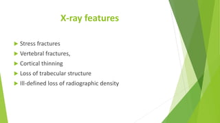
Metabolic and endocrine bone
disorders
Osteoporosis
Introduction
Patients with metabolic bone disorders usually appear to the
orthopaedic surgeon in one of the following guises:
• A child with bone deformities (rickets)
• An elderly with a fracture of femoral neck or vertebral body
• An elderly patient with bone pain and multiple compression fractures
• A middle-aged person with hypercalcaemia and pseudogout
• Someone with multiple fractures and history of prolonged
corticosteroid treatment
X-ray features
 Stress fractures
 Vertebral fractures,
 Cortical thinning
 Loss of trabecular structure
 Ill-defined loss of radiographic density
History
 Failure to thrive
 Below-normal growth
 Deformity of the lower limbs
 Back pain
 Generalized muscle weakness
Cont’d
 Patient’s sex and age
 Race
 Onset of menopause
 Nutritional background
 Level of physical activity
 Previous illnesses medication and operations
 Onset and duration of symptoms and their relationship to previous
disease
cont’d
 Retarded growth
 Malnutrition
 Dietary fads
 Intestinal malabsorption
 Alcohol abuse and cigarette smoking
Examination
 Moon face
 Cushingoid build
 Smooth, hairless skin of testicular atrophy
 Physical underdevelopment and bone deformities.
 Thoracic kyphosis
X-rays
 Decreased skeletal radiodensity
 Reduction in mineral or skeletal mass
 Presence of obvious fractures
 Small stress fractures
Measurement of bone mass
 Dual-energy X-ray absorptiometry (DXA) scan
Indications for bone densitometry
 Adults over the age of 50 who have experienced a low
trauma fracture
 All women over the age of 65
 All men over the age of 75
 To assess risk of future fracture
 To assess the degree and progress of bone loss
 To monitor the effect of treatment for osteoporosis.
Biochemical tests
 Serum calcium and phosphate concentrations should
be measured in the fasting state
 Raised in osteomalacia and in disorders associated with
high bone turnover
Bone biopsy
 Are obtained from the iliac crest
 Examined for histological bone volume, osteoid
formation and relative distribution of formation and
resorption surfaces
Osteoporosis
Definition
 Abnormally low bone mass
 Defects in bone structure, a combination which renders
the bone unusually fragile
Cause
 Predominant bone resorption, decreased bone
formation or a combination of the two
Risk factors for osteoporosis
 Age
 Female
 Previous fragility fracture
 Current use or frequent recent use of oral or systemic
glucocorticoids
 Family history of hip fracture
 Low body mass index (BMI) (less than 18.5 kg/m2)
 Smoking
 Alcohol intake of more than 14 units per week for
women and more than 21 units per week for men
X-rays
 Loss of trabecular definition,
 Thinning of the cortices
 Insufficiency fractures.
 Compression fractures of the vertebral bodies
Postmenopausal Osteoporosis
 It is an exaggerated form of the physiological bone
depletion that normally accompanies ageing and loss of
gonadal activity
Clinical features and investigations
 Osteoporosis is asymptomatic unless fractures occur.
 The fractures are classically low trauma
 Fracture of the distal radius (Colles’ fracture) is usually
the first fracture
 Osteoporotic vertebral fractures
 Significant height loss (often exceeding 4 cm) and
thoracic kyphosis
 Height loss and smaller kyphoses are most commonly
due to degenerative change.
Cont’d
 Assessment of fracture risk
 The rate of bone turnover is either normal or slightly
increased
Prevention
 Medications to reduce fracture risk.
 Primary screening for people who have not sustained
 Women should be advised on lifestyle choices to
maintain healthy bones.
Treatment
 The goal is to reduce risk of future fracture.
 Medications recommended for 3–5 years, after which
ongoing treatment should be reconsidered
 Bisphosphonates; taken orally
 Zoledronate can be given once per year intravenously.
 Denosumab subcutaneously injected every 6 months
 Parathyroid hormone Preotact and Teriparatide
 Selective oestrogen receptor modulators (SERMs)
Cont’d
 Strontium salt of ranelic acid, given as a sachet of
granules to be dissolved in water and drunk once per
day.
 Operative treatment.
 Analgesic treatment.
 Physiotherapy
 Postural training when symptoms allow.
 Spinal orthoses maybe needed for support and pain
relief
 Vertebral augmentation such as kyphoplasty or
vertebroplasty
Secondary causes of Osteoporosis
 Endocrine; Hypogonadism in either sex
 Treatment with aromatase inhibitors or androgen deprivation
therapy
 Hyperthyroidism
 Hyperparathyroidism
 Hyperprolactinaemia
 Cushing’s disease and Diabetes
Cont’d
 Respiratory; Cystic fibrosis and Smoking-related lung disease
 Metabolic; Homocystinuria
 Chronic renal disease
 Gastrointestinal; Coeliac disease, Inflammatory bowel disease,
Chronic liver disease
 Rheumatological; Rheumatoid arthritis and Other inflammatory
arthropathies
 Haematological; Multiple myeloma, Haemoglobinopathies and
Systemic mastocytosis
 Immobility; Neurological injury and Neurological disease

Metabolic and endocrine bone disorders.pdf

  • 1.
    Metabolic and endocrinebone disorders Osteoporosis
  • 2.
    Introduction Patients with metabolicbone disorders usually appear to the orthopaedic surgeon in one of the following guises: • A child with bone deformities (rickets) • An elderly with a fracture of femoral neck or vertebral body • An elderly patient with bone pain and multiple compression fractures • A middle-aged person with hypercalcaemia and pseudogout • Someone with multiple fractures and history of prolonged corticosteroid treatment
  • 3.
    X-ray features  Stressfractures  Vertebral fractures,  Cortical thinning  Loss of trabecular structure  Ill-defined loss of radiographic density
  • 4.
    History  Failure tothrive  Below-normal growth  Deformity of the lower limbs  Back pain  Generalized muscle weakness
  • 5.
    Cont’d  Patient’s sexand age  Race  Onset of menopause  Nutritional background  Level of physical activity  Previous illnesses medication and operations  Onset and duration of symptoms and their relationship to previous disease
  • 6.
    cont’d  Retarded growth Malnutrition  Dietary fads  Intestinal malabsorption  Alcohol abuse and cigarette smoking
  • 7.
    Examination  Moon face Cushingoid build  Smooth, hairless skin of testicular atrophy  Physical underdevelopment and bone deformities.  Thoracic kyphosis
  • 8.
    X-rays  Decreased skeletalradiodensity  Reduction in mineral or skeletal mass  Presence of obvious fractures  Small stress fractures
  • 9.
    Measurement of bonemass  Dual-energy X-ray absorptiometry (DXA) scan
  • 10.
    Indications for bonedensitometry  Adults over the age of 50 who have experienced a low trauma fracture  All women over the age of 65  All men over the age of 75  To assess risk of future fracture  To assess the degree and progress of bone loss  To monitor the effect of treatment for osteoporosis.
  • 11.
    Biochemical tests  Serumcalcium and phosphate concentrations should be measured in the fasting state  Raised in osteomalacia and in disorders associated with high bone turnover
  • 12.
    Bone biopsy  Areobtained from the iliac crest  Examined for histological bone volume, osteoid formation and relative distribution of formation and resorption surfaces
  • 13.
  • 14.
    Definition  Abnormally lowbone mass  Defects in bone structure, a combination which renders the bone unusually fragile
  • 15.
    Cause  Predominant boneresorption, decreased bone formation or a combination of the two
  • 16.
    Risk factors forosteoporosis  Age  Female  Previous fragility fracture  Current use or frequent recent use of oral or systemic glucocorticoids  Family history of hip fracture  Low body mass index (BMI) (less than 18.5 kg/m2)  Smoking  Alcohol intake of more than 14 units per week for women and more than 21 units per week for men
  • 17.
    X-rays  Loss oftrabecular definition,  Thinning of the cortices  Insufficiency fractures.  Compression fractures of the vertebral bodies
  • 18.
    Postmenopausal Osteoporosis  Itis an exaggerated form of the physiological bone depletion that normally accompanies ageing and loss of gonadal activity
  • 19.
    Clinical features andinvestigations  Osteoporosis is asymptomatic unless fractures occur.  The fractures are classically low trauma  Fracture of the distal radius (Colles’ fracture) is usually the first fracture  Osteoporotic vertebral fractures  Significant height loss (often exceeding 4 cm) and thoracic kyphosis  Height loss and smaller kyphoses are most commonly due to degenerative change.
  • 20.
    Cont’d  Assessment offracture risk  The rate of bone turnover is either normal or slightly increased
  • 21.
    Prevention  Medications toreduce fracture risk.  Primary screening for people who have not sustained  Women should be advised on lifestyle choices to maintain healthy bones.
  • 22.
    Treatment  The goalis to reduce risk of future fracture.  Medications recommended for 3–5 years, after which ongoing treatment should be reconsidered  Bisphosphonates; taken orally  Zoledronate can be given once per year intravenously.  Denosumab subcutaneously injected every 6 months  Parathyroid hormone Preotact and Teriparatide  Selective oestrogen receptor modulators (SERMs)
  • 23.
    Cont’d  Strontium saltof ranelic acid, given as a sachet of granules to be dissolved in water and drunk once per day.  Operative treatment.  Analgesic treatment.  Physiotherapy  Postural training when symptoms allow.  Spinal orthoses maybe needed for support and pain relief  Vertebral augmentation such as kyphoplasty or vertebroplasty
  • 24.
    Secondary causes ofOsteoporosis  Endocrine; Hypogonadism in either sex  Treatment with aromatase inhibitors or androgen deprivation therapy  Hyperthyroidism  Hyperparathyroidism  Hyperprolactinaemia  Cushing’s disease and Diabetes
  • 25.
    Cont’d  Respiratory; Cysticfibrosis and Smoking-related lung disease  Metabolic; Homocystinuria  Chronic renal disease  Gastrointestinal; Coeliac disease, Inflammatory bowel disease, Chronic liver disease  Rheumatological; Rheumatoid arthritis and Other inflammatory arthropathies  Haematological; Multiple myeloma, Haemoglobinopathies and Systemic mastocytosis  Immobility; Neurological injury and Neurological disease