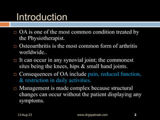
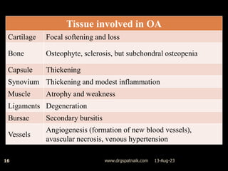
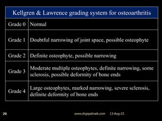
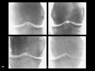
OSTEOARTHRITIS
OR
O = Old age,
A = Arthritis
By:
G S Patnaik
Introduction
 OA is one of the most common condition treated by
the Physiotherapist.
 Osteoarthritis is the most common form of arthritis
worldwide..
 It can occur in any synovial joint; the commonest
sites being the knees, hips & small hand joints.
 Consequences of OA include pain, reduced function,
& restriction in daily activities.
 Management is made complex because structural
changes can occur without the patient displaying any
symptoms.
13-Aug-23 www.drgspatnaik.com 2
Introduction cont…
13-Aug-23
www.drgspatnaik.com
3
 The word "arthritis," meaning "inflammation of a
joint," is a misnomer.
Definition
13-Aug-23
www.drgspatnaik.com
4
 Carol David, 1999 Definition of OA vary, but
considered to be a chronic degenerative &
progressive condition affecting synovial joint.
 John Ebnezar, 2003 It is a degenerative, non-
inflammatory joint disease characterized by
destruction of articular cartilage & formation of
new bone at the joint surface & margins.
 Royal College of Physician, 2008 OA refers to a
clinical syndrome of joint pain accompanied by
varying degrees of functional limitation & reduced
quality of life.
classification
 According to number of joint involved –
 Mono articular
 Oligo or Poly articular
 According to type of OA described –
 Inflammatory
 Erosive OA
 Generalized OA (GOA)
 Other classifications –
 Primary idiopathic OA
 Secondary OA
 Endemic OA
Cooper, 1994
13-Aug-23
5
www.drgspatnaik.com
Primary (idiopathic) oa
1. Localized - hands and feet, knee, hip, spine or
other joint
2. Generalized - three or more joint areas
 It occurs in old age, mainly in weight bearing joints
(Hip, knee)
 It is more common than secondary OA.
M. Sofue, N. Endo, 2007
13-Aug-23
6
www.drgspatnaik.com
Secondary oa
13-Aug-23
www.drgspatnaik.com
7
 There is an underlying primary disease of the joint
which leads to degeneration of the joint.
 It can occur at any age after adolescence.
 The predisposing factors are –
 Congenital mal development of joint
 Irregularity of joint surface from previous trauma
 Previous disease producing a damage to articular
cartilage
 Internal derangement of the knee
 Obesity & excessive weight
Examples of secondary oa
 Developmental
 Congenital hip dislocation
 Legg-Calves-Perthes disease
 Congenital hip dislocation
 Epiphyseal dysplasias
 Mechanical
 Hypermobility syndromes
 Leg length discrepancy
 Mal-alignment
 Trauma (acute or chronic)
 Accidental
 Sports injury
 Occupational
 Iatrogenic (post-surgical)
 Metabolic
 Hemachromatosis
 Mucopolysaccharidoses
 Gout
 Pseudogout
 Calcium crystal deposition
13-Aug-23
8
www.drgspatnaik.com
 Endocrine
 Acromegaly
 Hyperparathyroidism
 Hypothyroidism
 Inflammatory
 Any systemic
rheumatic disease
 Septic arthritis
 Miscellaneous
 Hemophilias
 Paget’s disease
 Osteonecrosis
 Neuropathic
arthropathy
13-Aug-23
9
www.drgspatnaik.com
Endemic oa
13-Aug-23
www.drgspatnaik.com
10
 Only found in a certain population or in a certain
region
(M. Sofue, N. Endo, 2007)
Pathology of oa
13-Aug-23
www.drgspatnaik.com
11
 OA is a multi-factorial, metabolically active
process usually begins in middle age.
 It was thought to be only degenerative, but it have
reparative features.
 The activity & behavior of chondrocytes provides
the key to progressive nature of joint degeneration.
Patho-mechanics
13-Aug-23
www.drgspatnaik.com
12
 Increased in water content in articular cartilage
 Changes in quality of collagen fibers, which
increased in diameter & disrupt collagen bundle.
 At molecular level – loss of proteoglycans in
cartilage & severity of lesions appear to be
proportional. (Lotts et al., 1987)
 Repeated weight bearing on such cartilage leads to
fibrillation.
 Cartilage gets abraded by the grinding mechanism
Patho-mechanics cont…
13-Aug-23
www.drgspatnaik.com
13
 Further rubbing – subchondral bone become hard
& glossy (eburnated)
 The bone at the margins of the joints hypertrophies
to form a rim of projecting spurs known as
osteophytes.
 The loose flakes of cartilage incite synovial
inflammation & thickening of capsule.
 These leads to stiffness & deformities of the joint.
Incidence
13-Aug-23
www.drgspatnaik.com
14
 Affected 44% - 70% of population of age 55years.
 Symptomatic OA increased with age & weight
 Weight bearing joints are more affected.
 Relationship between osteoporosis & OA is largely
increasing.
 Athletes involves in running does not reduce the
incidence of OA.
 Age, genetic & presence of other local articular
pathology affect the biomechanical structure of joint.
How common is arthritis?
13-Aug-23
www.drgspatnaik.com
15
 1 in 8 people have osteoporosis.
 1 in 10 people have osteoarthritis.
 1 in 33 people have fibromyalgia.
 1 in 100 people have rheumatoid arthritis.
 1 in 1,000 children have juvenile chronic arthritis.
 1 in 1,000 people have ankylosing spondylitis.
 1 in 2,000 people have systemic lupus erythematosus.
 1 in 10,000 people have scleroderma.
13-Aug-23
www.drgspatnaik.com
16
Tissue involved in OA
Cartilage Focal softening and loss
Bone Osteophyte, sclerosis, but subchondral osteopenia
Capsule Thickening
Synovium Thickening and modest inflammation
Muscle Atrophy and weakness
Ligaments Degeneration
Bursae Secondary bursitis
Vessels
Angiogenesis (formation of new blood vessels),
avascular necrosis, venous hypertension
Clinical features
13-Aug-23
www.drgspatnaik.com
17
 Pain
 Muscle spasm
 Stiffness
 Inflammation
 Loss of ROM
 Capsular pattern
 Muscular inhibition & atrophy
 Joint instability
 Crepitus
 Deformities
 Reduce function
pain
13-Aug-23
www.drgspatnaik.com
18
 It is often most immediate importance to the patient
 Worsen at night – due to raised pressure in subchondral bone
(Pinals, 1996)
 Often raised with movement & relive with rest.
 Many structure may give rise to pain in OA
 Periarticular soft tissue – capsular/ligament strain
 Periosteal elevation secondary to raised intraosseous pressure
 Muscular pain & weakness
 Inflamed & overstretched synovium
 Refer pain from spine
 Inability to cope
Muscle spasm
13-Aug-23
www.drgspatnaik.com
19
 It is a protective mechanism
 Movement cause pain so the body attempts to stop
movement
 But prolong spasm cause pain due to metabolic
accumulation & fatigue.
 Adaptive shortening may also occur in muscles.
stiffness
13-Aug-23
www.drgspatnaik.com
20
 Probably deprivation of normal movement
 Subchondral micro-fractures heal & callus forms,
this cause loss of joint mobility & stiffness
Inflammation & effusion
13-Aug-23
www.drgspatnaik.com
21
 It is not always present unless the joint is
underwent over activity
 Sign & symptoms includes are –
 Heat
 Erythema
 Tenderness
 Effusion
 Discomfort &
 Pain.
Loss of Range of motion
13-Aug-23
www.drgspatnaik.com
22
 Combination of joint pain, stiffness & possible
effusion will often cause limitation of end ROM
 Certain joint may develop capsular pattern with
restriction in certain ROM
13-Aug-23
www.drgspatnaik.com
23
CAPSULARPATTENS
Hip Adduction contracture – due to increase force in lateral margin of acetabulum
Knee Flexion contracture. 75% medial compartment, 25% lateral, 48% PF
Ankle Increase valgus force – limited inversion & supination
Great toe Hallux valgus – restricted abduction
Shoulder Adhesive capsulitis may develop – restricted abduction, lateral & medial rotation
Hands The small joints of fingers are often involved.
DIP Typically Heberden’s nodes – in 70% of OA hand
PIP Bouchard’s nodes – in 35% of patients
MCP In 10% of patients
CMC In 60% of patients
Muscle inhibition & atrophy
13-Aug-23
www.drgspatnaik.com
24
 Effusion will inhibit surrounding muscle of joint.
 This may be a safety mechanism as the intra
articular pressure becomes relatively positive.
 E.g. quadriceps contraction may lead to rupture of
knee joint capsule (Bland, 1994).
 Chronic muscle inhibition is often linked to chronic
pain & will lead to atrophy & ensuring weakness.
crepitus
13-Aug-23
www.drgspatnaik.com
25
 The flaked cartilage & eburnated bone end grate
against each other characterized sound.
 Mild creaking – indicate synovitis
 Loud cracking – indicate advance disease
Joint instability
13-Aug-23
www.drgspatnaik.com
26
 Surrounding muscle weaken & imbalance
 Pain episodes are unpredictable causing joint to give
away.
 These process together with chronic stretch of soft
tissue will alter joint alignment.
 These will lead to instability & possibly subluxation
deformities
13-Aug-23
www.drgspatnaik.com
27
 Osteophyte development reduce joint instability by
increasing the peripheral articular surface area.
 Such deformities are more profound in established
OA but may not developed equally on medial &
lateral.
 This may contribute to varus & valgus deformities
 Together with the soft tissue laxity, it will alter
normal joint biomechanics.
Radiographic finding
13-Aug-23
www.drgspatnaik.com
28
 X-ray changes –
 Loss of joint space
 Sclerosis
 Altered bone end shape
 Osteophytes
13-Aug-23
www.drgspatnaik.com
29
Kellgren & Lawrence grading system for osteoarthritis
Grade 0 Normal
Grade 1 Doubtful narrowing of joint space, possible osteophyte
Grade 2 Definite osteophyte, possible narrowing
Grade 3
Moderate multiple osteophytes, definite narrowing, some
sclerosis, possible deformity of bone ends
Grade 4
Large osteophytes, marked narrowing, severe sclerosis,
definite deformity of bone ends
13-Aug-23
www.drgspatnaik.com
30
Reduce function
13-Aug-23
www.drgspatnaik.com
31
 All the clinical features described above can result
in functional difficulty.
 Often described problems are – walking a distance,
climbing stairs, getting out of chair, writing,
opening jars etc.
 But most patients compensate by alternative ways
of achieving the task.
13-Aug-23
www.drgspatnaik.com
32
Inflammation
Pain
Loss of ROM
Muscle atrophy
Effusion
Muscle
Inhibition
Instability
Reduce Function
Inter-relationship of symptoms & sign in OA
ROLE OF KNEE LOADING IN
Oa
13-Aug-23
www.drgspatnaik.com
33
 Knee loading plays a major role in OA knee
development and progression.
 During the stance phase of gait, high loads are applied
to knee in both sagittal and frontal planes.
 The most relevant load is the external knee adduction
moment (AM) in the frontal plane generated because
the ground reaction force vector (GRFv) passes medial
to the joint center.
 This moment forces the knee laterally into varus & is
resisted by an internal abduction moment, resulting in
compression of the medial joint compartment &
stretching of the lateral structures.
ROLE OF KNEE LOADING IN
Oa
13-Aug-23
www.drgspatnaik.com
34
 The AM influences the load distribution between
the medial & lateral plateaus.
 The higher the AM the greater the load on the
medial plateau relative to the lateral plateau.
 Importantly, the AM during gait is a factors known
to predict OA progression in humans.
 A 20 to 30% increase in the AM is associated with a
2.8 to 6.5 time increase in the risk of progression.
13-Aug-23
www.drgspatnaik.com
35
LOCAL MECHANICAL FACTORS Influencing KNEE
LOADING & PHYSICAL THERAPY OUTCOMES
13-Aug-23
www.drgspatnaik.com
36
 The effectiveness of physical therapy interventions
in knee OA is likely to differ depending on local
mechanical factors.
 The main local mechanical factors are –
 Mal-alignment
 Laxity.
Mal-alignment
13-Aug-23
www.drgspatnaik.com
37
 The mechanical alignment of LL influences
distribution of loads across the medial and lateral knee
joint compartments.
 Pre-existing mal-alignment - contribute development
of OA
 Or mal-alignment may arise - consequence of OA
process due to cartilage loss, bony attrition, and
meniscal damage.
 Mal-alignment has been shown to be mediator for the
effects of other factors (such as obesity) on disease
progression.
13-Aug-23
www.drgspatnaik.com
38
 GRFv –
 In Neutrally Aligned Knee – passes slightly medial to
knee joint
 In Varus Knee – displaced more medially to knee
 In Valgus Knee – passes more laterally to knee
Laxity
13-Aug-23
www.drgspatnaik.com
39
 Passive knee laxity refers to abnormal motion of
tibia with respect to femur in unloaded state.
 It is determined by ligaments, joint capsule, other
soft tissues, and the joint surfaces.
 Varus-valgus laxity has been found to be greater in
people with knee OA.
 Dynamic stability relies on integrity of passive
structures with the coordinated activity of muscles
around the knee joint.
13-Aug-23
www.drgspatnaik.com
40
 Declines in joint stability can lead to a change in
load distribution.
 The cartilage may then be less able to withstand
applied loads and this may lead to degeneration.
Diagnostic Approach to Joint Pain
& OA
13-Aug-23
www.drgspatnaik.com
41
 Diagnosis of OA is made clinically based on –
 History
 Physical examination
 Laboratory and radiologic investigations
 To exclude inflammatory arthritis, secondary osteoarthritis,
and non-articular causes of joint pain.
13-Aug-23
www.drgspatnaik.com
42
 A practical diagnostic approach to a patient
presenting with joint pain, which is suspected to be
due to osteoarthritis is to ask 3 questions:
1. Is the source of pain articular or non-articular?
2. If articular, is the pathology osteoarthritis?
3. If osteoarthritis, is the pathogenesis idiopathic
(primary) or secondary?
Is it articular or non-articular pain?
 Peri-articular soft tissue pain:
 Ligament (tear/strain)
 Tendon (tendonitis, enthesitis)
 Muscle (myositis, myofascial pain, disuse
atrophy, tight
 hamstrings)
 Fascia (fasciitis, iliotibial band syndrome)
 Bursa (bursitis)
 Plica
 Fat pad (Hoffa’s syndrome)
 Blood vessel (aneurysm, varicose veins)
 Bone (avascular necrosis, tumour)
 Nerve (neuroma).
 Referred pain:, e.g. knee
pain due to:
 Hip pathology
 Myofascial piriformis pain
 Prolapsed lumbar disc with
sciatica.
 Central pain:
 Fibromyalgia
 Restless Leg Syndrome
 Complex regional pain
syndrome (Sudeck’s
dystrophy).
13-Aug-23
43
www.drgspatnaik.com
Palpation is key in evaluation. Non-articular sources of joint pain include:
Is it osteoarthritis ?
13-Aug-23
www.drgspatnaik.com
44
 As osteoarthritis has no specific clinical
characteristic or diagnostic laboratory test, and
radiographic findings may not correlate with
clinical severity, the diagnosis is made clinically
based on history and physical examination, with
laboratory and radiologic tests selectively
undertaken to exclude inflammatory arthritis,
secondary osteoarthritis, and non-articular causes
of joint pain.
“red-flags” to alert diagnosis of oa
13-Aug-23
www.drgspatnaik.com
45
Is it primary or secondary oa?
13-Aug-23
www.drgspatnaik.com
46
 Primary/idiopathic OA has a symmetrical predilection
for joints of fingers, hips, knees & spine.
 Involvement of other joints should prompt an evaluation
for secondary causes of osteoarthritis:
 Trauma, Charcot’s (neuropathic) joint, Avascular necrosis
 Inflammatory arthritis
 Crystal arthropathy
 Rheumatoid arthritis
 Septic arthritis
 Congenital/developmental
pattern of joint involvement
13-Aug-23
www.drgspatnaik.com
47
 Primary OA can be further
subdivided into localized or
generalized (involving 3 or more
sets of joints)
 The more common joints
involved in OA are shown
shaded in the figure:
 Pharmacological
 Conservative
 Surgical
Management of Oa
13-Aug-23
48
www.drgspatnaik.com
Pharmacologic Rx of OA
13-Aug-23
www.drgspatnaik.com
49
 Acetaminophen
 NSAIDs
 Non-selective NSAID
 COX-2 selective
 Tramadol, opioids
 Joint injection
 Supplements
 Glucosamine
 Chondroitin sulfate etc.
“Those who think they have not time
for bodily exercise will sooner or later
have to find time for illness”
—Edward Stanley,
British Prime Minister (1799-1869)
PHYSICAL THERAPY
INTERVENTIONS FOR KNEE
OA
13-Aug-23
50
www.drgspatnaik.com
Aims of physical therapy
13-Aug-23
www.drgspatnaik.com
51
 To educate the patient
 To reduce pain, inflammation & stiffness
 To eliminate aggravating factors
 To maintain or improvement of ROM
 To maintain or improvement, of muscle strength
 To restore muscle balance
 To reduce stress on the involved joints
 To retrain gait
 To maintain or improvement in functional
independence, including participation in a vocational
activities
Patient Education
13-Aug-23
www.drgspatnaik.com
52
 A major objective of education is to improve patient
knowledge in order to integrate him or her into the
decision-making team.
 Content should include information concerning OA
pathophysiology, clinical presentations, how the
disease is assessed, its natural course & the indications
and expected results of various Rx modalities.
 The route of administration include discussions with
health professionals, group discussion or self-reviewed
materials (e.g., booklets, web sites).
Exercise
13-Aug-23
www.drgspatnaik.com
53
 Goal of exercise –
 To prevent or delay disability.
 An exercise program should incorporate –
 To lessen pain during activity
 To increase or maintain joint ROM,
 To strength muscle,
 To stabilize joint &
 To improve aerobic capacity or level of conditioning.
 Exercise in OA should be adapted according to the
presence and severity of pain.
Exercise cont…
13-Aug-23
www.drgspatnaik.com
54
 In painful episodes –
 Isometric exercise
 Non weight-bearing exercise (OCK)
 e.g., biking, rowing with adapted tools or
 Partial weight-bearing exercises (CKC)
 e.g., aquatic exercises should be recommended.
 In painless (or less painful) periods –
 The exercise program may include progressive muscle
performance exercises.
Strengthening Specific Muscles
13-Aug-23
www.drgspatnaik.com
55
 Quadriceps Muscle Strengthening –
 A possible role for quadriceps-strengthening ex in
slowing disease progression was first explored in 1999.
 Muscle weakness (particularly quadriceps) is a well-
recognized impairment in people with knee OA.
 It has been associated with increased pain & a greater
deterioration in function over time.
 Quadriceps strengthening has formed the cornerstone
of traditional OA exercise therapy.
Strengthening Specific Muscles
cont…
13-Aug-23
www.drgspatnaik.com
56
 Quadriceps Muscle Strengthening – cont…
 Quadriceps strengthening ex have consistently found
significant reductions in pain & improvements in
physical function.
 Stronger quadriceps muscles reduced the risk of
developing radiographic knee OA.
 Quadriceps muscles play a large role in resisting the
abduction moment (AM).
 Women with a moderate to high isokinetic quadriceps
strength had respectively a 55% - 64% reduced risk of
developing hip or knee OA.
Strengthening Specific Muscles
cont…
13-Aug-23
www.drgspatnaik.com
57
 Hamstring Muscle Strengthening –
 Weakness of the hamstring muscles has been found in
patients with knee OA.
 Control of varus-valgus laxity is largely produced by
co-contraction of the quadriceps & hamstring muscles.
 An increase in hamstring strength was associated with
less deterioration in function in people with knee OA.
Strengthening Specific Muscles
cont…
13-Aug-23
www.drgspatnaik.com
58
 Hip Abductor Strengthening (Frontal plane mover)–
 Strengthening the hip abd muscles controlling pelvic
position in frontal plane may reduce knee loads and
slow disease progression.
 Weakness of hip abductor –
 Drop in the level of the pelvis,
 Shifting the center of mass (COM) and
 Increasing the knee AM.
 Strengthening abductor muscles could reduce knee
load by increasing toe-out during gait
Strengthening Specific Muscles
cont…
13-Aug-23
www.drgspatnaik.com
59
 Hip adductor muscles (Frontal plane mover) –
 Assist in resisting the knee AM – particularly in a
varus mal-aligned knee.
 Eccentrically restrain the tendency of the femur to
move into further varus
 Knee OA had stronger hip adductors compared with
age-matched controls group.
 Hip strengthening could be a novel intervention for
rehabilitation of knee OA patients.
13-Aug-23
www.drgspatnaik.com
60
Fig: Hip adductor muscles reduce knee varus by their distal
attachment to the proximal femur.
Strengthening Specific Muscles
cont…
13-Aug-23
www.drgspatnaik.com
61
 Strengthening of hip extensor (Sagittal plane mover) –
 Hip extensor muscle play an important role in
dynamically stabilizing hip & pelvic in sagittal plane.
 The gluteus maximus act as a restraint for forward
progression during gait.
 It also helps to minimize deformity in sagittal plane.
 E.g. hip & knee flexion deformity
 Strengthening should consider both short & long lever
Exercise – stretching
13-Aug-23
www.drgspatnaik.com
62
 Stretching ex for hip flexor, hamstring & calf
musculature helps improving ROM, pain &
flexibility of knee OA.
 It should be made as a routine part of Rx.
Recommendations for
musculoskeletal flexibility
13-Aug-23
www.drgspatnaik.com
63
 Mode: Gentle static stretching
 Frequency: Minimum 2–3 days/week
 Intensity: Stretch to a position of mild
tension/discomfort
 Duration: Hold position for 10–30 seconds
 Repetitions: 3–4 repetitions for each stretch
Muscles imbalance in bow-leg
 Hip –
 Flexors
 Knee –
 Medial hamstring
 Q,ceps
 Ankle –
 Gastrocnemius (medial
head)
 Hip –
 Abductors
 Extensors
 Knee –
 Lateral hamstring
 Q,ceps (VMO)
 Ankle –
 Gastrocnemius (lateral
head)
13-Aug-23
64
www.drgspatnaik.com
Tight or Short Muscles Weak or Elongated Muscles
Muscles imbalance in knock -knee
 Hip –
 Flexors
 Adductors
 Knee –
 Lateral hamstring
 Q,ceps
 Ankle –
 Gastrocnemius (lateral
head)
 Hip –
 Abductors
 Extensors
 Knee –
 Medial hamstring
 Q,ceps (VMO)
 Ankle –
 Gastrocnemius (medial
head)
13-Aug-23
65
www.drgspatnaik.com
Tight or Short Muscles Weak or Elongated Muscles
Gait Retraining
13-Aug-23
www.drgspatnaik.com
66
 Gait patterns can influence loading at the knee joint,
and thus changing them through gait retraining could
slow disease progression.
 Parameters altering include – toe-out angle, walking
speed & location of loading under foot during stance.
 Although patients may be able to alter their gait pattern
when instructed in clinic, use of biofeedback devices,
leg/foot taping, or other strategies may be necessary to
allow the pattern to become habitual.
Gait Retraining cont…
13-Aug-23
www.drgspatnaik.com
67
 Degree of toe-out
 It represents the angle of foot placement (FP)
 It is the measure of angle formed by each foot’s line of
progression & a line intersecting the center of the heel
and the 2nd toe.
 Normal angle for male 70
 The degree of toe-out decreases as the speed of walking
increases in normal men.
 Toe-out angle –
 There was 10% reduction in odds of structural
disease progression per additional 10 of toe-out angle.
 Thus, small alterations in toe-out angle may have
clinically relevant effects.
Gait Retraining cont…
13-Aug-23
www.drgspatnaik.com
68
 Walking speed –
 It is another factor associated with knee load, with
faster walking speeds increasing all knee loads
(including the knee AM).
 Indeed, people with knee OA often walk more slowly
than the average, which is thought to be an adaptive
mechanism in reducing knee loads.
Aerobic Exercise
13-Aug-23
www.drgspatnaik.com
69
 Aerobic exercise – including cycling, swimming,
and walking has been found to be effective for
relieving symptoms in knee OA.
 Such exercise could also have benefits for longer-
term joint health by assisting with weight
reduction.
 the combination of dietary weight loss and exercise
(including both aerobic and resistance components)
was more effective in improving function and pain
in people with knee OA
Orthoses/ knee bracing
13-Aug-23
www.drgspatnaik.com
70
 Supports, braces & corrective devices may assist in
relieving pain & improving function of affected
joints.
 They are used –
 To reduce vertical forces applied to skeleton at heel
strike
 Realign unstable or structurally deficient joints with
restoration of normal force distribution
 Improve proprioception; and improve stability and
patient perception of instability.
13-Aug-23
www.drgspatnaik.com
71
Patellofemoral joint brace Unloader knee brace
FOOTWEAR AND INSOLES
13-Aug-23
www.drgspatnaik.com
72
 Lateral Wedges (LW) –
 Wedged insoles were first proposed as a treatment for
knee OA in the 1980s by Japanese researchers.
 Wedged insoles exert a mechanical effect on the lower
limb by altering the magnitude, temporal pattern, and
plantar location of GRFv acting on the foot during gait.
 LW increase the subtalar joint valgus moment thereby
reducing the moment arm of the knee AM arm in the
frontal plane.
13-Aug-23
www.drgspatnaik.com
73
Shock-absorbing Insoles
13-Aug-23
www.drgspatnaik.com
74
 Viscoelastic materials used in footwear or in insoles
augment body tissues (particularly the heel pad) in
reducing the magnitude of the heel-strike transient.
 With age, heel pad structure alters and results in a
loss of shock absorbing capacity.
 Viscoelastic insoles can attenuate transient forces
incurred during walking, running, stair climbing,
and jumping activities.
Electrotherapy for pain
13-Aug-23
www.drgspatnaik.com
75
 Electrotherapeutic modalities are widely used in PT
departments to decrease pain associated with OA.
 Popular Rx include - US, IFT, SWD, LASER &
TENS.
 The proposed physiological effects of these
modalities include deep heating, increased blood
flow, reduced muscle-spasm, promotion of
inflammatory response, and pain relief.
13-Aug-23
www.drgspatnaik.com
76
 There are many laboratory-based studies that
demonstrate the physiological effects of electrotherapy
modalities that should theoretically produce therapeutic
effects.
 Until clinical trials replicate laboratory findings,
electrotherapy cannot be considered an efficacious,
cost-effective, evidence-based intervention for OA.
 However, it should be noted that patients generally like
electrotherapy Rx & considerable placebo effects could
be used to enhance other aspects of a Rx package.
Thermotherapy
13-Aug-23
www.drgspatnaik.com
77
 Heat applied through various heated packs, relieves
pain
 Heat ‘close the pain gate’, improved local circulation,
increased collagen extensibility, reduced muscle spasm,
and improved ROM.
 Similarly, cold therapy applied through ice packs or
baths may relieve pain via the ‘pain-gate’ mechanism,
reduced peripheral nerve excitability, and reduction in
joint effusions and oedema.
 Thermotherapy appears to be a simple, cost-effective,
means of assisting pain control & therefore is an
appropriate tool in patient self-management regimes.
13-Aug-23
www.drgspatnaik.com
78
Ultrasound (US)
13-Aug-23
www.drgspatnaik.com
79
 Ultrasound (US) is probably the most commonly
used electrotherapy modality, especially for hip,
knee, and vertebral OA.
 It is claimed to alters cell function, vascularity, and
collagen extensibility, resulting in a
proinflammatory effect.
 A meta-analysis of US in musculoskeletal
conditions concluded that it has no role in the relief
of pain.
Transcutaneous electrical nerve
stimulation (TENS)
13-Aug-23
www.drgspatnaik.com
80
 TENS receives widespread use in many acute &
chronic pain conditions.
 The main theoretical rationale for pain relief is that
electrical stimulation of large diameter neural fibres
‘closes the pain gate’.
 Alternatively, counter-irritant stimulation may
facilitate release of endogenous opioid substances.
 TENS can effect pain relief when used at high
frequency or strong burst mode for more than four
weeks.
Interferential therapy (IFT)
13-Aug-23
www.drgspatnaik.com
81
 Physiological effects of this modality differ according to
level of stimulation & type of nerves fibres stimulated.
 Stimulation of motor nerves – Leads to muscle
contraction, as a result increases circulation in the area.
 This is of limited use in OA where active exercise is of
proven benefit
 Sensory nerve stimulation – Facilitating opioid
production and ‘closing the pain-gate’.
 However, there is no evidence for its benefit in
stimulating healing & only limited evidence supporting
analgesic effects.
SWD
13-Aug-23
www.drgspatnaik.com
82
 SWD have been used in a variety of orthopaedic
and musculoskeletal conditions with varied
success.
 Pulsed or continuous delivery results in tissue
heating and subsequent increased circulation of
treated area.
 Cell membrane potentials may also be effected
although this theory remains contentious.
 Study suggested that pulsed Rx relieved pain in
subjects with knee OA.
Low-level laser therapy (LLLT)
13-Aug-23
www.drgspatnaik.com
83
 LLLT has evolved as a therapeutic intervention for
OA over the last decade.
 Therapeutic doses are too low to induce thermal
effects within the tissues and the physiological
benefits are thought to derive from photochemical
reactions at cellular level, which produce an anti-
inflammatory effect.
 A recent review failed to conclude whether LLLT
was beneficial in Rx of OA.
Balneotherapy (hydrotherapy or spa
therapy)
13-Aug-23
www.drgspatnaik.com
84
 Balneotherapy is one of the oldest recorded
treatments for rheumatic conditions.
 It utilizes buoyancy—the assistant and resistant
properties offered by water- in combination with
the ‘healing’ effects of warm, mineral rich waters.
 The aim is to relieve muscle spasm, increase joint
ROM and muscle strength, with subsequent
improvement in function.
Spa Therapy
13-Aug-23
www.drgspatnaik.com
85
 Spa Therapy is normally delivered on a 2–3 week
residential basis at spa resorts.
 It consists of daily thermal bathing, exercise
sessions, mudpacks, and jet massage.
Hydrotherapy
13-Aug-23
www.drgspatnaik.com
86
 Hydrotherapy consisting of heated pool is popular with
patients, and effective in relieving pain, improving
joint ROM & patient function & quality of life.
 Due to demand and limited resources, Rx are normally
of short duration with little possibility of follow-up Rx.
 Patients with a variety of rheumatic conditions benefit
from balneotherapy, with reductions in pain and muscle
spasm, and accompanying improvements in functional
activities.
 At present it is an expensive intervention based on
scientific evidence.
Walking aids
13-Aug-23
www.drgspatnaik.com
87
 Sticks & crutches are supplied to reduce the stress
applied to weight bearing joints and to improve
patient stability during ambulation.
 Unfortunately, walking aids are not always popular
with patients, who perceive them as being for the
elderly and infirm.
 They can also be impractical when performing
other functional activities.
Walking aids cont…
13-Aug-23
www.drgspatnaik.com
88
 Historically, patients have been encouraged to use
walking aids on the contralateral side to the
problematic joint, thus encouraging improved
weight distribution, and an energy efficient gait
pattern.
 For knee patients walking aids function as a
vertical load-sharing implement and cannot effect
forces in the frontal plane.
Manual therapy
13-Aug-23
www.drgspatnaik.com
89
 Physiotherapists, osteopaths and chiropractors use
manual techniques, to reduce joint pain and
stiffness, and increase ROM.
 Manual therapy applied to knee together with an ex
programme may be used to improve knee function
& pain relief for patients with OA of the knee.
 Manipulation often gain short-term benefit.
 Studies suggest minimal efficacy in relieving pain,
improving ROM and function.
Joint Mobilization cont…
13-Aug-23
www.drgspatnaik.com
90
 Despite, it is still commonly used in outpatient
departments in conjunction with other modalities
such as electrotherapy and exercise.
 Further work is necessary to determine the efficacy
of these interventions especially at different stages
of disease progression, as there is a possibility that
benefits will differ accordingly.
Massage
13-Aug-23
www.drgspatnaik.com
91
 Patients frequently report that rubbing or massaging a
joint temporarily relieves pain, probably because the
mechanical stimulus excites large diameter nerve fibres
closing the pain gate.
 The additional application of topical agents may
enhance the benefits of massage.
 However, one back pain study reported that massage
was no better than manipulation, but was inferior to
TENS, in relieving pain.
 Massage is likely to be used by patients and
encouraged by practitioners.
Patellar taping
13-Aug-23
www.drgspatnaik.com
92
 Aim is to control patellar tracking and minimize
contact stress
 Most common method is medially directed taping
to offload lateral compartment of PFJ
 Significant improvements in pain and physical
function
 Direct effect on pain not attributable to placebo or
cutaneous stimulation
 No research on long-term effects of taping or role
in disease pathogenesis
Physical activity recommendations
for health
13-Aug-23
www.drgspatnaik.com
93
 Activity: Daily activity (walking, yard work, etc.)
 Frequency: Most days of the week
 Intensity: Moderate; 55–70% of age-predicted
maximal heart rate; RPE 2–4
 Duration: Accumulate at least 30 minutes of
activity (e.g., three 10-minute bouts)
Recommendations for physical
fitness (cv fitness)
13-Aug-23
www.drgspatnaik.com
94
 Mode: Rhythmic, aerobic exercise (walking,
jogging, cycling, swimming, etc.)
 Frequency: 3–5 days/week
 Intensity: 70–85% age-predicted maximal heart
rate; RPE 4–5
 Duration: 20–30 minutes continuous
Recommendations for physical fitness
(muscular fitness)
13-Aug-23
www.drgspatnaik.com
95
 Mode: Dynamic, resistance exercise for major
muscle groups
 Frequency: 2–3 days/week on alternate days
 Volume:
 8–10 exercises; resistance adequate to induce
moderate, volitional fatigue after 8–12 repetitions.
 If the subject is more than 50–60 years of age or frail,
or the primary goal is to improve endurance, choose a
level of resistance that will produce moderate fatigue
after 10–15 repetitions.
references
13-Aug-23
www.drgspatnaik.com
96
 M. Sofue, N. Endo, Treatment of Osteoarthritic
Change in the Hip Joint Preservation or Joint
Replacement?, 2007
 J. Maheshwari, Essential Orthopaedics, 3rd edition,
2008
 John Ebnezar, Essential of Orthopaedics for
Physiotherapists, 1st edition, 2003
 Carol David, Jtll Lloyd, Rheumatological
Physiotherapy, 1999
13-Aug-23
www.drgspatnaik.com
97
 Dr Marwan Bukhari, The NICE guideline on
osteoarthritis: treatment and management in primary
care, 2008
 ROYAL COLLEGE OF PHYSICIANS,
OSTEOARTHRITIS National Clinical Guideline For
Care & Management In Adults, 2008

Osteoarthritis.pptx

  • 1.
    OSTEOARTHRITIS OR O = Oldage, A = Arthritis By: G S Patnaik
  • 2.
    Introduction  OA isone of the most common condition treated by the Physiotherapist.  Osteoarthritis is the most common form of arthritis worldwide..  It can occur in any synovial joint; the commonest sites being the knees, hips & small hand joints.  Consequences of OA include pain, reduced function, & restriction in daily activities.  Management is made complex because structural changes can occur without the patient displaying any symptoms. 13-Aug-23 www.drgspatnaik.com 2
  • 3.
    Introduction cont… 13-Aug-23 www.drgspatnaik.com 3  Theword "arthritis," meaning "inflammation of a joint," is a misnomer.
  • 4.
    Definition 13-Aug-23 www.drgspatnaik.com 4  Carol David,1999 Definition of OA vary, but considered to be a chronic degenerative & progressive condition affecting synovial joint.  John Ebnezar, 2003 It is a degenerative, non- inflammatory joint disease characterized by destruction of articular cartilage & formation of new bone at the joint surface & margins.  Royal College of Physician, 2008 OA refers to a clinical syndrome of joint pain accompanied by varying degrees of functional limitation & reduced quality of life.
  • 5.
    classification  According tonumber of joint involved –  Mono articular  Oligo or Poly articular  According to type of OA described –  Inflammatory  Erosive OA  Generalized OA (GOA)  Other classifications –  Primary idiopathic OA  Secondary OA  Endemic OA Cooper, 1994 13-Aug-23 5 www.drgspatnaik.com
  • 6.
    Primary (idiopathic) oa 1.Localized - hands and feet, knee, hip, spine or other joint 2. Generalized - three or more joint areas  It occurs in old age, mainly in weight bearing joints (Hip, knee)  It is more common than secondary OA. M. Sofue, N. Endo, 2007 13-Aug-23 6 www.drgspatnaik.com
  • 7.
    Secondary oa 13-Aug-23 www.drgspatnaik.com 7  Thereis an underlying primary disease of the joint which leads to degeneration of the joint.  It can occur at any age after adolescence.  The predisposing factors are –  Congenital mal development of joint  Irregularity of joint surface from previous trauma  Previous disease producing a damage to articular cartilage  Internal derangement of the knee  Obesity & excessive weight
  • 8.
    Examples of secondaryoa  Developmental  Congenital hip dislocation  Legg-Calves-Perthes disease  Congenital hip dislocation  Epiphyseal dysplasias  Mechanical  Hypermobility syndromes  Leg length discrepancy  Mal-alignment  Trauma (acute or chronic)  Accidental  Sports injury  Occupational  Iatrogenic (post-surgical)  Metabolic  Hemachromatosis  Mucopolysaccharidoses  Gout  Pseudogout  Calcium crystal deposition 13-Aug-23 8 www.drgspatnaik.com
  • 9.
     Endocrine  Acromegaly Hyperparathyroidism  Hypothyroidism  Inflammatory  Any systemic rheumatic disease  Septic arthritis  Miscellaneous  Hemophilias  Paget’s disease  Osteonecrosis  Neuropathic arthropathy 13-Aug-23 9 www.drgspatnaik.com
  • 10.
    Endemic oa 13-Aug-23 www.drgspatnaik.com 10  Onlyfound in a certain population or in a certain region (M. Sofue, N. Endo, 2007)
  • 11.
    Pathology of oa 13-Aug-23 www.drgspatnaik.com 11 OA is a multi-factorial, metabolically active process usually begins in middle age.  It was thought to be only degenerative, but it have reparative features.  The activity & behavior of chondrocytes provides the key to progressive nature of joint degeneration.
  • 12.
    Patho-mechanics 13-Aug-23 www.drgspatnaik.com 12  Increased inwater content in articular cartilage  Changes in quality of collagen fibers, which increased in diameter & disrupt collagen bundle.  At molecular level – loss of proteoglycans in cartilage & severity of lesions appear to be proportional. (Lotts et al., 1987)  Repeated weight bearing on such cartilage leads to fibrillation.  Cartilage gets abraded by the grinding mechanism
  • 13.
    Patho-mechanics cont… 13-Aug-23 www.drgspatnaik.com 13  Furtherrubbing – subchondral bone become hard & glossy (eburnated)  The bone at the margins of the joints hypertrophies to form a rim of projecting spurs known as osteophytes.  The loose flakes of cartilage incite synovial inflammation & thickening of capsule.  These leads to stiffness & deformities of the joint.
  • 14.
    Incidence 13-Aug-23 www.drgspatnaik.com 14  Affected 44%- 70% of population of age 55years.  Symptomatic OA increased with age & weight  Weight bearing joints are more affected.  Relationship between osteoporosis & OA is largely increasing.  Athletes involves in running does not reduce the incidence of OA.  Age, genetic & presence of other local articular pathology affect the biomechanical structure of joint.
  • 15.
    How common isarthritis? 13-Aug-23 www.drgspatnaik.com 15  1 in 8 people have osteoporosis.  1 in 10 people have osteoarthritis.  1 in 33 people have fibromyalgia.  1 in 100 people have rheumatoid arthritis.  1 in 1,000 children have juvenile chronic arthritis.  1 in 1,000 people have ankylosing spondylitis.  1 in 2,000 people have systemic lupus erythematosus.  1 in 10,000 people have scleroderma.
  • 16.
    13-Aug-23 www.drgspatnaik.com 16 Tissue involved inOA Cartilage Focal softening and loss Bone Osteophyte, sclerosis, but subchondral osteopenia Capsule Thickening Synovium Thickening and modest inflammation Muscle Atrophy and weakness Ligaments Degeneration Bursae Secondary bursitis Vessels Angiogenesis (formation of new blood vessels), avascular necrosis, venous hypertension
  • 17.
    Clinical features 13-Aug-23 www.drgspatnaik.com 17  Pain Muscle spasm  Stiffness  Inflammation  Loss of ROM  Capsular pattern  Muscular inhibition & atrophy  Joint instability  Crepitus  Deformities  Reduce function
  • 18.
    pain 13-Aug-23 www.drgspatnaik.com 18  It isoften most immediate importance to the patient  Worsen at night – due to raised pressure in subchondral bone (Pinals, 1996)  Often raised with movement & relive with rest.  Many structure may give rise to pain in OA  Periarticular soft tissue – capsular/ligament strain  Periosteal elevation secondary to raised intraosseous pressure  Muscular pain & weakness  Inflamed & overstretched synovium  Refer pain from spine  Inability to cope
  • 19.
    Muscle spasm 13-Aug-23 www.drgspatnaik.com 19  Itis a protective mechanism  Movement cause pain so the body attempts to stop movement  But prolong spasm cause pain due to metabolic accumulation & fatigue.  Adaptive shortening may also occur in muscles.
  • 20.
    stiffness 13-Aug-23 www.drgspatnaik.com 20  Probably deprivationof normal movement  Subchondral micro-fractures heal & callus forms, this cause loss of joint mobility & stiffness
  • 21.
    Inflammation & effusion 13-Aug-23 www.drgspatnaik.com 21 It is not always present unless the joint is underwent over activity  Sign & symptoms includes are –  Heat  Erythema  Tenderness  Effusion  Discomfort &  Pain.
  • 22.
    Loss of Rangeof motion 13-Aug-23 www.drgspatnaik.com 22  Combination of joint pain, stiffness & possible effusion will often cause limitation of end ROM  Certain joint may develop capsular pattern with restriction in certain ROM
  • 23.
    13-Aug-23 www.drgspatnaik.com 23 CAPSULARPATTENS Hip Adduction contracture– due to increase force in lateral margin of acetabulum Knee Flexion contracture. 75% medial compartment, 25% lateral, 48% PF Ankle Increase valgus force – limited inversion & supination Great toe Hallux valgus – restricted abduction Shoulder Adhesive capsulitis may develop – restricted abduction, lateral & medial rotation Hands The small joints of fingers are often involved. DIP Typically Heberden’s nodes – in 70% of OA hand PIP Bouchard’s nodes – in 35% of patients MCP In 10% of patients CMC In 60% of patients
  • 24.
    Muscle inhibition &atrophy 13-Aug-23 www.drgspatnaik.com 24  Effusion will inhibit surrounding muscle of joint.  This may be a safety mechanism as the intra articular pressure becomes relatively positive.  E.g. quadriceps contraction may lead to rupture of knee joint capsule (Bland, 1994).  Chronic muscle inhibition is often linked to chronic pain & will lead to atrophy & ensuring weakness.
  • 25.
    crepitus 13-Aug-23 www.drgspatnaik.com 25  The flakedcartilage & eburnated bone end grate against each other characterized sound.  Mild creaking – indicate synovitis  Loud cracking – indicate advance disease
  • 26.
    Joint instability 13-Aug-23 www.drgspatnaik.com 26  Surroundingmuscle weaken & imbalance  Pain episodes are unpredictable causing joint to give away.  These process together with chronic stretch of soft tissue will alter joint alignment.  These will lead to instability & possibly subluxation
  • 27.
    deformities 13-Aug-23 www.drgspatnaik.com 27  Osteophyte developmentreduce joint instability by increasing the peripheral articular surface area.  Such deformities are more profound in established OA but may not developed equally on medial & lateral.  This may contribute to varus & valgus deformities  Together with the soft tissue laxity, it will alter normal joint biomechanics.
  • 28.
    Radiographic finding 13-Aug-23 www.drgspatnaik.com 28  X-raychanges –  Loss of joint space  Sclerosis  Altered bone end shape  Osteophytes
  • 29.
    13-Aug-23 www.drgspatnaik.com 29 Kellgren & Lawrencegrading system for osteoarthritis Grade 0 Normal Grade 1 Doubtful narrowing of joint space, possible osteophyte Grade 2 Definite osteophyte, possible narrowing Grade 3 Moderate multiple osteophytes, definite narrowing, some sclerosis, possible deformity of bone ends Grade 4 Large osteophytes, marked narrowing, severe sclerosis, definite deformity of bone ends
  • 30.
  • 31.
    Reduce function 13-Aug-23 www.drgspatnaik.com 31  Allthe clinical features described above can result in functional difficulty.  Often described problems are – walking a distance, climbing stairs, getting out of chair, writing, opening jars etc.  But most patients compensate by alternative ways of achieving the task.
  • 32.
    13-Aug-23 www.drgspatnaik.com 32 Inflammation Pain Loss of ROM Muscleatrophy Effusion Muscle Inhibition Instability Reduce Function Inter-relationship of symptoms & sign in OA
  • 33.
    ROLE OF KNEELOADING IN Oa 13-Aug-23 www.drgspatnaik.com 33  Knee loading plays a major role in OA knee development and progression.  During the stance phase of gait, high loads are applied to knee in both sagittal and frontal planes.  The most relevant load is the external knee adduction moment (AM) in the frontal plane generated because the ground reaction force vector (GRFv) passes medial to the joint center.  This moment forces the knee laterally into varus & is resisted by an internal abduction moment, resulting in compression of the medial joint compartment & stretching of the lateral structures.
  • 34.
    ROLE OF KNEELOADING IN Oa 13-Aug-23 www.drgspatnaik.com 34  The AM influences the load distribution between the medial & lateral plateaus.  The higher the AM the greater the load on the medial plateau relative to the lateral plateau.  Importantly, the AM during gait is a factors known to predict OA progression in humans.  A 20 to 30% increase in the AM is associated with a 2.8 to 6.5 time increase in the risk of progression.
  • 35.
  • 36.
    LOCAL MECHANICAL FACTORSInfluencing KNEE LOADING & PHYSICAL THERAPY OUTCOMES 13-Aug-23 www.drgspatnaik.com 36  The effectiveness of physical therapy interventions in knee OA is likely to differ depending on local mechanical factors.  The main local mechanical factors are –  Mal-alignment  Laxity.
  • 37.
    Mal-alignment 13-Aug-23 www.drgspatnaik.com 37  The mechanicalalignment of LL influences distribution of loads across the medial and lateral knee joint compartments.  Pre-existing mal-alignment - contribute development of OA  Or mal-alignment may arise - consequence of OA process due to cartilage loss, bony attrition, and meniscal damage.  Mal-alignment has been shown to be mediator for the effects of other factors (such as obesity) on disease progression.
  • 38.
    13-Aug-23 www.drgspatnaik.com 38  GRFv – In Neutrally Aligned Knee – passes slightly medial to knee joint  In Varus Knee – displaced more medially to knee  In Valgus Knee – passes more laterally to knee
  • 39.
    Laxity 13-Aug-23 www.drgspatnaik.com 39  Passive kneelaxity refers to abnormal motion of tibia with respect to femur in unloaded state.  It is determined by ligaments, joint capsule, other soft tissues, and the joint surfaces.  Varus-valgus laxity has been found to be greater in people with knee OA.  Dynamic stability relies on integrity of passive structures with the coordinated activity of muscles around the knee joint.
  • 40.
    13-Aug-23 www.drgspatnaik.com 40  Declines injoint stability can lead to a change in load distribution.  The cartilage may then be less able to withstand applied loads and this may lead to degeneration.
  • 41.
    Diagnostic Approach toJoint Pain & OA 13-Aug-23 www.drgspatnaik.com 41  Diagnosis of OA is made clinically based on –  History  Physical examination  Laboratory and radiologic investigations  To exclude inflammatory arthritis, secondary osteoarthritis, and non-articular causes of joint pain.
  • 42.
    13-Aug-23 www.drgspatnaik.com 42  A practicaldiagnostic approach to a patient presenting with joint pain, which is suspected to be due to osteoarthritis is to ask 3 questions: 1. Is the source of pain articular or non-articular? 2. If articular, is the pathology osteoarthritis? 3. If osteoarthritis, is the pathogenesis idiopathic (primary) or secondary?
  • 43.
    Is it articularor non-articular pain?  Peri-articular soft tissue pain:  Ligament (tear/strain)  Tendon (tendonitis, enthesitis)  Muscle (myositis, myofascial pain, disuse atrophy, tight  hamstrings)  Fascia (fasciitis, iliotibial band syndrome)  Bursa (bursitis)  Plica  Fat pad (Hoffa’s syndrome)  Blood vessel (aneurysm, varicose veins)  Bone (avascular necrosis, tumour)  Nerve (neuroma).  Referred pain:, e.g. knee pain due to:  Hip pathology  Myofascial piriformis pain  Prolapsed lumbar disc with sciatica.  Central pain:  Fibromyalgia  Restless Leg Syndrome  Complex regional pain syndrome (Sudeck’s dystrophy). 13-Aug-23 43 www.drgspatnaik.com Palpation is key in evaluation. Non-articular sources of joint pain include:
  • 44.
    Is it osteoarthritis? 13-Aug-23 www.drgspatnaik.com 44  As osteoarthritis has no specific clinical characteristic or diagnostic laboratory test, and radiographic findings may not correlate with clinical severity, the diagnosis is made clinically based on history and physical examination, with laboratory and radiologic tests selectively undertaken to exclude inflammatory arthritis, secondary osteoarthritis, and non-articular causes of joint pain.
  • 45.
    “red-flags” to alertdiagnosis of oa 13-Aug-23 www.drgspatnaik.com 45
  • 46.
    Is it primaryor secondary oa? 13-Aug-23 www.drgspatnaik.com 46  Primary/idiopathic OA has a symmetrical predilection for joints of fingers, hips, knees & spine.  Involvement of other joints should prompt an evaluation for secondary causes of osteoarthritis:  Trauma, Charcot’s (neuropathic) joint, Avascular necrosis  Inflammatory arthritis  Crystal arthropathy  Rheumatoid arthritis  Septic arthritis  Congenital/developmental
  • 47.
    pattern of jointinvolvement 13-Aug-23 www.drgspatnaik.com 47  Primary OA can be further subdivided into localized or generalized (involving 3 or more sets of joints)  The more common joints involved in OA are shown shaded in the figure:
  • 48.
     Pharmacological  Conservative Surgical Management of Oa 13-Aug-23 48 www.drgspatnaik.com
  • 49.
    Pharmacologic Rx ofOA 13-Aug-23 www.drgspatnaik.com 49  Acetaminophen  NSAIDs  Non-selective NSAID  COX-2 selective  Tramadol, opioids  Joint injection  Supplements  Glucosamine  Chondroitin sulfate etc.
  • 50.
    “Those who thinkthey have not time for bodily exercise will sooner or later have to find time for illness” —Edward Stanley, British Prime Minister (1799-1869) PHYSICAL THERAPY INTERVENTIONS FOR KNEE OA 13-Aug-23 50 www.drgspatnaik.com
  • 51.
    Aims of physicaltherapy 13-Aug-23 www.drgspatnaik.com 51  To educate the patient  To reduce pain, inflammation & stiffness  To eliminate aggravating factors  To maintain or improvement of ROM  To maintain or improvement, of muscle strength  To restore muscle balance  To reduce stress on the involved joints  To retrain gait  To maintain or improvement in functional independence, including participation in a vocational activities
  • 52.
    Patient Education 13-Aug-23 www.drgspatnaik.com 52  Amajor objective of education is to improve patient knowledge in order to integrate him or her into the decision-making team.  Content should include information concerning OA pathophysiology, clinical presentations, how the disease is assessed, its natural course & the indications and expected results of various Rx modalities.  The route of administration include discussions with health professionals, group discussion or self-reviewed materials (e.g., booklets, web sites).
  • 53.
    Exercise 13-Aug-23 www.drgspatnaik.com 53  Goal ofexercise –  To prevent or delay disability.  An exercise program should incorporate –  To lessen pain during activity  To increase or maintain joint ROM,  To strength muscle,  To stabilize joint &  To improve aerobic capacity or level of conditioning.  Exercise in OA should be adapted according to the presence and severity of pain.
  • 54.
    Exercise cont… 13-Aug-23 www.drgspatnaik.com 54  Inpainful episodes –  Isometric exercise  Non weight-bearing exercise (OCK)  e.g., biking, rowing with adapted tools or  Partial weight-bearing exercises (CKC)  e.g., aquatic exercises should be recommended.  In painless (or less painful) periods –  The exercise program may include progressive muscle performance exercises.
  • 55.
    Strengthening Specific Muscles 13-Aug-23 www.drgspatnaik.com 55 Quadriceps Muscle Strengthening –  A possible role for quadriceps-strengthening ex in slowing disease progression was first explored in 1999.  Muscle weakness (particularly quadriceps) is a well- recognized impairment in people with knee OA.  It has been associated with increased pain & a greater deterioration in function over time.  Quadriceps strengthening has formed the cornerstone of traditional OA exercise therapy.
  • 56.
    Strengthening Specific Muscles cont… 13-Aug-23 www.drgspatnaik.com 56 Quadriceps Muscle Strengthening – cont…  Quadriceps strengthening ex have consistently found significant reductions in pain & improvements in physical function.  Stronger quadriceps muscles reduced the risk of developing radiographic knee OA.  Quadriceps muscles play a large role in resisting the abduction moment (AM).  Women with a moderate to high isokinetic quadriceps strength had respectively a 55% - 64% reduced risk of developing hip or knee OA.
  • 57.
    Strengthening Specific Muscles cont… 13-Aug-23 www.drgspatnaik.com 57 Hamstring Muscle Strengthening –  Weakness of the hamstring muscles has been found in patients with knee OA.  Control of varus-valgus laxity is largely produced by co-contraction of the quadriceps & hamstring muscles.  An increase in hamstring strength was associated with less deterioration in function in people with knee OA.
  • 58.
    Strengthening Specific Muscles cont… 13-Aug-23 www.drgspatnaik.com 58 Hip Abductor Strengthening (Frontal plane mover)–  Strengthening the hip abd muscles controlling pelvic position in frontal plane may reduce knee loads and slow disease progression.  Weakness of hip abductor –  Drop in the level of the pelvis,  Shifting the center of mass (COM) and  Increasing the knee AM.  Strengthening abductor muscles could reduce knee load by increasing toe-out during gait
  • 59.
    Strengthening Specific Muscles cont… 13-Aug-23 www.drgspatnaik.com 59 Hip adductor muscles (Frontal plane mover) –  Assist in resisting the knee AM – particularly in a varus mal-aligned knee.  Eccentrically restrain the tendency of the femur to move into further varus  Knee OA had stronger hip adductors compared with age-matched controls group.  Hip strengthening could be a novel intervention for rehabilitation of knee OA patients.
  • 60.
    13-Aug-23 www.drgspatnaik.com 60 Fig: Hip adductormuscles reduce knee varus by their distal attachment to the proximal femur.
  • 61.
    Strengthening Specific Muscles cont… 13-Aug-23 www.drgspatnaik.com 61 Strengthening of hip extensor (Sagittal plane mover) –  Hip extensor muscle play an important role in dynamically stabilizing hip & pelvic in sagittal plane.  The gluteus maximus act as a restraint for forward progression during gait.  It also helps to minimize deformity in sagittal plane.  E.g. hip & knee flexion deformity  Strengthening should consider both short & long lever
  • 62.
    Exercise – stretching 13-Aug-23 www.drgspatnaik.com 62 Stretching ex for hip flexor, hamstring & calf musculature helps improving ROM, pain & flexibility of knee OA.  It should be made as a routine part of Rx.
  • 63.
    Recommendations for musculoskeletal flexibility 13-Aug-23 www.drgspatnaik.com 63 Mode: Gentle static stretching  Frequency: Minimum 2–3 days/week  Intensity: Stretch to a position of mild tension/discomfort  Duration: Hold position for 10–30 seconds  Repetitions: 3–4 repetitions for each stretch
  • 64.
    Muscles imbalance inbow-leg  Hip –  Flexors  Knee –  Medial hamstring  Q,ceps  Ankle –  Gastrocnemius (medial head)  Hip –  Abductors  Extensors  Knee –  Lateral hamstring  Q,ceps (VMO)  Ankle –  Gastrocnemius (lateral head) 13-Aug-23 64 www.drgspatnaik.com Tight or Short Muscles Weak or Elongated Muscles
  • 65.
    Muscles imbalance inknock -knee  Hip –  Flexors  Adductors  Knee –  Lateral hamstring  Q,ceps  Ankle –  Gastrocnemius (lateral head)  Hip –  Abductors  Extensors  Knee –  Medial hamstring  Q,ceps (VMO)  Ankle –  Gastrocnemius (medial head) 13-Aug-23 65 www.drgspatnaik.com Tight or Short Muscles Weak or Elongated Muscles
  • 66.
    Gait Retraining 13-Aug-23 www.drgspatnaik.com 66  Gaitpatterns can influence loading at the knee joint, and thus changing them through gait retraining could slow disease progression.  Parameters altering include – toe-out angle, walking speed & location of loading under foot during stance.  Although patients may be able to alter their gait pattern when instructed in clinic, use of biofeedback devices, leg/foot taping, or other strategies may be necessary to allow the pattern to become habitual.
  • 67.
    Gait Retraining cont… 13-Aug-23 www.drgspatnaik.com 67 Degree of toe-out  It represents the angle of foot placement (FP)  It is the measure of angle formed by each foot’s line of progression & a line intersecting the center of the heel and the 2nd toe.  Normal angle for male 70  The degree of toe-out decreases as the speed of walking increases in normal men.  Toe-out angle –  There was 10% reduction in odds of structural disease progression per additional 10 of toe-out angle.  Thus, small alterations in toe-out angle may have clinically relevant effects.
  • 68.
    Gait Retraining cont… 13-Aug-23 www.drgspatnaik.com 68 Walking speed –  It is another factor associated with knee load, with faster walking speeds increasing all knee loads (including the knee AM).  Indeed, people with knee OA often walk more slowly than the average, which is thought to be an adaptive mechanism in reducing knee loads.
  • 69.
    Aerobic Exercise 13-Aug-23 www.drgspatnaik.com 69  Aerobicexercise – including cycling, swimming, and walking has been found to be effective for relieving symptoms in knee OA.  Such exercise could also have benefits for longer- term joint health by assisting with weight reduction.  the combination of dietary weight loss and exercise (including both aerobic and resistance components) was more effective in improving function and pain in people with knee OA
  • 70.
    Orthoses/ knee bracing 13-Aug-23 www.drgspatnaik.com 70 Supports, braces & corrective devices may assist in relieving pain & improving function of affected joints.  They are used –  To reduce vertical forces applied to skeleton at heel strike  Realign unstable or structurally deficient joints with restoration of normal force distribution  Improve proprioception; and improve stability and patient perception of instability.
  • 71.
  • 72.
    FOOTWEAR AND INSOLES 13-Aug-23 www.drgspatnaik.com 72 Lateral Wedges (LW) –  Wedged insoles were first proposed as a treatment for knee OA in the 1980s by Japanese researchers.  Wedged insoles exert a mechanical effect on the lower limb by altering the magnitude, temporal pattern, and plantar location of GRFv acting on the foot during gait.  LW increase the subtalar joint valgus moment thereby reducing the moment arm of the knee AM arm in the frontal plane.
  • 73.
  • 74.
    Shock-absorbing Insoles 13-Aug-23 www.drgspatnaik.com 74  Viscoelasticmaterials used in footwear or in insoles augment body tissues (particularly the heel pad) in reducing the magnitude of the heel-strike transient.  With age, heel pad structure alters and results in a loss of shock absorbing capacity.  Viscoelastic insoles can attenuate transient forces incurred during walking, running, stair climbing, and jumping activities.
  • 75.
    Electrotherapy for pain 13-Aug-23 www.drgspatnaik.com 75 Electrotherapeutic modalities are widely used in PT departments to decrease pain associated with OA.  Popular Rx include - US, IFT, SWD, LASER & TENS.  The proposed physiological effects of these modalities include deep heating, increased blood flow, reduced muscle-spasm, promotion of inflammatory response, and pain relief.
  • 76.
    13-Aug-23 www.drgspatnaik.com 76  There aremany laboratory-based studies that demonstrate the physiological effects of electrotherapy modalities that should theoretically produce therapeutic effects.  Until clinical trials replicate laboratory findings, electrotherapy cannot be considered an efficacious, cost-effective, evidence-based intervention for OA.  However, it should be noted that patients generally like electrotherapy Rx & considerable placebo effects could be used to enhance other aspects of a Rx package.
  • 77.
    Thermotherapy 13-Aug-23 www.drgspatnaik.com 77  Heat appliedthrough various heated packs, relieves pain  Heat ‘close the pain gate’, improved local circulation, increased collagen extensibility, reduced muscle spasm, and improved ROM.  Similarly, cold therapy applied through ice packs or baths may relieve pain via the ‘pain-gate’ mechanism, reduced peripheral nerve excitability, and reduction in joint effusions and oedema.  Thermotherapy appears to be a simple, cost-effective, means of assisting pain control & therefore is an appropriate tool in patient self-management regimes.
  • 78.
  • 79.
    Ultrasound (US) 13-Aug-23 www.drgspatnaik.com 79  Ultrasound(US) is probably the most commonly used electrotherapy modality, especially for hip, knee, and vertebral OA.  It is claimed to alters cell function, vascularity, and collagen extensibility, resulting in a proinflammatory effect.  A meta-analysis of US in musculoskeletal conditions concluded that it has no role in the relief of pain.
  • 80.
    Transcutaneous electrical nerve stimulation(TENS) 13-Aug-23 www.drgspatnaik.com 80  TENS receives widespread use in many acute & chronic pain conditions.  The main theoretical rationale for pain relief is that electrical stimulation of large diameter neural fibres ‘closes the pain gate’.  Alternatively, counter-irritant stimulation may facilitate release of endogenous opioid substances.  TENS can effect pain relief when used at high frequency or strong burst mode for more than four weeks.
  • 81.
    Interferential therapy (IFT) 13-Aug-23 www.drgspatnaik.com 81 Physiological effects of this modality differ according to level of stimulation & type of nerves fibres stimulated.  Stimulation of motor nerves – Leads to muscle contraction, as a result increases circulation in the area.  This is of limited use in OA where active exercise is of proven benefit  Sensory nerve stimulation – Facilitating opioid production and ‘closing the pain-gate’.  However, there is no evidence for its benefit in stimulating healing & only limited evidence supporting analgesic effects.
  • 82.
    SWD 13-Aug-23 www.drgspatnaik.com 82  SWD havebeen used in a variety of orthopaedic and musculoskeletal conditions with varied success.  Pulsed or continuous delivery results in tissue heating and subsequent increased circulation of treated area.  Cell membrane potentials may also be effected although this theory remains contentious.  Study suggested that pulsed Rx relieved pain in subjects with knee OA.
  • 83.
    Low-level laser therapy(LLLT) 13-Aug-23 www.drgspatnaik.com 83  LLLT has evolved as a therapeutic intervention for OA over the last decade.  Therapeutic doses are too low to induce thermal effects within the tissues and the physiological benefits are thought to derive from photochemical reactions at cellular level, which produce an anti- inflammatory effect.  A recent review failed to conclude whether LLLT was beneficial in Rx of OA.
  • 84.
    Balneotherapy (hydrotherapy orspa therapy) 13-Aug-23 www.drgspatnaik.com 84  Balneotherapy is one of the oldest recorded treatments for rheumatic conditions.  It utilizes buoyancy—the assistant and resistant properties offered by water- in combination with the ‘healing’ effects of warm, mineral rich waters.  The aim is to relieve muscle spasm, increase joint ROM and muscle strength, with subsequent improvement in function.
  • 85.
    Spa Therapy 13-Aug-23 www.drgspatnaik.com 85  SpaTherapy is normally delivered on a 2–3 week residential basis at spa resorts.  It consists of daily thermal bathing, exercise sessions, mudpacks, and jet massage.
  • 86.
    Hydrotherapy 13-Aug-23 www.drgspatnaik.com 86  Hydrotherapy consistingof heated pool is popular with patients, and effective in relieving pain, improving joint ROM & patient function & quality of life.  Due to demand and limited resources, Rx are normally of short duration with little possibility of follow-up Rx.  Patients with a variety of rheumatic conditions benefit from balneotherapy, with reductions in pain and muscle spasm, and accompanying improvements in functional activities.  At present it is an expensive intervention based on scientific evidence.
  • 87.
    Walking aids 13-Aug-23 www.drgspatnaik.com 87  Sticks& crutches are supplied to reduce the stress applied to weight bearing joints and to improve patient stability during ambulation.  Unfortunately, walking aids are not always popular with patients, who perceive them as being for the elderly and infirm.  They can also be impractical when performing other functional activities.
  • 88.
    Walking aids cont… 13-Aug-23 www.drgspatnaik.com 88 Historically, patients have been encouraged to use walking aids on the contralateral side to the problematic joint, thus encouraging improved weight distribution, and an energy efficient gait pattern.  For knee patients walking aids function as a vertical load-sharing implement and cannot effect forces in the frontal plane.
  • 89.
    Manual therapy 13-Aug-23 www.drgspatnaik.com 89  Physiotherapists,osteopaths and chiropractors use manual techniques, to reduce joint pain and stiffness, and increase ROM.  Manual therapy applied to knee together with an ex programme may be used to improve knee function & pain relief for patients with OA of the knee.  Manipulation often gain short-term benefit.  Studies suggest minimal efficacy in relieving pain, improving ROM and function.
  • 90.
    Joint Mobilization cont… 13-Aug-23 www.drgspatnaik.com 90 Despite, it is still commonly used in outpatient departments in conjunction with other modalities such as electrotherapy and exercise.  Further work is necessary to determine the efficacy of these interventions especially at different stages of disease progression, as there is a possibility that benefits will differ accordingly.
  • 91.
    Massage 13-Aug-23 www.drgspatnaik.com 91  Patients frequentlyreport that rubbing or massaging a joint temporarily relieves pain, probably because the mechanical stimulus excites large diameter nerve fibres closing the pain gate.  The additional application of topical agents may enhance the benefits of massage.  However, one back pain study reported that massage was no better than manipulation, but was inferior to TENS, in relieving pain.  Massage is likely to be used by patients and encouraged by practitioners.
  • 92.
    Patellar taping 13-Aug-23 www.drgspatnaik.com 92  Aimis to control patellar tracking and minimize contact stress  Most common method is medially directed taping to offload lateral compartment of PFJ  Significant improvements in pain and physical function  Direct effect on pain not attributable to placebo or cutaneous stimulation  No research on long-term effects of taping or role in disease pathogenesis
  • 93.
    Physical activity recommendations forhealth 13-Aug-23 www.drgspatnaik.com 93  Activity: Daily activity (walking, yard work, etc.)  Frequency: Most days of the week  Intensity: Moderate; 55–70% of age-predicted maximal heart rate; RPE 2–4  Duration: Accumulate at least 30 minutes of activity (e.g., three 10-minute bouts)
  • 94.
    Recommendations for physical fitness(cv fitness) 13-Aug-23 www.drgspatnaik.com 94  Mode: Rhythmic, aerobic exercise (walking, jogging, cycling, swimming, etc.)  Frequency: 3–5 days/week  Intensity: 70–85% age-predicted maximal heart rate; RPE 4–5  Duration: 20–30 minutes continuous
  • 95.
    Recommendations for physicalfitness (muscular fitness) 13-Aug-23 www.drgspatnaik.com 95  Mode: Dynamic, resistance exercise for major muscle groups  Frequency: 2–3 days/week on alternate days  Volume:  8–10 exercises; resistance adequate to induce moderate, volitional fatigue after 8–12 repetitions.  If the subject is more than 50–60 years of age or frail, or the primary goal is to improve endurance, choose a level of resistance that will produce moderate fatigue after 10–15 repetitions.
  • 96.
    references 13-Aug-23 www.drgspatnaik.com 96  M. Sofue,N. Endo, Treatment of Osteoarthritic Change in the Hip Joint Preservation or Joint Replacement?, 2007  J. Maheshwari, Essential Orthopaedics, 3rd edition, 2008  John Ebnezar, Essential of Orthopaedics for Physiotherapists, 1st edition, 2003  Carol David, Jtll Lloyd, Rheumatological Physiotherapy, 1999
  • 97.
    13-Aug-23 www.drgspatnaik.com 97  Dr MarwanBukhari, The NICE guideline on osteoarthritis: treatment and management in primary care, 2008  ROYAL COLLEGE OF PHYSICIANS, OSTEOARTHRITIS National Clinical Guideline For Care & Management In Adults, 2008