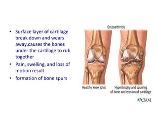
Osteoarthritis O
Knee
OA KNEE
• Chronic, Non inflammatory, degenerative
disease.
• characterised by loss of articular cartilage
and periarticular bone remodelling,
particularly large weight-bearing joints
• Common in older patients but can occur in
younger patients ( genetic mechanism ,
previous joint trauma )
Pathophysiology
• Degenerative alterations primarily begin in the
articular cartilage
• External forces accelerate the catabolic effects of the
chondrocytes and disrupt the cartilaginous matrix
• Enzymatic destruction increases cartilage
degradation ↓ proteoglycans and collagen
synthesis
• Decreased strength of the cartilage is compounded
by adverse alterations of the collagen
• Reduced contact area of the cartilage
Pathophysiology
• Loss of cartilage results in the loss of the joint
space
• Progressive erosion of the damaged cartilage
occurs until the underlying bone is exposed
• Subchondral bone responds with vascular
invasion and increased cellularity, at areas of
pressure
Pathophysiology
• The traumatized subchondral bone may
undergo cystic degeneration
• At nonpressure areas along the articular
margin → irregular outgrowth of new bone
(osteophytes)
• Normal joint
• hinge joint formed
• Surface layer of cartilage
break down and wears
away,causes the bones
under the cartilage to rub
together
• Pain, swelling, and loss of
motion result
• formation of bone spurs
Incidence
• Incidence increases with age
• USA approximately 80-90% of individuals
older than 65 years have evidence of primary
osteoarthritis
• After age 55 years, the prevalence increases in
women in comparison with men
Incidence
• Equivalent prevalence occurs in men and
women aged 45-55 years (↑dramatically after
the age of 50 years)
• Most adults older than 55 years show
radiographic evidence of osteoarthritis
• No significant correlation exists between
incidence of OA and race
Causes
Primary OA
• Idiopathic
• Defective gene
Causes
Secondary OA
– Obesity
– Repetitive use (ie, jobs requiring heavy labor and
bending)
– Previous trauma (ie, posttraumatic OA)
– Infection
Causes
– Crystal deposition
– Acromegaly
– Previous rheumatoid arthritis (ie, burnt-out
rheumatoid arthritis)
– Heritable metabolic causes (eg, alkaptonuria,
hemochromatosis, Wilson disease)
Causes
– Hemoglobinopathies (eg, sickle cell disease,
thalassemia)
– Underlying orthopedic disorders (eg, congenital
hip dislocation, slipped femoral capital epiphysis)
– Disorders of bone (eg, Paget disease, avascular
necrosis)
History
• Insidious throbbing arthralgias with activity
• Initially, resting relieves the pain
• Eventually, the pain occurs even at rest
• Morning stiffness ≥ 30 minutes
• Intermittent joint swelling
Symptoms
• Pain
• Stiffness
• Gelling
• Instability
Signs
• Pain
• Tenderness
• Swelling
• Effusion
• Crepitus
• Limitation of movement and muscle wasting
Physical
• Early
– Joints may appear normal
– Gait may be antalgic if weight-bearing joints are
involved
Physical
• Later
– Visible osteophytes may be noted
– Joints may be warm to palpation
– Palpable osteophytes frequently are noted
– Joint effusion frequently is evidenced in
superficial joints
Physical
– Range-of-motion limitations, because of bony
restrictions and/or soft tissue contractures, are
characteristic
– Crepitus with range of motion is not uncommon
classification
1. Ahlbäck classification system
2. Kellgren and Lawrence system
1. Ahlbäck classification system
• This classification was proposed by Ahlback et al in
1968.
• grade 1: joint space narrowing (less than 3 mm)
• grade 2: joint space obliteration
• grade 3: minor bone attrition (0-5 mm)
• grade 4: moderate bone attrition (5-10 mm)
• grade 5: severe bone attrition (more than 10 mm)
2. Kellgren and Lawrence system
• This classification was proposed by Kellgren et al. in 1957 2
and later accepted by WHO in 1961.
• grade 0: no radiographic features of OA are present
• grade 1: doubtful joint space narrowing (JSN) and possible
osteophytic lipping
• grade 2: definite osteophytes and possible JSN on
anteroposterior weight-bearing radiograph
• grade 3: multiple osteophytes, definite JSN, sclerosis, possible
bony deformity
• grade 4: large osteophytes, marked JSN, severe sclerosis and
definite bony deformity
Imaging
• Plain radiographs
• Bone scans may be helpful in early diagnosis
of OA of the hand
• The space between the
bones of the upper and
lower leg is smaller
• Bony spurs
(osteophytes)
• Increase bone density
at the margin of the
joint
x-ray findings
–Joint space narrowing
–Osteophytes
–Subchondral sclerosis : ↑ bone density,
frequently found adjacent to joint space
–Subchondral cysts : fluid-filled sacs which
extrude from the joint
Diagnosis
• On the basis of the initial history and
examination
• X-rays
PROGRESS
• Osteoarthritis begins when the joint
cartilage starts to become worn down →
decreases the ability of the cartilage to
work as a shock-absorber to reduce the
impact of stress on the joints
• The remaining cartilage wears down
faster→ bones to grind against one
another
• Bone spurs may form
Treatment
Goals of managing OA
• Controlling pain
• Maintaining and improving the range of
movement and stability of affected joints
• Limiting functional impairment
Treatment
• Education and behavioural intervention
- Aim is to provide patients with an
understanding of the disease process, its
prognosis and the rationale and implications
of managing their condition
• Weight loss
- Weight loss (< 5 kg) has significant short-term
and long-term reduction in symptoms of OA
Treatment
• Mechanical aids
- Wear shock-absorbing footwear with good
mediolateral support, adequate arch support and
calcaneal cushion
• Exercise
• Aim of exercise is to reduce pain and disability by
strengthening muscle, improving joint stability,
increasing the range of movement and improving
aerobic fitness
Treatment
• Medication
- Acetaminophen (Tylenol®) is a mild pain
reliever with few side effects
• Anti-inflammatory medication, such as
ibuprofen and aspirin
• COX-2 inhibitors
• Glucosamine and Chondroitin sulfate
Treatment
• Intra-articular injection
- Glucocorticoids injection
- Hyaluronic Acid (HA) and similar
hyaluronan preparations (eg, Synvisc)
Treatment
• Surgery
• Arthroscopy (including debridement,and
lavage/irrigation)
• Proximal Tibial Osteotomy
• Artificial Knee Replacement
• Osteotomy
• Arthroplasty or Joint Replacement

Oa knee (shravan)[1]

  • 1.
  • 2.
    OA KNEE • Chronic,Non inflammatory, degenerative disease. • characterised by loss of articular cartilage and periarticular bone remodelling, particularly large weight-bearing joints • Common in older patients but can occur in younger patients ( genetic mechanism , previous joint trauma )
  • 3.
    Pathophysiology • Degenerative alterationsprimarily begin in the articular cartilage • External forces accelerate the catabolic effects of the chondrocytes and disrupt the cartilaginous matrix • Enzymatic destruction increases cartilage degradation ↓ proteoglycans and collagen synthesis • Decreased strength of the cartilage is compounded by adverse alterations of the collagen • Reduced contact area of the cartilage
  • 4.
    Pathophysiology • Loss ofcartilage results in the loss of the joint space • Progressive erosion of the damaged cartilage occurs until the underlying bone is exposed • Subchondral bone responds with vascular invasion and increased cellularity, at areas of pressure
  • 5.
    Pathophysiology • The traumatizedsubchondral bone may undergo cystic degeneration • At nonpressure areas along the articular margin → irregular outgrowth of new bone (osteophytes)
  • 7.
    • Normal joint •hinge joint formed
  • 8.
    • Surface layerof cartilage break down and wears away,causes the bones under the cartilage to rub together • Pain, swelling, and loss of motion result • formation of bone spurs
  • 9.
    Incidence • Incidence increaseswith age • USA approximately 80-90% of individuals older than 65 years have evidence of primary osteoarthritis • After age 55 years, the prevalence increases in women in comparison with men
  • 10.
    Incidence • Equivalent prevalenceoccurs in men and women aged 45-55 years (↑dramatically after the age of 50 years) • Most adults older than 55 years show radiographic evidence of osteoarthritis • No significant correlation exists between incidence of OA and race
  • 11.
  • 12.
    Causes Secondary OA – Obesity –Repetitive use (ie, jobs requiring heavy labor and bending) – Previous trauma (ie, posttraumatic OA) – Infection
  • 13.
    Causes – Crystal deposition –Acromegaly – Previous rheumatoid arthritis (ie, burnt-out rheumatoid arthritis) – Heritable metabolic causes (eg, alkaptonuria, hemochromatosis, Wilson disease)
  • 14.
    Causes – Hemoglobinopathies (eg,sickle cell disease, thalassemia) – Underlying orthopedic disorders (eg, congenital hip dislocation, slipped femoral capital epiphysis) – Disorders of bone (eg, Paget disease, avascular necrosis)
  • 15.
    History • Insidious throbbingarthralgias with activity • Initially, resting relieves the pain • Eventually, the pain occurs even at rest • Morning stiffness ≥ 30 minutes • Intermittent joint swelling
  • 16.
    Symptoms • Pain • Stiffness •Gelling • Instability
  • 17.
    Signs • Pain • Tenderness •Swelling • Effusion • Crepitus • Limitation of movement and muscle wasting
  • 18.
    Physical • Early – Jointsmay appear normal – Gait may be antalgic if weight-bearing joints are involved
  • 19.
    Physical • Later – Visibleosteophytes may be noted – Joints may be warm to palpation – Palpable osteophytes frequently are noted – Joint effusion frequently is evidenced in superficial joints
  • 20.
    Physical – Range-of-motion limitations,because of bony restrictions and/or soft tissue contractures, are characteristic – Crepitus with range of motion is not uncommon
  • 21.
    classification 1. Ahlbäck classificationsystem 2. Kellgren and Lawrence system
  • 22.
    1. Ahlbäck classificationsystem • This classification was proposed by Ahlback et al in 1968. • grade 1: joint space narrowing (less than 3 mm) • grade 2: joint space obliteration • grade 3: minor bone attrition (0-5 mm) • grade 4: moderate bone attrition (5-10 mm) • grade 5: severe bone attrition (more than 10 mm)
  • 23.
    2. Kellgren andLawrence system • This classification was proposed by Kellgren et al. in 1957 2 and later accepted by WHO in 1961. • grade 0: no radiographic features of OA are present • grade 1: doubtful joint space narrowing (JSN) and possible osteophytic lipping • grade 2: definite osteophytes and possible JSN on anteroposterior weight-bearing radiograph • grade 3: multiple osteophytes, definite JSN, sclerosis, possible bony deformity • grade 4: large osteophytes, marked JSN, severe sclerosis and definite bony deformity
  • 24.
    Imaging • Plain radiographs •Bone scans may be helpful in early diagnosis of OA of the hand
  • 25.
    • The spacebetween the bones of the upper and lower leg is smaller • Bony spurs (osteophytes) • Increase bone density at the margin of the joint
  • 26.
    x-ray findings –Joint spacenarrowing –Osteophytes –Subchondral sclerosis : ↑ bone density, frequently found adjacent to joint space –Subchondral cysts : fluid-filled sacs which extrude from the joint
  • 27.
    Diagnosis • On thebasis of the initial history and examination • X-rays
  • 28.
    PROGRESS • Osteoarthritis beginswhen the joint cartilage starts to become worn down → decreases the ability of the cartilage to work as a shock-absorber to reduce the impact of stress on the joints • The remaining cartilage wears down faster→ bones to grind against one another • Bone spurs may form
  • 29.
  • 30.
    Goals of managingOA • Controlling pain • Maintaining and improving the range of movement and stability of affected joints • Limiting functional impairment
  • 31.
    Treatment • Education andbehavioural intervention - Aim is to provide patients with an understanding of the disease process, its prognosis and the rationale and implications of managing their condition • Weight loss - Weight loss (< 5 kg) has significant short-term and long-term reduction in symptoms of OA
  • 32.
    Treatment • Mechanical aids -Wear shock-absorbing footwear with good mediolateral support, adequate arch support and calcaneal cushion • Exercise • Aim of exercise is to reduce pain and disability by strengthening muscle, improving joint stability, increasing the range of movement and improving aerobic fitness
  • 34.
    Treatment • Medication - Acetaminophen(Tylenol®) is a mild pain reliever with few side effects • Anti-inflammatory medication, such as ibuprofen and aspirin • COX-2 inhibitors • Glucosamine and Chondroitin sulfate
  • 35.
    Treatment • Intra-articular injection -Glucocorticoids injection - Hyaluronic Acid (HA) and similar hyaluronan preparations (eg, Synvisc)
  • 36.
    Treatment • Surgery • Arthroscopy(including debridement,and lavage/irrigation) • Proximal Tibial Osteotomy • Artificial Knee Replacement • Osteotomy • Arthroplasty or Joint Replacement