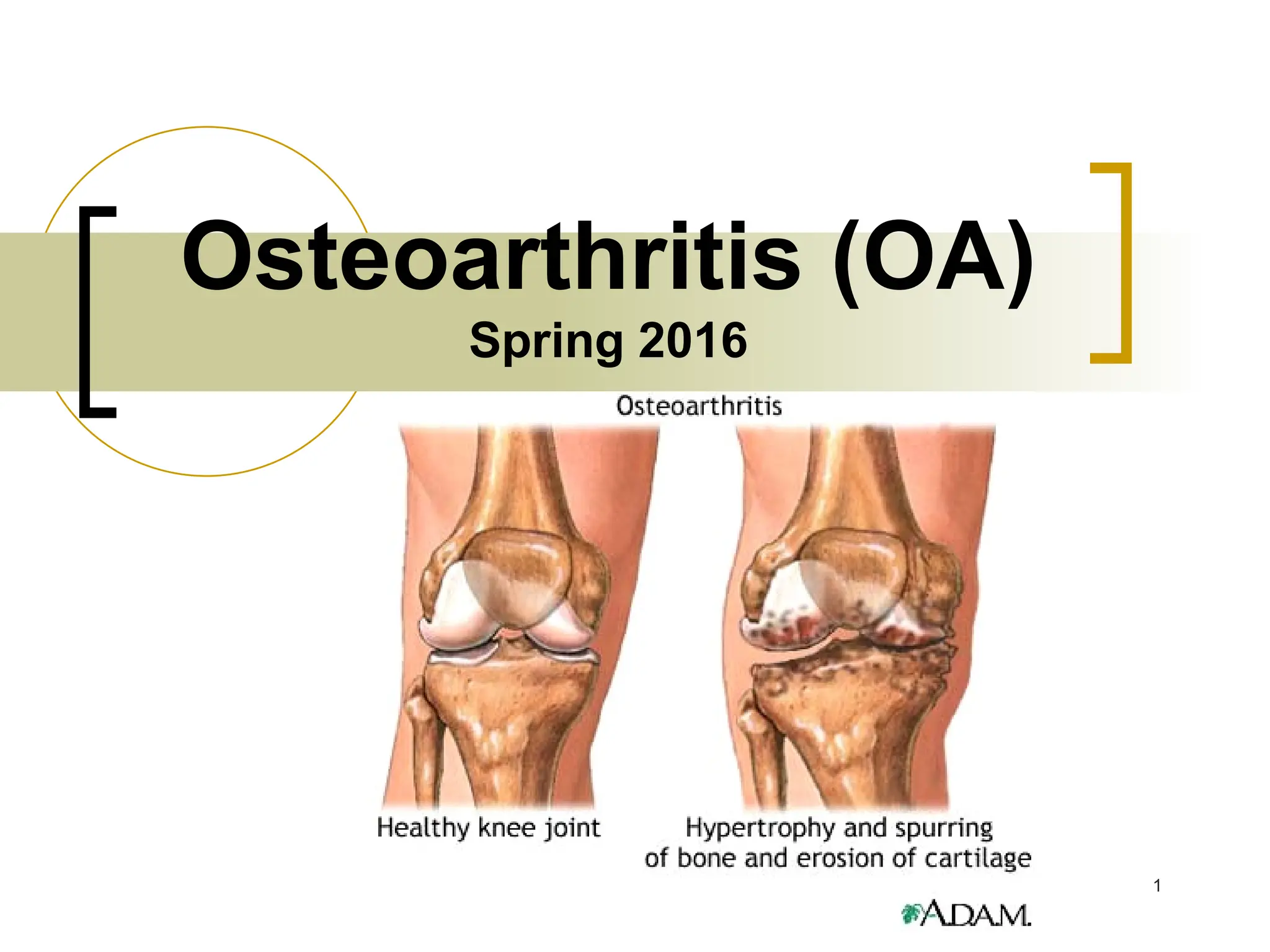
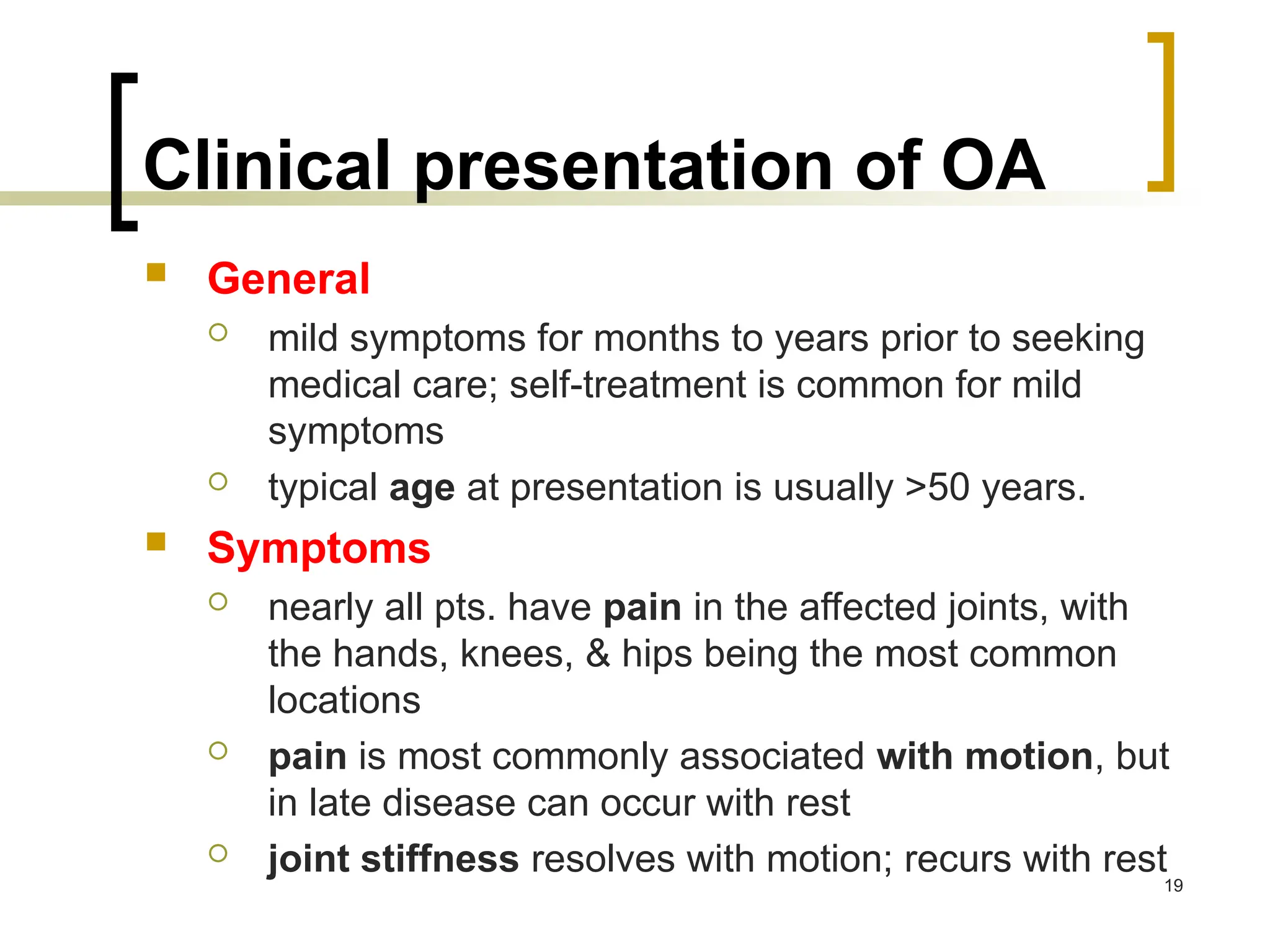
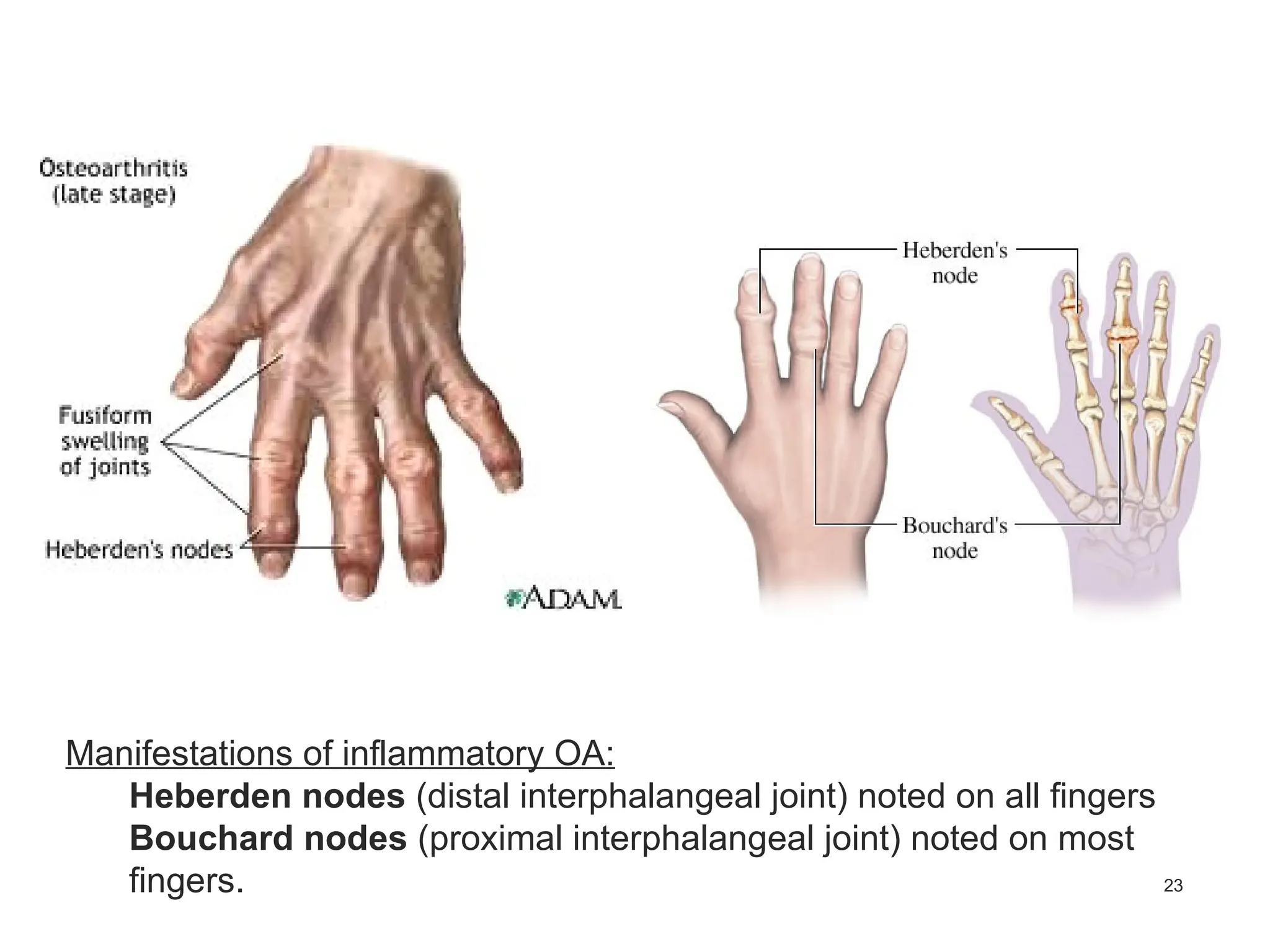
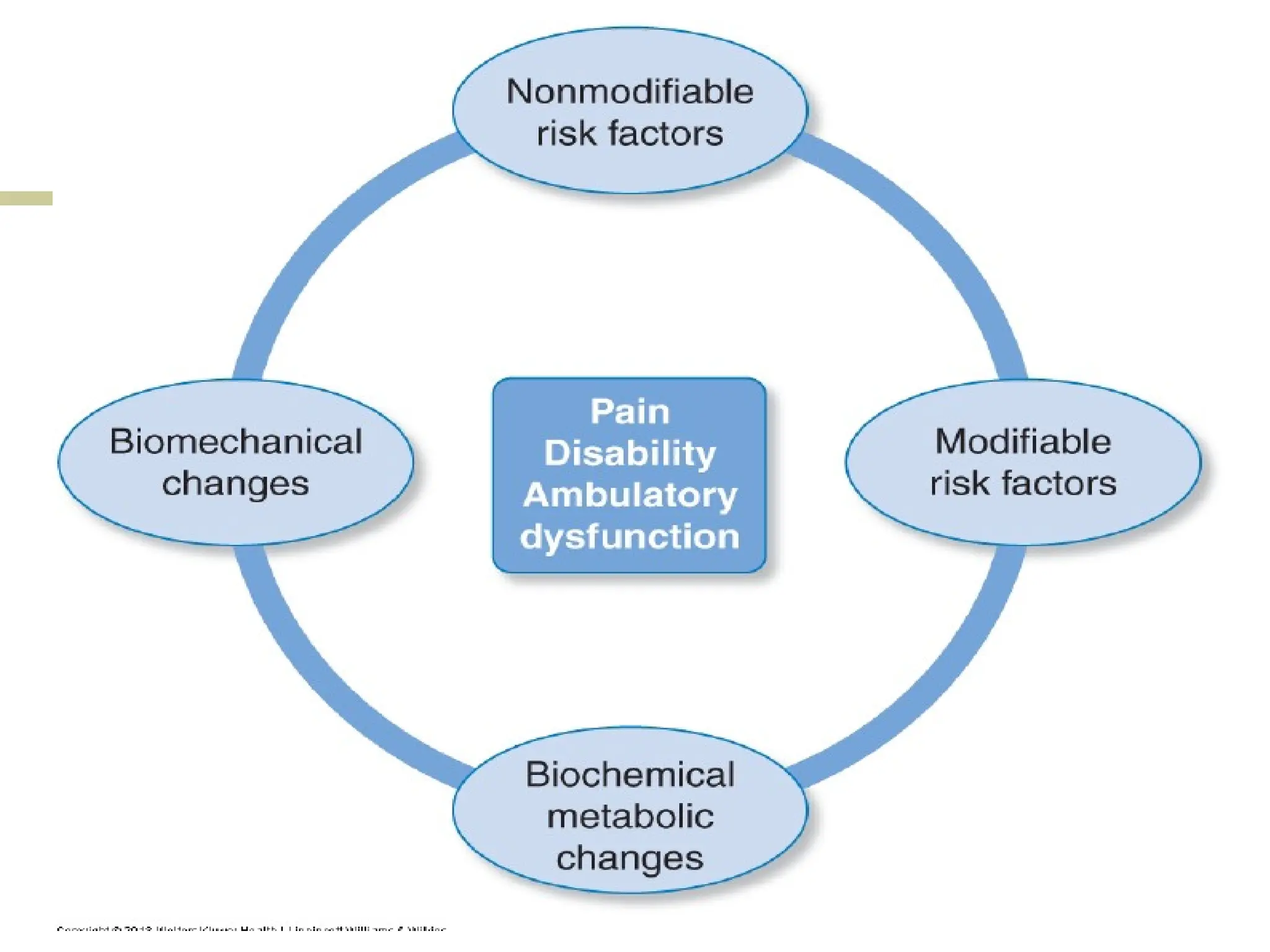
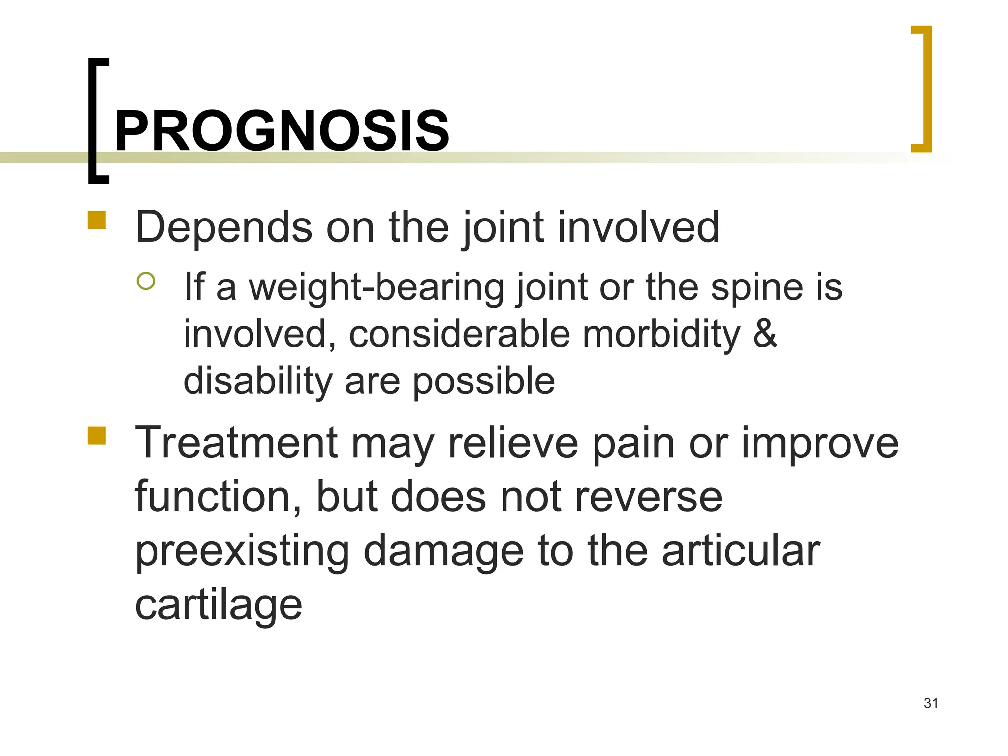
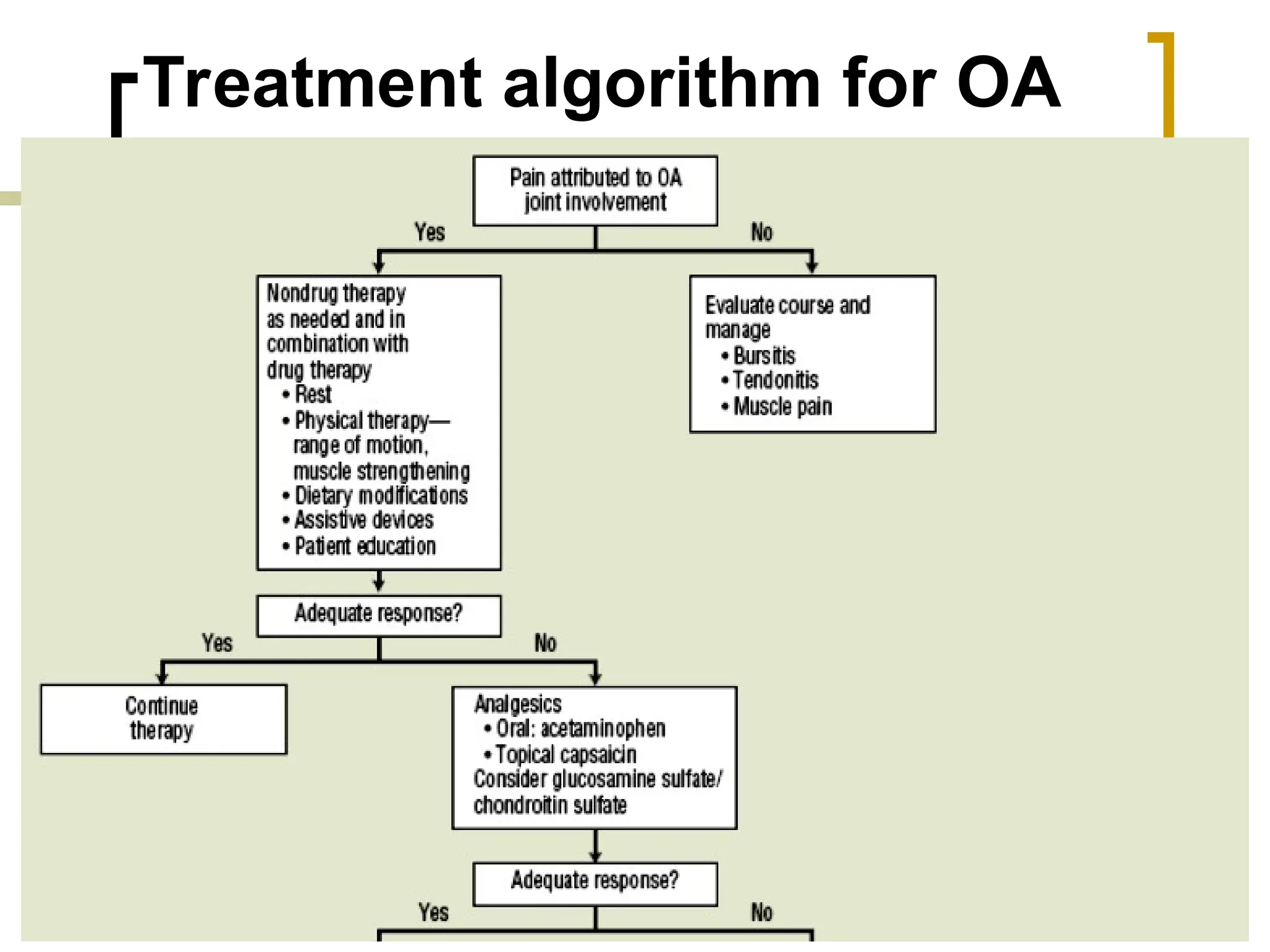
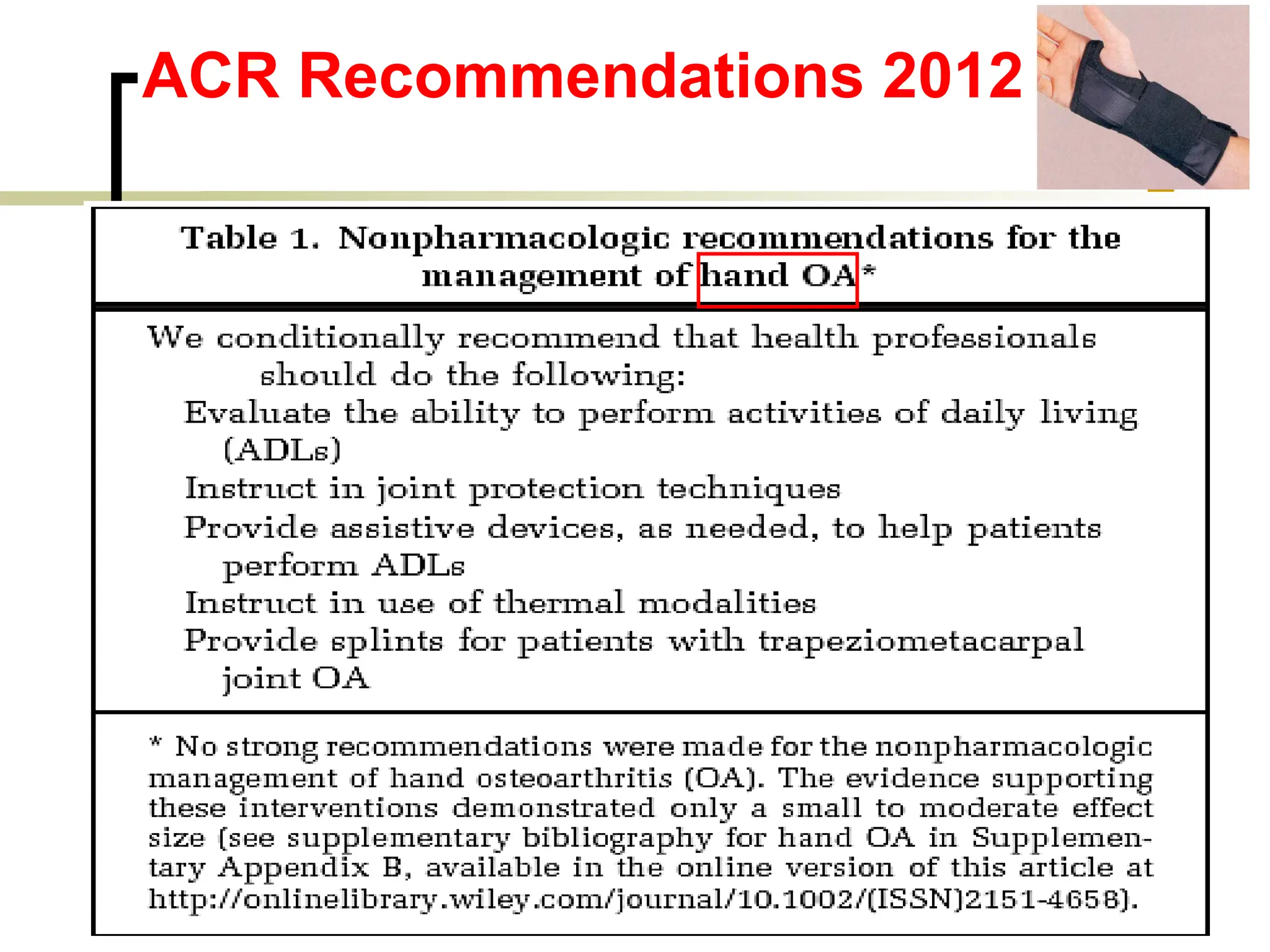
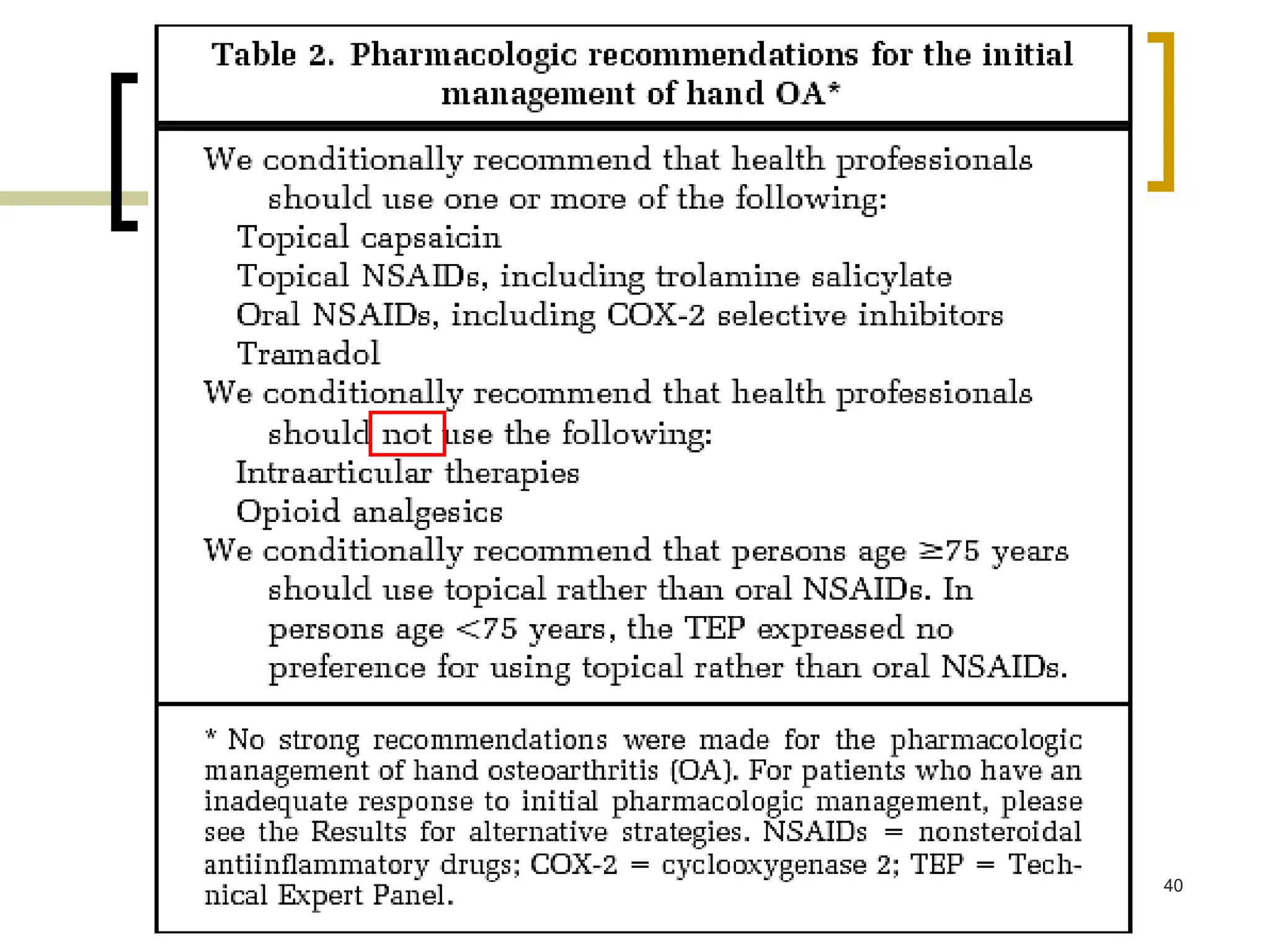
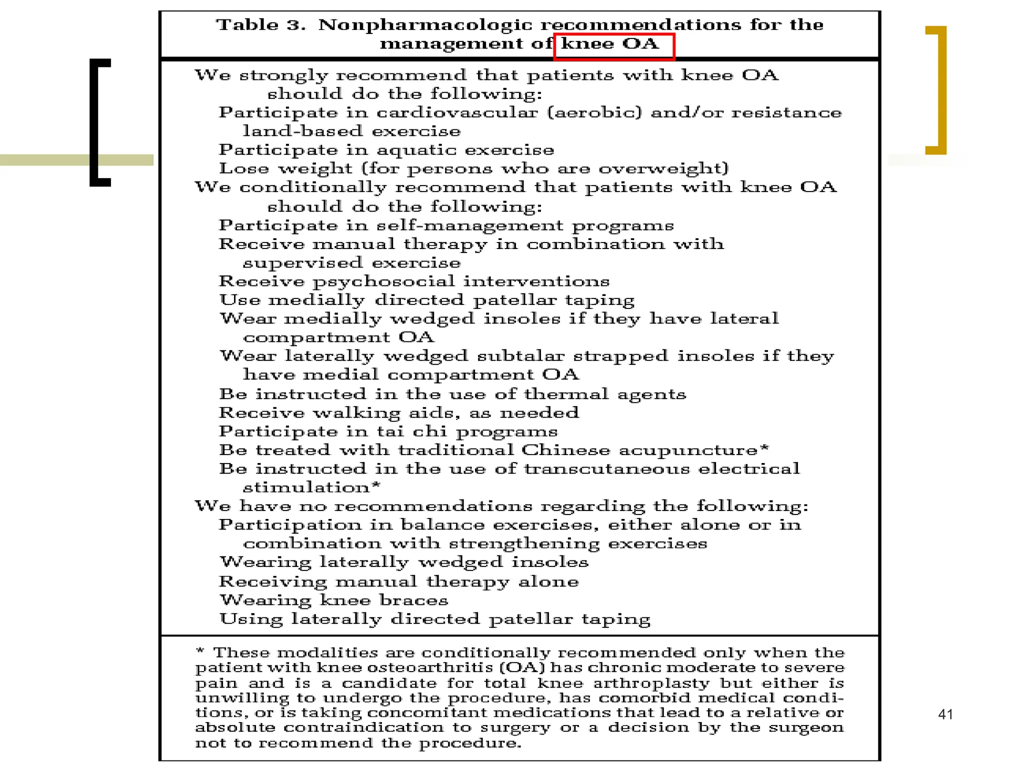
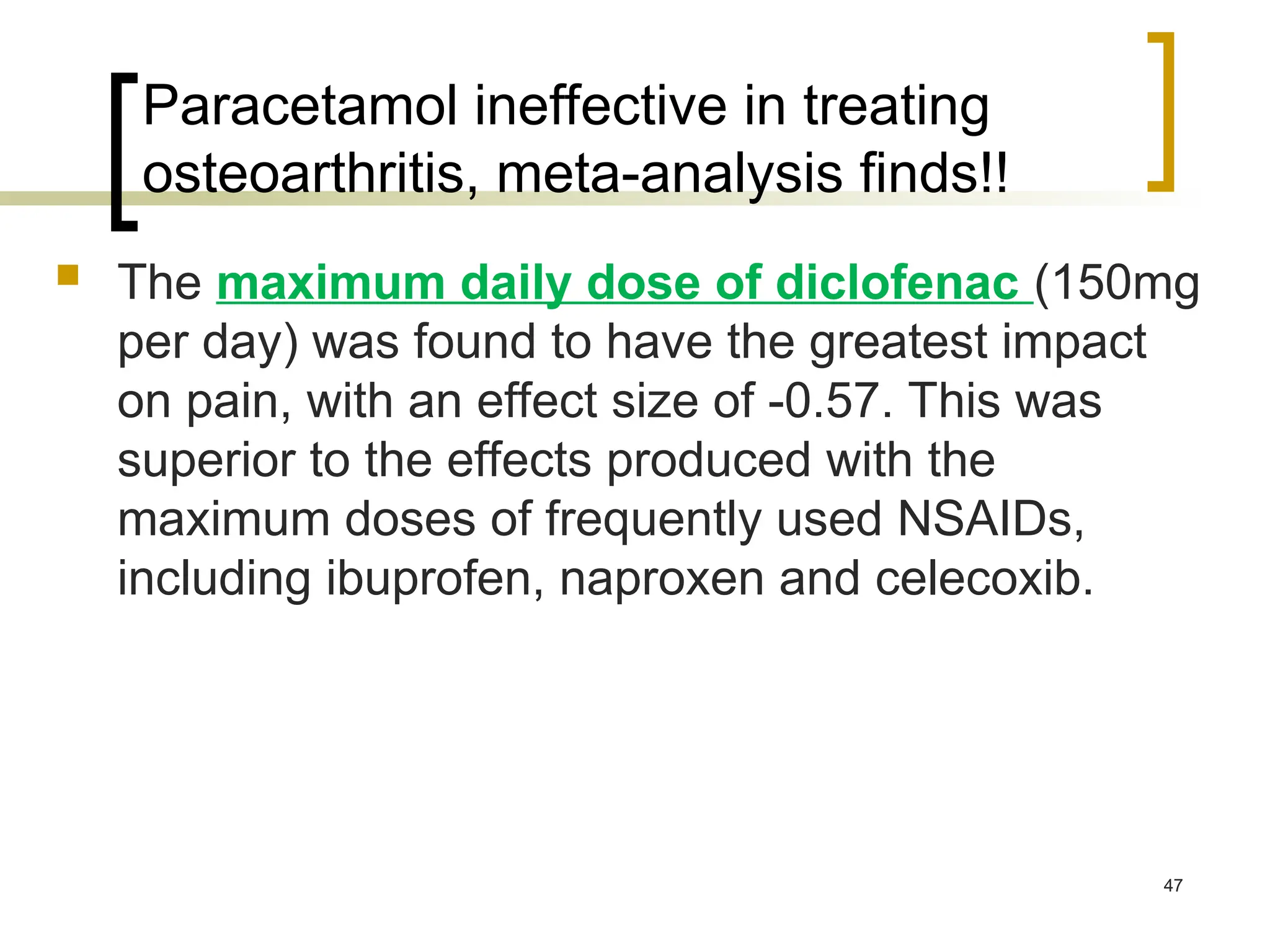
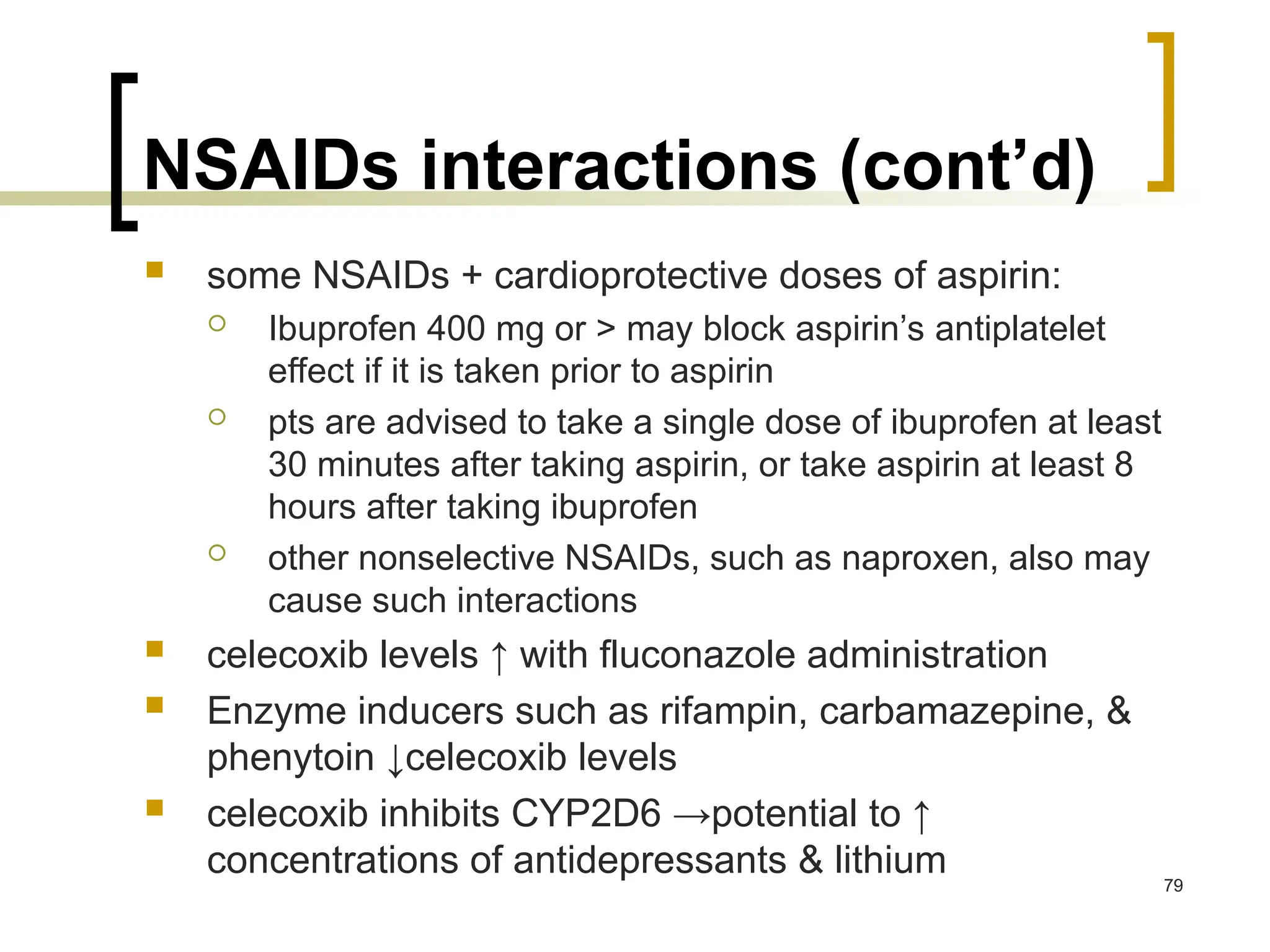
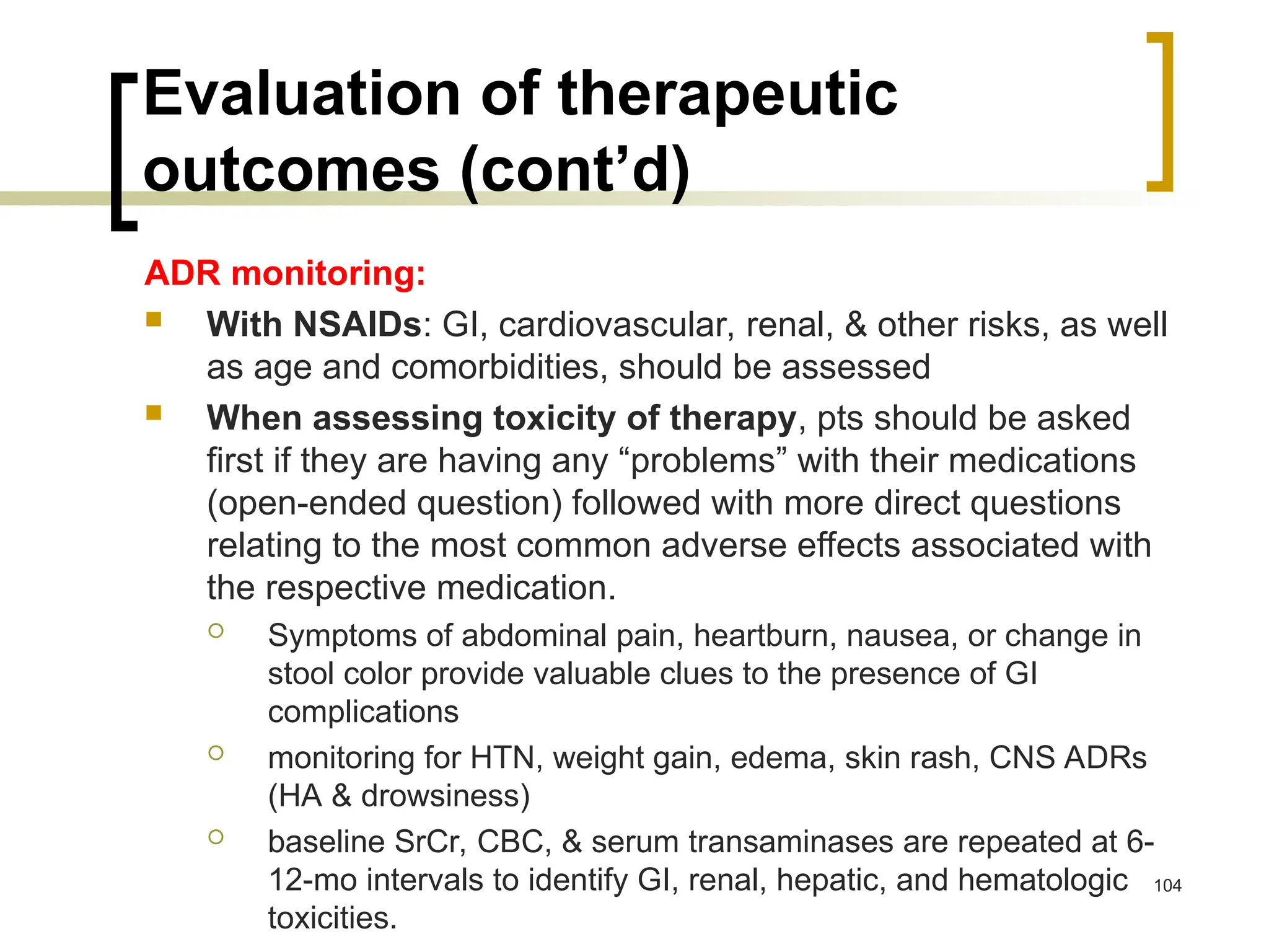
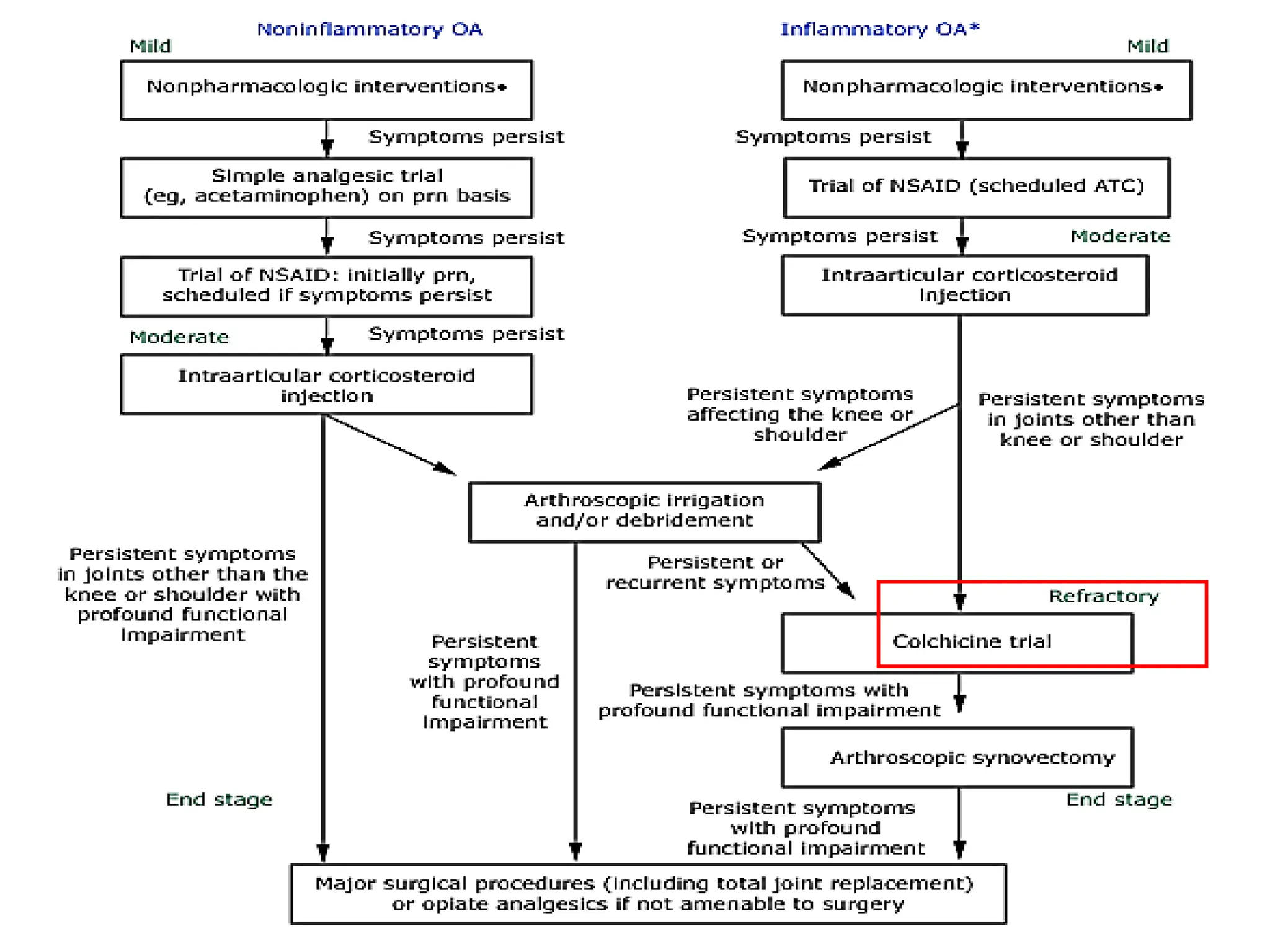
1
Osteoarthritis (OA)
Spring 2016
References
 NICE Clinical Guideline: Osteoarthritis
Care and management in adults Issued:
February 2014
 DiPiro Ch. 95: Osteoarthritis
 Koda-Kimble K et al. Applied Therapeutics,
10th edition, (2013); Ch. 43
 American College of Rheumatology 2012
Recommendations for the Use of
Nonpharmacologic and Pharmacologic
Therapies in Osteoarthritis of the Hand,
Hip, and Knee.
http://www.rheumatology.org
 American Academy of Orthopaedic
Surgeons 2013 guidelines for the
treatment of knee osteoarithritis. 2
Introduction
 Osteoarthritis is a disease characterized by degeneration
of cartilage and its underlying bone within a joint as well as
bony overgrowth.
 The breakdown of these tissues eventually leads to pain
and joint stiffness. The joints most commonly affected are
the knees, hips, and those in the hands and spine.
 The specific causes of osteoarthritis are unknown, but are
believed to be a result of both mechanical and molecular
events in the affected joint.
3
Introduction (cont’d)
 Disease onset is gradual and usually begins
after the age of 40.
 There is currently no cure for OA.
Treatment for OA focuses on relieving
symptoms and improving function, and
can include a combination of:
 patient education,
 physical therapy,
 weight control,
 and use of medications.
4
Before we start!
5
DEFINITION
 Osteoarthritis (OA), which is also known as
osteoarthrosis or degenerative joint disease
(DJD), is a progressive disorder of the joints
caused by gradual loss of cartilage and resulting
in the development of bony spurs and cysts at
the margins of the joints. The name
osteoarthritis comes from three Greek words
meaning bone, joint, and inflammation.
6
EPIDEMIOLOGY
 OA is the most prevalent (3-6%) of the rheumatic diseases & is
responsible for enormous disability & loss of productivity
 The prevalence of OA is > in older age groups
 In those < age 45 yrs, ~1/5 have OA of the hands, while of those ages
75-79 yrs, 85% have OA of the hands
 OA of the knee occurs in less than 0.1% of those aged 25 to 34 years,
but in 10% to 20% of those aged 65 to 74 years.
 OA severity also ↑ with age
 Women are > often affected by OA,
 older women being twice as likely as men
to have OA of the knee & hands
 Women are also more likely to have
inflammatory OA of the proximal & distal
interphalangeal joints of the hands.
 OA of the hip occurs more frequently in men. 8
ETIOLOGY
 Multifactorial
 Many patients have >1 risk factor
 The most common risk factors:
 obesity
 number one preventable risk factor for OA, and a predictor for eventual
prosthetic joint replacement
 previous occupation,
 participation in certain sports,
 history of joint trauma,
 genetic predisposition to OA (e.g., genes for IL-1, calmodulin)
 Patients with OA are < likely to have osteoporosis because
heavy individuals have > bone density as a result of weight-
bearing, but ↑ risk of OA as a consequence of excessive joint
loading 10
PATHOPHYSIOLOGY
OA falls into 2 etiologic classes:
 Primary (idiopathic) OA - the most common type, has no
identifiable cause. Subclasses:
 localized OA, involving 1 or 2 sites
 generalized OA, affecting 3 or > sites
 Erosive osteoarthritis: erosion & marked proliferation in the
proximal & distal interphalangeal joints of the hands
 Secondary OA - associated with a known cause such as
rheumatoid or another inflammatory arthritis, trauma,
metabolic or endocrine disorders, & congenital factors
 Some changes in the OA joint may reflect compensatory
processes to maintain function in the face of ongoing joint
destruction 11
PATHOPHYSIOLOGY
 OA is a multifactorial disease typified by:
 progressive destruction of joint cartilage,
 erratic new bone formation,
 thickening of subchondral bone and the
joint capsule,
 bony remodeling,
 development of osteophytes,
 variable degrees of mild synovitis, and
other changes.
12
Cartilage in OA
 OA most commonly begins with damage to
articular cartilage, through trauma, joint
loading from obesity, etc.
 damage to cartilage →↑ metabolic activity of
chondrocytes →↑ synthesis of matrix
constituents, with swelling of cartilage
 this hypertrophic response does not restore
the cartilage to normal
 →↑ turnover (↑ collagen synthesis &
destruction), but with destruction outpacing
formation →loss of cartilage. 13
Characteristics of osteoarthritis in the
diarthrodial joint.
14
Cartilage in OA (cont’d)
 Key players in cartilage destruction are the
matrix metalloproteinases (MMPs)
 In normal cartilage, activities of these enzymes
are blocked by tissue inhibitors of
metalloproteinases
 In OA cartilage: ↑ expression & synthesis of
MMPs by chondrocytes in response to
inflammatory mediators present in OA (IL-1 &
TNF-α)
 Chondrocytes in OA cartilage undergo apoptosis,
likely as a result of induction of nitric oxide
synthase & production of toxic metabolites 15
Cartilage in OA (cont’d)
 Subchondral bone undergoes pathologic changes that may
precede, coincide with, or follow damage to the articular cartilage
 In OA, subchondral bone releases vasoactive peptides & MMPs
 Neovascularization & subsequent ↑ permeability → further
cartilage loss
 → joint space narrowing & painful, deformed joint
 the remaining cartilage softens & develops fibrillations (vertical
clefts into the cartilage), with splitting & exposure of underlying
bone
 → cartilage is eroded completely → subchondral bone becomes
dense, smooth (eburnation) →
 ↓ weight-bearing ability & microfractures →
 new bone formations, or osteophytes
16
17
Cartilage in OA (cont’d)
 There is evidence that osteophytes can help stabilize
osteoarthritic joints
 local inflammatory changes & pathologic changes can occur
in the joint capsule & synovium: infiltration with T-helper type 1
phenotype occurs + immune complexes
 Inflammation is due to crystals or cartilage shards in synovial
fluid + inflammatory mediators (IL-1, PG E2, TNF-α, & NO)
 effusions & synovial thickening occur
 pain of OA is not related to destruction of cartilage, but due to
activation of nociceptive nerve endings within the joint by
mechanical & chemical irritants: distension of the synovial
capsule by ↑ joint fluid, microfracture, or damage to ligaments
or synovium 18
Clinical presentation of OA
 General
 mild symptoms for months to years prior to seeking
medical care; self-treatment is common for mild
symptoms
 typical age at presentation is usually >50 years.
 Symptoms
 nearly all pts. have pain in the affected joints, with
the hands, knees, & hips being the most common
locations
 pain is most commonly associated with motion, but
in late disease can occur with rest
 joint stiffness resolves with motion; recurs with rest
19
Clinical presentation of OA
(cont’d)
 Signs
 joint stiffness with or without joint enlargement
 crepitus, a crackling or grating sound heard with joint
movement caused by irregularity of joint surfaces may be
present
 limited range of motion that may be accompanied by
joint instability
 late-stage disease: joint deformity
 Diagnostic Tests
 no specific laboratory tests useful in the dx
 Plain Radiographic Films:
 joint space narrowing, appearance of osteophytes in moderate
disease
 abnormal alignment of joints & joint effusion in late disease 20
21
b. Plain x-ray films of
the knee demonstrating
joint space narrowing
a. Physical findings of
joint enlargement &
genu varum (bandiness)
of the knees
22
23
Manifestations of inflammatory OA:
Heberden nodes (distal interphalangeal joint) noted on all fingers
Bouchard nodes (proximal interphalangeal joint) noted on most
fingers.
Heberden's nodes
(osteophytes)
Clinical manifestations of osteoarthritis
Age of onset Usually after age 40
Commonly affected
joints
Cervical and lumbar spine, first carpometacarpal
joint, proximal interphalangeal joint, distal
interphalangeal joint, hip, knee, subtalar joint, first
metarsophalangeal joint
Uncommonly affected
joints
Shoulder, wrist, elbow, metacarpophalangeal joint
Symptoms Pain, stiffness, gelling
Findings on physical
examination
Crepitus, bony enlargement, decreased range of
motion, malalignment, tenderness to palpation
Synovial fluid analysis Clear fluid, WBC <2000/mm3, normal viscosity
Radiographic features Joint space narrowing, subchondral sclerosis,
marginal osteophytes, subchondral cysts
Patterns of
presentation
Monoarticular in young adult; pauciarticular, large-
joint in middle age; polyarticular generalized; rapidly
progressive; secondary to trauma, congenital
abnormality, or systemic disease
Prognosis Variable, generally slowly progressive 25
26
DIAGNOSIS
 Diagnosis depends on:
 history
 physical examination
 characteristic radiographic findings
 laboratory testing
 The major diagnostic goals:
 to discriminate between primary & secondary
OA
 to clarify the joints involved, severity of joint
involvement, & response to prior therapies 27
NICE 2014 guideline
28
Distinction between rheumatoid arthritis
and osteoarthritis
Feature Rheumatoid arthritis Osteoarthritis
Primary joints affected Metacarpophalangeal Distal interphalangeal
Proximal interphalangeal Carpometacarpal
Heberden's nodes Absent Frequently present
Joint characteristics Soft, warm, and tender Hard and bony
Stiffness Worse after resting (eg,
morning stiffness)
If present, worse after
effort, may be described
as evening stiffness
Laboratory findings Positive rheumatoid factor Rheumatoid factor
negative
Positive anti-CCP
antibody
Anti-CCP antibody
negative
Elevated ESR and C
reactive protein
Normal ESR and C
reactive protein 29
CCP: cyclic citrullinated peptide
30
PROGNOSIS
 Depends on the joint involved
 If a weight-bearing joint or the spine is
involved, considerable morbidity &
disability are possible
 Treatment may relieve pain or improve
function, but does not reverse
preexisting damage to the articular
cartilage
31
TREATMENT
 Desired outcome
 to educate the patient, caregivers, &
relatives
 to relieve pain & stiffness
 to maintain or improve joint mobility
 to limit functional impairment
 to maintain or improve quality of life
32
General approach to treatment
 depends on:
 the distribution & severity of joint involvement,
 comorbid disease states,
 concomitant medications,
 allergies
 management for all individuals with OA
should begin with:
 patient education,
 physical &/or occupational therapy,
 weight loss or assistive devices if appropriate
33
General approach to treatment
(cont’d)
 The primary objective of medication is to alleviate pain
 scheduled acetaminophen, up to 4 g/day initially →
NSAIDs COX-2 selective inhibitor (celecoxib)
 capsaicin or methylsalicylate topical creams over
specific joints as adjuncts
 glucosamine & chondroitin in combination for
moderate to severe arthritis
 joint aspiration followed by glucocorticoid or
hyaluronate injection to relieve pain concomitantly with
oral analgesics or after their lack of efficacy
 opioid analgesics if other therapies are unsuccessful
 when symptoms are intractable or there is significant
loss of function → joint replacement 34
35
Homework:
 Summary of Recommendations for
Osteoarthritis (table 95-1 in DiPiro)
(by Third Canadian Consensus Conference
Group. An evidence-based approach to
prescribing nonsteroidal antiinflammatory
drugs. Third Canadian Consensus
Conference. J Rheumatol 2006;33:140–157)
Treatment algorithm for OA
36
37
38
ACR Recommendations 2012
39
40
41
42
A: lateral wedge insole (conventional)
B: lateral wedge insole with the strapping (strapping
insole)
Fig. 3 Biomechanical effect of both insoles to correct lower leg alignment. (A) Barefoot; (B) wearing conventional insole; (C) wearing
strapping insole. Conventional insoles move the weight bearing axis and reduce knee joint load in the medial compartment,...
Y. Kuroyanagi , T. Nagura , H. Matsumoto , T. Otani , Y. Suda , T. Nakamura , Y. Toyama
The lateral wedged insole with subtalar strapping significantly reduces dynamic knee load in the medial compartment :
Gait analysis on patients with medial knee osteoarthritis
Osteoarthritis and Cartilage, Volume 15, Issue 8, 2007, 932 - 936
http://dx.doi.org/10.1016/j.joca.2007.02.004
44
AAOS 2013-symptomatic
knee OA
 A 2013 clinical practice guideline from the American Academy of
Orthopaedic Surgeons (AAOS) recommends the following
 Oral NSAIDs
 Topical NSAIDs
 Tramadol
 The AAOS was unable to recommend for or against the use of the
following:
 Acetaminophen
 Opioids
 Pain patches
 Intra-articular corticosteroid injections
 Growth factor injections and/or platelet rich plasma
 The recommendation on acetaminophen is a downgrade from the
previous AAOS guideline, and reflects the selection of only one study,
which found no statistical significance or minimum clinically important
improvement with acetaminophen compared with placebo. 45
Paracetamol ineffective in treating
osteoarthritis, meta-analysis finds!!
 17-3-2016:
 da Costa BR, Reichenbach S, Keller N et al. Effectiveness of non-
steroidal anti-inflammatory drugs for the treatment of pain in knee
and hip osteoarthritis: a network meta-analysis. The Lancet 2016.
 The researchers pooled data from 74 randomised trials published
between 1980 and 2015, which involved 58,556 patients with
osteoarthritis and compared the effects of 22 treatments and placebo
on pain intensity and physical activity.
 All 22 preparations of medications, irrespective of dose, improved
symptoms of pain compared with placebo, but paracetamol did not
achieve the minimum clinically important difference. Its effect size
was -0·17 and to be deemed clinically important a difference of -0.37 is
required, the researchers say, meaning that paracetamol had “nearly
a null effect on pain symptoms at various doses”.
46
Paracetamol ineffective in treating
osteoarthritis, meta-analysis finds!!
 The maximum daily dose of diclofenac (150mg
per day) was found to have the greatest impact
on pain, with an effect size of -0.57. This was
superior to the effects produced with the
maximum doses of frequently used NSAIDs,
including ibuprofen, naproxen and celecoxib.
47
AAOS 2013-symptomatic
knee OA
 The AAOS does not recommend
treatment with any of the following:
 Intra-articular hyaluronic acid
 Glucosamine and/or chondroitin sulfate
or hydrochloride
48
49
50
Nonpharmacologic therapy for
patients with osteoarthritis
 Patient education
 Self-management programs
 Personalized social support through telephone contact
 Weight loss (if overweight)
 Aerobic exercise programs
 Physical therapy
 Range-of-motion exercises
 Muscle-strengthening exercises (as quadriceps strengthening
exercise)
 Assistive devices for ambulation (as canes, crutches, or walkers)
 Patellar taping (for patients with OA of the knee who have
symptomatic patellofemoral compartment involvement)
 Appropriate footwear
 Lateral-wedged insoles (for genu varum)
 Bracing
 Occupational therapy
 Joint protection and energy conservation
 Assistive devices for activities of daily living
ACR 2000
52
53
Foot, ankle brace Wrist, hand brace Neck brace
Spinal brace Join Supports
Physical & Occupational
Therapy
 Physical therapy – with heat or cold treatments & an
exercise program – helps to maintain & restore joint
range of motion & to ↓ pain & muscle spasms.
 Pts should be warned not to fall asleep on the heat
source or to lie on it for more than brief periods to avoid
burns
 Exercise programs & quadriceps strengthening can
improve physical functioning & ↓ disability, pain, &
analgesic use
 Exercises should be taught & then observed before the
pt exercises at home, ideally 3-4 times daily
 The pt should be instructed to ↓ the number of
repetitions if severe pain develops with exercise 54
Physical & Occupational
Therapy (cont’d)
 Walking:
 With weak or deconditioned muscles, load is transmitted
excessively to joints → weight-bearing activities can
exacerbate symptoms
 But: avoidance of activity by those with hip or knee OA
leads to further deconditioning or weight gain
 Pt education, muscle stretching & strengthening, &
supervised walking can improve physical function &↓ pain
in patients with knee OA
 Occupational therapist can recommend exercises &
methods of protecting the affected joint from excessive
forces + provide assistive & orthotic devices, such as
canes, walkers, braces, heel cups, splints, or insoles for
use during exercise or daily activities 55
Pharmacologic Therapy for OA
56
57
Acetaminophen
Place in Therapy
 ACR: first-line drug therapy for pain management in OA (of knee and hip,
but not OA of the hand) because of its relative safety, efficacy, & < cost
compared to NSAIDs
 Pain relief with acetaminophen has been reported as similar to that obtained
with NSAIDs, although many patients respond better to NSAIDs
 The European League Against Rheumatism also stresses the importance
of acetaminophen as first-line drug therapy for OA
 It is a weak COX-1 & COX-2 inhibitor in peripheral tissues & possesses no
significant anti-inflammatory effects. Recent evidence suggests that
acetaminophen may inhibit a third enzyme, COX-3, in the CNS.
 The AAOS 2013 was not able to recommend for or against the use of
acetaminophen in the management of knee arithritis!
58
59
MOA
Acetaminophen (cont’d)
ADRs
 is one of the safest analgesics
 risk of hepatotoxicity, & possibly renal toxicity with long-
term use
 overdose: serious hepatotoxicity, including fatalities
 maximum dose of acetaminophen should be not be
exceeded in any patient population, & chronic use of
even the maximum 4 g/day can affect the liver
 should be used cautiously in patients with liver disease
or in those who abuse alcohol: acute liver failure can
develop at doses < 4 g/daily
60
Acetaminophen (cont’d)
 FDA: chronic alcohol users (3 & > drinks daily)
should be warned regarding ↑ risk of liver damage
or GI bleeding with acetaminophen
 The National Kidney Foundation strongly
discourages the use of OTC combination
analgesic products (e.g., acetaminophen &
NSAIDs) because this is associated with ↑
prevalence of renal failure
 Pts should be warned about potential toxicity if
they inadvertently ingest > the recommended
dose when using both nonprescription and
prescription products containing acetaminophen 61
Acetaminophen (cont’d)
Drug–Drug Interactions
 isoniazid can ↑ risk of hepatotoxicity
 chronic ingestion of maximal doses of acetaminophen may
intensify the anticoagulant effect in patients taking warfarin →
closer monitoring
Dosing & Administration
 for chronic OA, acetaminophen should be administered in a
scheduled manner (around the clock – ATC)
 may be taken with or without food
 can be taken at 325-650 mg every 4-6 hours, but total dose
must not exceed 4 g daily
 in chronic alcohol intake or in underlying disease, duration
should be ↓ & dose should not exceed 2 g daily 62
NSAIDs
Place in Therapy
 ACR: consider NSAIDs for OA patients in whom acetaminophen is
ineffective
 All NSAIDs display comparable analgesic & antiinflammatory
efficacy & are similarly beneficial in OA
 Because nonspecific NSAIDs & COX-2 selective inhibitors have similar
efficacy, drug selection often depends on toxicity & cost
 Prostaglandins made by COX-2 enzyme, contribute to pain sensations
in OA & other conditions + implicated in some physiologic processes,
including renal function, tissue repair, reproduction, & development
 COX-2 inhibitors (“coxibs”) are efficacious in relieving OA & other
pain & some do have improved GI safety
63
MoA
64
NSAIDs (cont’d)
 COX-2 enzyme found in blood vessel endothelial cells leads to
production of prostaglandin I2 (prostacyclin), which has
antithrombotic effects
 COX-1 enzyme found in platelets forms thromboxane A2
(prothrombotic)
 blocking COX-2 alone could upset hemostatic balance, in favor
of thromboxane A2, with prothrombotic events possible
 whether the “prothrombotic imbalance” explanation accounts for
↑ cardiovascular risk seen with COX-2 selective inhibitors is
unknown
 Rofecoxib was withdrawn from the market in 2004 because of ↑
cardiovascular events, & celecoxib is less often used now &
carries a black box warning for cardiovascular & GI risks, as do
other NSAIDs at this time
65
NSAIDs (cont’d)
 The most important pharmacokinetic difference in NSAIDs is a
serum t1/2: 1 hour for tolmetin to 50 hours for piroxicam
 elimination largely hepatic, with a small fraction renally
excreted
 penetrate joint fluid: ~ 60% of blood levels
 individual patient response differs among NSAIDs
 trial adequate in time (2 to 3 weeks) & dose is needed
 pts must understand this approach, appreciate need of
adherence to medication therapy in the trial period
 combining 2 NSAIDs ↑ ADRs without providing additional
benefit
 coxibs: similar analgesic benefits to traditional NSAIDs & to
each other
66
NSAIDs ADRs
GI Effects
 are the most common ADRs of nonselective NSAIDs
 minor complaints: nausea, dyspepsia, anorexia, abdominal pain,
flatulence, & diarrhea  affect 0-60% of pts
 to ↓them, NSAIDs should be taken with food or milk, except for enteric-
coated products, which should not be taken with milk or antacids
 potential to cause GI bleeding: the most common sites of GI injury
are the gastric & duodenal mucosae
 incidence of gastric ulcers with NSAID use is ~11-13%, & duodenal
ulcers 7-10%
 NSAIDs may cause perforations, gastric outlet obstruction, &
bleeding (1.5-4% of pts/yr)
 risk ↑ to 9%/yr for pts with risk factors
 For pts taking NSAIDs, there is a poor correlation between GI
ulceration & symptoms
67
NSAIDs ADRs (cont’d)
 a key part of the clinician’s decision to start NSAID therapy for OA pt is his risk
for GI toxicity (Homework: Patients at high risk of GI toxicity from NSAIDs?)
 ACR recommends CBC yearly to detect a silent bleeding ulcer characterized by
an asymptomatic ↓Hct
 Fecal occult blood is an unreliable predictor of complications
 Misoprostol protects against both gastric & duodenal NSAID-induced ulcers &
their associated serious GI complications but frequently causes diarrhea &
abdominal cramps + is contraindicated in pregnancy & in women of childbearing
age who are not maintaining adequate contraception
 PPIs are also protective, but not sucralfate or H2RAs
 At present, use of either a coxib, or an NSAID in combination with either a PPI or
misoprostol is recommended for treatment of OA patients who are at high risk for
GI complications
 GI Effects of coxibs.
 fewer endoscopically observed ulcers compared to traditional NSAIDs
 data regarding perforation, obstruction, or bleeding are more difficult to obtain as very large
numbers of patients are required for such studies 68
NSAIDs ADRs (cont’d)
 With lumiracoxib, there was a significant ↓ risk of serious
GI events (perforation, gastric outlet obstruction,
bleeding) compared to naproxen, but in those taking
aspirin, there was no difference in risk
 (withdrawn because of increased risk of hepatotoxicity)
 etoricoxib: significantly fewer upper GI uncomplicated
clinical events with etoricoxib than with diclofenac, but
no ↓in more serious complicated events
 etoricoxib is currently sold in 63 countries but received an
FDA nonapproval (further data necessary to demonstrate
a favorable risk-to-benefit ratio)
69
NSAIDs ADRs (cont’d)
Cardiovascular Risk of COX-2 Inhibitors & Traditional NSAIDs.
 In 2004, rofecoxib was withdrawn from the market after analysis
of the Adenomatous Polyp Prevention on Vioxx (APPROVe)
trial, where rofecoxib appeared to double the risk of
cardiovascular events compared to placebo
 VIGOR trial also showed ↑risk
 celecoxib use in the Adenoma Prevention with Celecoxib (APC)
trial also ↑ CV risk
 CLASS trial showed a small but not significantly ↑ risk
 Prevention of Spontaneous Polyps (PreSAP) study, with
celecoxib used at 400 mg/day, & Alzheimer’s Disease
Antiinflammatory Prevention Trial (ADAPT) study, using 200 mg
twice daily, did not show ↑ risk
70
NSAIDs ADRs (cont’d)
 More recently, in a meta-analysis of randomized trials (> 100,000)
pts, rofecoxib ↑ risk of arrhythmias, relative to placebo and to other
NSAIDs, & also in comparison to celecoxib, which did not significantly
↑ arrhythmia
 → new FDA regulations & new labeling on all NSAIDs & meta-
analyses of randomized controlled trials of coxibs & of NSAIDs
 Much remains to be defined about these risks, regarding class
effects, effect of dose & duration, & the population studied
 COX-2 selectivity is relative: Some traditional NSAIDs, such as
diclofenac, possess some COX-2 selectivity
 rofecoxib is more COX-2 selective than celecoxib → better GI
protection & ↑ cardiovascular risk (TxA2 prostacyclin)
 selective COX-2 inhibitor may tilt the balance in favor of TxA2, thus
promoting platelet aggregation (beneficial for preventing GI bleeds,
but posing a prothrombotic risk to the cardiovascular system)
71
NSAIDs ADRs (cont’d)
Considerations for Pts at Risk for Both GI & CV Events
 for those with ↑ GI risk but not on aspirin consider COX-
2 selective inhibitor or a traditional NSAID + PPI
 for those taking regular low-dose aspirin for CV risk
(with or without ↑ GI risk), a traditional NSAID + PPI,
or a COX-2 selective inhibitor + PPI can be
considered
 treatment with the lowest dose possible for the shortest
duration possible is warranted
72
73
ACG 2009: Prevention of NSAID-Induced
Ulcers
NSAIDs ADRs (cont’d)
Other Toxicities
kidney diseases:
 acute renal insufficiency, tubulointerstitial nephropathy,
hyperkalemia, & renal papillary necrosis
 Clinical feature: ↑ SrCr & BUN, hyperkalemia, ↑ BP, peripheral
edema, & weight gain
 Mechanisms: direct toxicity, & inhibition of local
prostaglandins that promote vasodilation of renal blood
vessels & preserve renal blood flow
 Pts at high risk:
 conditions with ↓ renal blood flow - chronic renal insufficiency,
CHF, severe hepatic disease, nephrotic syndrome, advanced
age
 medications - diuretics, ACEIs, cyclosporine, or aminoglycosides
74
NSAIDs ADRs (cont’d)
 Such pts need close monitoring of SrCr at baseline & 3-7
days of drug initiation
 For those with impaired renal fxn, the NKF recommends
acetaminophen as the drug of choice
 Coxibs can also pose renal risks, and there is no evidence
that they are safer than nonspecific NSAIDs
 it is prudent to prescribe all coxibs & other NSAIDs with
caution in pts who are at ↑ risk for renal dysfunction
 Coxibs & NSAIDs rarely cause drug-induced hepatitis; most
frequently diclofenac & sulindac
 Pt monitoring: should periodic liver enzymes (AST & ALT),
with cessation of therapy if these values exceed 2-3 times the
normal range 75
NSAIDs ADRs (cont’d)
 Other ADRs: hypersensitivity reactions, rash, & CNS (drowsiness,
dizziness, headaches, depression, confusion, & tinnitus)
 NSAIDs are generally avoided in patients with asthma who are
aspirin-intolerant, studies indicate that celecoxib is well tolerated
 Celecoxib is a sulfonamide → C/I for those with sulfa allergies
 However, some pts with sulfa allergies have shown no reaction to celecoxib
 All nonspecific NSAIDs inhibit COX-1–dependent TXA production
reversibly in platelets → ↑ risk of bleeding → normalization of
platelet function 1-3 days after the drug is stopped
 Nonacetylated salicylates & nabumetone, which have partial COX-
2 selectivity, may be preferable to nonspecific NSAIDs
 Coxibs do not block TXA synthesis & should pose even less bleeding
risk
 Pts receiving warfarin & coxibs should be followed closely (Coxibs
inhibit CYP2C9)
76
NSAIDs ADRs (cont’d)
 NSAIDs should be used with great caution & only if
definitely necessary during pregnancy: risk of
bleeding in fetus
 In late pregnancy, all NSAIDs should be avoided
because they may enhance premature closure of the
ductus arteriosus (Homework: what will be the result?)
 NSAIDs have a pregnancy risk factor of C/D (third
trimester)
 Aspirin is also listed as C/D (D in third trimester if used
at full dose)
 Acetaminophen has a pregnancy risk factor of B
77
NSAIDs: Drug–Drug and
Drug–Food Interactions
 NSAIDs are prone to drug interactions due to high
protein binding, detrimental renal effects, and
antiplatelet activity.
 The most potentially serious
 lithium,
 Warfarin,
 oral hypoglycemics,
 high-dose methotrexate,
 antihypertensives,
 ACEIs,
 β-blockers,
 diuretics
78
NSAIDs interactions (cont’d)
 some NSAIDs + cardioprotective doses of aspirin:
 Ibuprofen 400 mg or > may block aspirin’s antiplatelet
effect if it is taken prior to aspirin
 pts are advised to take a single dose of ibuprofen at least
30 minutes after taking aspirin, or take aspirin at least 8
hours after taking ibuprofen
 other nonselective NSAIDs, such as naproxen, also may
cause such interactions
 celecoxib levels ↑ with fluconazole administration
 Enzyme inducers such as rifampin, carbamazepine, &
phenytoin ↓celecoxib levels
 celecoxib inhibits CYP2D6 →potential to ↑
concentrations of antidepressants & lithium 79
80
81
Topical Therapies
 Topical products can be used alone or in combination with oral
analgesics or NSAIDs
Capsaicin
 releases & ultimately depletes substance P from afferent
nociceptive nerve fibers  decreasing pain trasmission.
 Is an OTC product available as a cream, gel, or lotion in conc.
from 0.025% to 0.075%
 ADRs are primarily local, with 1 in 3 patients experiencing
burning, stinging, &/or erythema that usually subsides with
repeated application
 Some pts - coughing
 must be used regularly, & it may take up to 2 wks to take effect 
not for acute pain.
 the recommended use is 4 t.d., a 2 t.d. application may enhance
long-term adherence & still provide adequate pain relief
82
Topical therapies (cont)
Topical diclofenac
 in a DMSO carrier is safe & effective
Topical rubefacients
 containing methylsalicylate, trolamine salicylate, &
other salicylates, may have modest, short-term efficacy in
the treatment of acute pain associated with
musculoskeletal conditions, including OA.
 They act as topical counterirritants, rather than by local
inhibition of COX-2 enzymes
 Chronic OA pain may respond less favorably than acute
pain
 Clinically significant ADR: rare local skin reactions
 NICE guideline recommends NOT to use rubefacients
for people with OA.
Glucosamine & Chondroitin
 stimulate proteoglycan synthesis from articular cartilage in
vitro
 A recent large, well-controlled study: no significant
clinical response to glucosamine therapy alone,
chondroitin therapy alone, or combination glucosamine–
chondroitin therapy when compared to placebo
 But in subgroup analyses pts with moderate to severe knee
pain showed a response to combination glucosamine–
chondroitin (did not reach the predetermined threshold for
pain reduction)
 Safety of glucosamine & chondroitin was similar to that of
placebo
 Because of relative safety of these agents, a trial of
glucosamine–chondroitin may be reasonable in patients
considering alternatives to traditional OA treatments
84
Glucosamine & Chondroitin
(cont’d)
 Dosing should be at least 1,500 mg/day of glucosamine &
1,200 mg/day of chondroitin
 Glucosamine component should be the sulfate salt rather than
the hydrochloride salt (better absorbed)
 Glucosamine ADRs:
 GI (gas, bloating, cramps).
 If made from shellfish should not be used in pts with shellfish
allergies
 Does not significantly ↑ blood glucose
 Chondroitin ADRs:
 the most common ADR is nausea
 Depending on the source of chondroitin (cattle, pig, or shark),
may be a risk to persons who are allergic to shark 85
MOA of corticosteroids
86
87
88
Intraarticular Therapy
 Intraarticular injection of corticosteroids or hyaluronan represents
an alternative to oral agents for the treatment of joint pain.
 These modalities usually are reserved for patients unresponsive
to other treatments because of the relative invasiveness of
intraarticular injections compared with oral drugs, the small risk of
infection, and the cost of the procedure.
89
2 method for intra-articular injection
90
Corticosteroids (CSs)
intraarticular injections
 Intraarticular glucocorticoid injections can provide
excellent pain relief, particularly when a joint effusion is
present
 Alone as monotherapy or adjunctive with analgesic
 Aspiration of effusion & injection of glucocorticoid are
carried out aseptically, with examination of aspirate
recommended to exclude crystalline arthritis or infection
 After injection, pt should minimize activity & stress on
the joint for several days
 Initial pain relief may be seen within 24-72 hrs after
injection, with peak about 1 wk after injection & lasting
up to 4-8 wks
91
CSs (cont’d)
 equipotent doses of methylprednisolone acetate &
triamcinolone hexacetonide have similar efficacy
 Average doses for injection of large joints in adults are:
 10-20 mg of triamcinolone hexacetonide
 20-40 mg of methylprednisolone acetate
 Usually 3-4 injections per year because of the potential
systemic effects of steroids, & because the need for
more frequent injections indicates little response to the
therapy
 ADRs associated with intraarticular injection of
corticosteroids can be local or systemic (Homework)
 potential risk of cartilage destruction with steroid
injections has not been established.
Hyaluronate (HA)
intraarticular injections
 High-molecular-weight HA is an important
constituent of synovial fluid
 may also have anti-inflammatory effects
 concentration & molecular size of synovial HA ↓ in
OA, administration of exogenous HA products could
reconstitute synovial fluid & ↓ symptoms & can
temporarily & modestly ↑ viscosity.
 Hyaluronan provides greater pain relief for a
longer time than intraarticular corticosteroids,
but corticosteroids work more rapidly.
93
NICE 2014 Update
 Do not offer intra-articular hyaluronan
injections for the management of
osteoarthritis. [2014]
 Efficacy is modest and inconsistent.
Prices of hyaluronan injections are
expensive.
94
95
Opioid Analgesics
 At low doses can be useful in pts who experience
no relief with acetaminophen, NSAIDs,
intraarticular injections, or topical therapy.
 are particularly useful in pts who cannot take
NSAIDs because of renal failure, & for pts in
whom all other treatment options have failed &
who are at high surgical risk, precluding joint
arthroplasty
 Low-dose opioids are the initial intervention,
usually given in combination with
acetaminophen
96
Opioid analgesics (cont)
 Oxycodone is the most extensively studied of the
agents recommended for OA. However, other narcotic
analgesics such as morphine, hydromorphone,
methadone, and transdermal fentanyl are also
effective.
 SR compounds usually offer better pain control
throughout the day, & are used when simple opioids are
ineffective.
 typical opioid-related ADRs:
 nausea, somnolence, constipation, & dizziness
 Should be used cautiously in elderly patients
 If pain is intolerable & limits activities of daily living, & the
pt has sufficiently good cardiopulmonary health to
undergo major surgery, joint replacement may be
preferable to continued reliance on opioids
Tramadol
 Tramadol is a centrally acting synthetic opioid oral
analgesic that also weakly inhibits the reuptake of serotonin
and norepinephrine.
 with or without acetaminophen has modest analgesic
effects in pts with OA & can be add-on therapy in pts taking
concomitant NSAIDs or coxibs
 As with opioid analgesics, may be helpful for pts who
cannot take NSAIDs or coxibs
 should be initiated at a lower dose (100 mg/d) & may be
titrated as needed for pain control to a dose of 200 mg/d
 available in combination tablet with acetaminophen & as a SR
tablet
 Opioid-like ADRs: N&V, dizziness, constipation, HA, &
somnolence are common with tramadol 98
99
Alternatives
 In addition to pharmacologic
agents, acupuncture has
been examined in OA.
However, NICE guideline
recommends NOT to offer
acupuncture to OA patients
 Electrotherapy, laser,
transcutaneous electrical
nerve stimulation or TENS
can be used.
NICE 2008
TENS
101
Surgery
 Types of procedures vary according to the site
and the degree of involvement.
 Various surgical interventions include the
following:
1. Surgical interventions for OA of the knee
 Arthroscopic lavage
 Using a saline lavage to wash out the joint
 Joint realignment (realignment osteotomy)
 Joint fusion (arthrodesis)
 Surgically fusing the joint to eliminate motion
 Joint replacement (arthroplasty)
2. Surgical interventions for OA of the hip
 Joint realignment (realignment osteotomy)
 Joint fusion
 Joint replacement (arthroplasty)
102
Evaluation of therapeutic
outcomes
 Pt monitoring in OA is patient-specific: pain or ↓ function, pt’s
risk of ADRs
To monitor efficacy
 baseline pain can be assessed with a visual analog scale
 baseline range of motion for affected joints
 baseline radiographs
 additional tests of OA severity may include:
 measurement of grip strength,
 50-foot walking time,
 patient & physician global assessment of OA severity
 ↓ use of analgesics or NSAIDs would suggest a beneficial
effect of nonpharmacologic interventions
 disease-specific QoL is valuable in assessing clinical
response to interventions 103
Evaluation of therapeutic
outcomes (cont’d)
ADR monitoring:
 With NSAIDs: GI, cardiovascular, renal, & other risks, as well
as age and comorbidities, should be assessed
 When assessing toxicity of therapy, pts should be asked
first if they are having any “problems” with their medications
(open-ended question) followed with more direct questions
relating to the most common adverse effects associated with
the respective medication.
 Symptoms of abdominal pain, heartburn, nausea, or change in
stool color provide valuable clues to the presence of GI
complications
 monitoring for HTN, weight gain, edema, skin rash, CNS ADRs
(HA & drowsiness)
 baseline SrCr, CBC, & serum transaminases are repeated at 6-
12-mo intervals to identify GI, renal, hepatic, and hematologic
toxicities.
104
Evaluation of therapeutic
outcomes (cont’d)
 intraarticular corticosteroids: improvement should begin
with 2-3 days & last 4-8 wks
Pts should be advised about possible injection site reactions,
& systemic effects, especially for those with HTN & DM (more
likely for higher doses given more frequently)
 For pts receiving intraarticular HA, improvement can begin
within 3-4 wks & can last several months, & pts should be
advised about possible injection-site reactions & allergic
reactions
 For pts receiving opioids or tramadol, relief from pain is
expected to occur rapidly
Pts should be monitored carefully & cautioned about sedation,
dysphoria, nausea, risk of falls, constipation, & development
of tolerance
105
Clinical case
 G.R., a 190-lb, 60-yo school teacher, developed painful,
tender swelling in the right knee ~1 year ago.
Since then, she has experienced intermittent pain in the right
knee & hip.
She now has moderate morning stiffness in the hip & knee &
some joint stiffness after inactivity.
Her pain is ↑ significantly by ambulation.
Examination of her joints revealed Heberden's nodes in both
hands, limitation of flexion of the right hip to 45 degrees,
patellar crepitus, & mild tenderness at the joint margin of the
right knee.
Laboratory studies were all normal, including negative RF.
 What S&S of OA are present in G.R.?
 How should G.R. be treated? 106
Clinical case (cont’d)
 G.R. starts a regimen of acetaminophen 1 g QID PRN &
returns to the clinic 4 wks later.
She states that most joint-related symptomatically & general
functioning have improved; however, she claims that the pain
& stiffness in her right knee is more bothersome now than 1
month ago.
 What is the best treatment option for G.R. at this visit?
 What is the role of the NSAIDs and/or COX-2 inhibitors in
managing G.R.'s OA?
107
108

Topic#3-Osteoarthritis_2017_finalize.ppt

  • 1.
  • 2.
    References  NICE ClinicalGuideline: Osteoarthritis Care and management in adults Issued: February 2014  DiPiro Ch. 95: Osteoarthritis  Koda-Kimble K et al. Applied Therapeutics, 10th edition, (2013); Ch. 43  American College of Rheumatology 2012 Recommendations for the Use of Nonpharmacologic and Pharmacologic Therapies in Osteoarthritis of the Hand, Hip, and Knee. http://www.rheumatology.org  American Academy of Orthopaedic Surgeons 2013 guidelines for the treatment of knee osteoarithritis. 2
  • 3.
    Introduction  Osteoarthritis isa disease characterized by degeneration of cartilage and its underlying bone within a joint as well as bony overgrowth.  The breakdown of these tissues eventually leads to pain and joint stiffness. The joints most commonly affected are the knees, hips, and those in the hands and spine.  The specific causes of osteoarthritis are unknown, but are believed to be a result of both mechanical and molecular events in the affected joint. 3
  • 4.
    Introduction (cont’d)  Diseaseonset is gradual and usually begins after the age of 40.  There is currently no cure for OA. Treatment for OA focuses on relieving symptoms and improving function, and can include a combination of:  patient education,  physical therapy,  weight control,  and use of medications. 4
  • 5.
  • 6.
    DEFINITION  Osteoarthritis (OA),which is also known as osteoarthrosis or degenerative joint disease (DJD), is a progressive disorder of the joints caused by gradual loss of cartilage and resulting in the development of bony spurs and cysts at the margins of the joints. The name osteoarthritis comes from three Greek words meaning bone, joint, and inflammation. 6
  • 7.
    EPIDEMIOLOGY  OA isthe most prevalent (3-6%) of the rheumatic diseases & is responsible for enormous disability & loss of productivity  The prevalence of OA is > in older age groups  In those < age 45 yrs, ~1/5 have OA of the hands, while of those ages 75-79 yrs, 85% have OA of the hands  OA of the knee occurs in less than 0.1% of those aged 25 to 34 years, but in 10% to 20% of those aged 65 to 74 years.  OA severity also ↑ with age  Women are > often affected by OA,  older women being twice as likely as men to have OA of the knee & hands  Women are also more likely to have inflammatory OA of the proximal & distal interphalangeal joints of the hands.  OA of the hip occurs more frequently in men. 8
  • 8.
    ETIOLOGY  Multifactorial  Manypatients have >1 risk factor  The most common risk factors:  obesity  number one preventable risk factor for OA, and a predictor for eventual prosthetic joint replacement  previous occupation,  participation in certain sports,  history of joint trauma,  genetic predisposition to OA (e.g., genes for IL-1, calmodulin)  Patients with OA are < likely to have osteoporosis because heavy individuals have > bone density as a result of weight- bearing, but ↑ risk of OA as a consequence of excessive joint loading 10
  • 9.
    PATHOPHYSIOLOGY OA falls into2 etiologic classes:  Primary (idiopathic) OA - the most common type, has no identifiable cause. Subclasses:  localized OA, involving 1 or 2 sites  generalized OA, affecting 3 or > sites  Erosive osteoarthritis: erosion & marked proliferation in the proximal & distal interphalangeal joints of the hands  Secondary OA - associated with a known cause such as rheumatoid or another inflammatory arthritis, trauma, metabolic or endocrine disorders, & congenital factors  Some changes in the OA joint may reflect compensatory processes to maintain function in the face of ongoing joint destruction 11
  • 10.
    PATHOPHYSIOLOGY  OA isa multifactorial disease typified by:  progressive destruction of joint cartilage,  erratic new bone formation,  thickening of subchondral bone and the joint capsule,  bony remodeling,  development of osteophytes,  variable degrees of mild synovitis, and other changes. 12
  • 11.
    Cartilage in OA OA most commonly begins with damage to articular cartilage, through trauma, joint loading from obesity, etc.  damage to cartilage →↑ metabolic activity of chondrocytes →↑ synthesis of matrix constituents, with swelling of cartilage  this hypertrophic response does not restore the cartilage to normal  →↑ turnover (↑ collagen synthesis & destruction), but with destruction outpacing formation →loss of cartilage. 13
  • 12.
    Characteristics of osteoarthritisin the diarthrodial joint. 14
  • 13.
    Cartilage in OA(cont’d)  Key players in cartilage destruction are the matrix metalloproteinases (MMPs)  In normal cartilage, activities of these enzymes are blocked by tissue inhibitors of metalloproteinases  In OA cartilage: ↑ expression & synthesis of MMPs by chondrocytes in response to inflammatory mediators present in OA (IL-1 & TNF-α)  Chondrocytes in OA cartilage undergo apoptosis, likely as a result of induction of nitric oxide synthase & production of toxic metabolites 15
  • 14.
    Cartilage in OA(cont’d)  Subchondral bone undergoes pathologic changes that may precede, coincide with, or follow damage to the articular cartilage  In OA, subchondral bone releases vasoactive peptides & MMPs  Neovascularization & subsequent ↑ permeability → further cartilage loss  → joint space narrowing & painful, deformed joint  the remaining cartilage softens & develops fibrillations (vertical clefts into the cartilage), with splitting & exposure of underlying bone  → cartilage is eroded completely → subchondral bone becomes dense, smooth (eburnation) →  ↓ weight-bearing ability & microfractures →  new bone formations, or osteophytes 16
  • 15.
  • 16.
    Cartilage in OA(cont’d)  There is evidence that osteophytes can help stabilize osteoarthritic joints  local inflammatory changes & pathologic changes can occur in the joint capsule & synovium: infiltration with T-helper type 1 phenotype occurs + immune complexes  Inflammation is due to crystals or cartilage shards in synovial fluid + inflammatory mediators (IL-1, PG E2, TNF-α, & NO)  effusions & synovial thickening occur  pain of OA is not related to destruction of cartilage, but due to activation of nociceptive nerve endings within the joint by mechanical & chemical irritants: distension of the synovial capsule by ↑ joint fluid, microfracture, or damage to ligaments or synovium 18
  • 17.
    Clinical presentation ofOA  General  mild symptoms for months to years prior to seeking medical care; self-treatment is common for mild symptoms  typical age at presentation is usually >50 years.  Symptoms  nearly all pts. have pain in the affected joints, with the hands, knees, & hips being the most common locations  pain is most commonly associated with motion, but in late disease can occur with rest  joint stiffness resolves with motion; recurs with rest 19
  • 18.
    Clinical presentation ofOA (cont’d)  Signs  joint stiffness with or without joint enlargement  crepitus, a crackling or grating sound heard with joint movement caused by irregularity of joint surfaces may be present  limited range of motion that may be accompanied by joint instability  late-stage disease: joint deformity  Diagnostic Tests  no specific laboratory tests useful in the dx  Plain Radiographic Films:  joint space narrowing, appearance of osteophytes in moderate disease  abnormal alignment of joints & joint effusion in late disease 20
  • 19.
    21 b. Plain x-rayfilms of the knee demonstrating joint space narrowing a. Physical findings of joint enlargement & genu varum (bandiness) of the knees
  • 20.
  • 21.
    23 Manifestations of inflammatoryOA: Heberden nodes (distal interphalangeal joint) noted on all fingers Bouchard nodes (proximal interphalangeal joint) noted on most fingers.
  • 22.
  • 23.
    Clinical manifestations ofosteoarthritis Age of onset Usually after age 40 Commonly affected joints Cervical and lumbar spine, first carpometacarpal joint, proximal interphalangeal joint, distal interphalangeal joint, hip, knee, subtalar joint, first metarsophalangeal joint Uncommonly affected joints Shoulder, wrist, elbow, metacarpophalangeal joint Symptoms Pain, stiffness, gelling Findings on physical examination Crepitus, bony enlargement, decreased range of motion, malalignment, tenderness to palpation Synovial fluid analysis Clear fluid, WBC <2000/mm3, normal viscosity Radiographic features Joint space narrowing, subchondral sclerosis, marginal osteophytes, subchondral cysts Patterns of presentation Monoarticular in young adult; pauciarticular, large- joint in middle age; polyarticular generalized; rapidly progressive; secondary to trauma, congenital abnormality, or systemic disease Prognosis Variable, generally slowly progressive 25
  • 24.
  • 25.
    DIAGNOSIS  Diagnosis dependson:  history  physical examination  characteristic radiographic findings  laboratory testing  The major diagnostic goals:  to discriminate between primary & secondary OA  to clarify the joints involved, severity of joint involvement, & response to prior therapies 27
  • 26.
  • 27.
    Distinction between rheumatoidarthritis and osteoarthritis Feature Rheumatoid arthritis Osteoarthritis Primary joints affected Metacarpophalangeal Distal interphalangeal Proximal interphalangeal Carpometacarpal Heberden's nodes Absent Frequently present Joint characteristics Soft, warm, and tender Hard and bony Stiffness Worse after resting (eg, morning stiffness) If present, worse after effort, may be described as evening stiffness Laboratory findings Positive rheumatoid factor Rheumatoid factor negative Positive anti-CCP antibody Anti-CCP antibody negative Elevated ESR and C reactive protein Normal ESR and C reactive protein 29 CCP: cyclic citrullinated peptide
  • 28.
  • 29.
    PROGNOSIS  Depends onthe joint involved  If a weight-bearing joint or the spine is involved, considerable morbidity & disability are possible  Treatment may relieve pain or improve function, but does not reverse preexisting damage to the articular cartilage 31
  • 30.
    TREATMENT  Desired outcome to educate the patient, caregivers, & relatives  to relieve pain & stiffness  to maintain or improve joint mobility  to limit functional impairment  to maintain or improve quality of life 32
  • 31.
    General approach totreatment  depends on:  the distribution & severity of joint involvement,  comorbid disease states,  concomitant medications,  allergies  management for all individuals with OA should begin with:  patient education,  physical &/or occupational therapy,  weight loss or assistive devices if appropriate 33
  • 32.
    General approach totreatment (cont’d)  The primary objective of medication is to alleviate pain  scheduled acetaminophen, up to 4 g/day initially → NSAIDs COX-2 selective inhibitor (celecoxib)  capsaicin or methylsalicylate topical creams over specific joints as adjuncts  glucosamine & chondroitin in combination for moderate to severe arthritis  joint aspiration followed by glucocorticoid or hyaluronate injection to relieve pain concomitantly with oral analgesics or after their lack of efficacy  opioid analgesics if other therapies are unsuccessful  when symptoms are intractable or there is significant loss of function → joint replacement 34
  • 33.
    35 Homework:  Summary ofRecommendations for Osteoarthritis (table 95-1 in DiPiro) (by Third Canadian Consensus Conference Group. An evidence-based approach to prescribing nonsteroidal antiinflammatory drugs. Third Canadian Consensus Conference. J Rheumatol 2006;33:140–157)
  • 34.
  • 35.
  • 36.
  • 37.
  • 38.
  • 39.
  • 40.
    42 A: lateral wedgeinsole (conventional) B: lateral wedge insole with the strapping (strapping insole)
  • 41.
    Fig. 3 Biomechanicaleffect of both insoles to correct lower leg alignment. (A) Barefoot; (B) wearing conventional insole; (C) wearing strapping insole. Conventional insoles move the weight bearing axis and reduce knee joint load in the medial compartment,... Y. Kuroyanagi , T. Nagura , H. Matsumoto , T. Otani , Y. Suda , T. Nakamura , Y. Toyama The lateral wedged insole with subtalar strapping significantly reduces dynamic knee load in the medial compartment : Gait analysis on patients with medial knee osteoarthritis Osteoarthritis and Cartilage, Volume 15, Issue 8, 2007, 932 - 936 http://dx.doi.org/10.1016/j.joca.2007.02.004
  • 42.
  • 43.
    AAOS 2013-symptomatic knee OA A 2013 clinical practice guideline from the American Academy of Orthopaedic Surgeons (AAOS) recommends the following  Oral NSAIDs  Topical NSAIDs  Tramadol  The AAOS was unable to recommend for or against the use of the following:  Acetaminophen  Opioids  Pain patches  Intra-articular corticosteroid injections  Growth factor injections and/or platelet rich plasma  The recommendation on acetaminophen is a downgrade from the previous AAOS guideline, and reflects the selection of only one study, which found no statistical significance or minimum clinically important improvement with acetaminophen compared with placebo. 45
  • 44.
    Paracetamol ineffective intreating osteoarthritis, meta-analysis finds!!  17-3-2016:  da Costa BR, Reichenbach S, Keller N et al. Effectiveness of non- steroidal anti-inflammatory drugs for the treatment of pain in knee and hip osteoarthritis: a network meta-analysis. The Lancet 2016.  The researchers pooled data from 74 randomised trials published between 1980 and 2015, which involved 58,556 patients with osteoarthritis and compared the effects of 22 treatments and placebo on pain intensity and physical activity.  All 22 preparations of medications, irrespective of dose, improved symptoms of pain compared with placebo, but paracetamol did not achieve the minimum clinically important difference. Its effect size was -0·17 and to be deemed clinically important a difference of -0.37 is required, the researchers say, meaning that paracetamol had “nearly a null effect on pain symptoms at various doses”. 46
  • 45.
    Paracetamol ineffective intreating osteoarthritis, meta-analysis finds!!  The maximum daily dose of diclofenac (150mg per day) was found to have the greatest impact on pain, with an effect size of -0.57. This was superior to the effects produced with the maximum doses of frequently used NSAIDs, including ibuprofen, naproxen and celecoxib. 47
  • 46.
    AAOS 2013-symptomatic knee OA The AAOS does not recommend treatment with any of the following:  Intra-articular hyaluronic acid  Glucosamine and/or chondroitin sulfate or hydrochloride 48
  • 47.
  • 48.
  • 49.
    Nonpharmacologic therapy for patientswith osteoarthritis  Patient education  Self-management programs  Personalized social support through telephone contact  Weight loss (if overweight)  Aerobic exercise programs  Physical therapy  Range-of-motion exercises  Muscle-strengthening exercises (as quadriceps strengthening exercise)  Assistive devices for ambulation (as canes, crutches, or walkers)  Patellar taping (for patients with OA of the knee who have symptomatic patellofemoral compartment involvement)  Appropriate footwear  Lateral-wedged insoles (for genu varum)  Bracing  Occupational therapy  Joint protection and energy conservation  Assistive devices for activities of daily living ACR 2000
  • 50.
  • 51.
    53 Foot, ankle braceWrist, hand brace Neck brace Spinal brace Join Supports
  • 52.
    Physical & Occupational Therapy Physical therapy – with heat or cold treatments & an exercise program – helps to maintain & restore joint range of motion & to ↓ pain & muscle spasms.  Pts should be warned not to fall asleep on the heat source or to lie on it for more than brief periods to avoid burns  Exercise programs & quadriceps strengthening can improve physical functioning & ↓ disability, pain, & analgesic use  Exercises should be taught & then observed before the pt exercises at home, ideally 3-4 times daily  The pt should be instructed to ↓ the number of repetitions if severe pain develops with exercise 54
  • 53.
    Physical & Occupational Therapy(cont’d)  Walking:  With weak or deconditioned muscles, load is transmitted excessively to joints → weight-bearing activities can exacerbate symptoms  But: avoidance of activity by those with hip or knee OA leads to further deconditioning or weight gain  Pt education, muscle stretching & strengthening, & supervised walking can improve physical function &↓ pain in patients with knee OA  Occupational therapist can recommend exercises & methods of protecting the affected joint from excessive forces + provide assistive & orthotic devices, such as canes, walkers, braces, heel cups, splints, or insoles for use during exercise or daily activities 55
  • 54.
  • 55.
  • 56.
    Acetaminophen Place in Therapy ACR: first-line drug therapy for pain management in OA (of knee and hip, but not OA of the hand) because of its relative safety, efficacy, & < cost compared to NSAIDs  Pain relief with acetaminophen has been reported as similar to that obtained with NSAIDs, although many patients respond better to NSAIDs  The European League Against Rheumatism also stresses the importance of acetaminophen as first-line drug therapy for OA  It is a weak COX-1 & COX-2 inhibitor in peripheral tissues & possesses no significant anti-inflammatory effects. Recent evidence suggests that acetaminophen may inhibit a third enzyme, COX-3, in the CNS.  The AAOS 2013 was not able to recommend for or against the use of acetaminophen in the management of knee arithritis! 58
  • 57.
  • 58.
    Acetaminophen (cont’d) ADRs  isone of the safest analgesics  risk of hepatotoxicity, & possibly renal toxicity with long- term use  overdose: serious hepatotoxicity, including fatalities  maximum dose of acetaminophen should be not be exceeded in any patient population, & chronic use of even the maximum 4 g/day can affect the liver  should be used cautiously in patients with liver disease or in those who abuse alcohol: acute liver failure can develop at doses < 4 g/daily 60
  • 59.
    Acetaminophen (cont’d)  FDA:chronic alcohol users (3 & > drinks daily) should be warned regarding ↑ risk of liver damage or GI bleeding with acetaminophen  The National Kidney Foundation strongly discourages the use of OTC combination analgesic products (e.g., acetaminophen & NSAIDs) because this is associated with ↑ prevalence of renal failure  Pts should be warned about potential toxicity if they inadvertently ingest > the recommended dose when using both nonprescription and prescription products containing acetaminophen 61
  • 60.
    Acetaminophen (cont’d) Drug–Drug Interactions isoniazid can ↑ risk of hepatotoxicity  chronic ingestion of maximal doses of acetaminophen may intensify the anticoagulant effect in patients taking warfarin → closer monitoring Dosing & Administration  for chronic OA, acetaminophen should be administered in a scheduled manner (around the clock – ATC)  may be taken with or without food  can be taken at 325-650 mg every 4-6 hours, but total dose must not exceed 4 g daily  in chronic alcohol intake or in underlying disease, duration should be ↓ & dose should not exceed 2 g daily 62
  • 61.
    NSAIDs Place in Therapy ACR: consider NSAIDs for OA patients in whom acetaminophen is ineffective  All NSAIDs display comparable analgesic & antiinflammatory efficacy & are similarly beneficial in OA  Because nonspecific NSAIDs & COX-2 selective inhibitors have similar efficacy, drug selection often depends on toxicity & cost  Prostaglandins made by COX-2 enzyme, contribute to pain sensations in OA & other conditions + implicated in some physiologic processes, including renal function, tissue repair, reproduction, & development  COX-2 inhibitors (“coxibs”) are efficacious in relieving OA & other pain & some do have improved GI safety 63
  • 62.
  • 63.
    NSAIDs (cont’d)  COX-2enzyme found in blood vessel endothelial cells leads to production of prostaglandin I2 (prostacyclin), which has antithrombotic effects  COX-1 enzyme found in platelets forms thromboxane A2 (prothrombotic)  blocking COX-2 alone could upset hemostatic balance, in favor of thromboxane A2, with prothrombotic events possible  whether the “prothrombotic imbalance” explanation accounts for ↑ cardiovascular risk seen with COX-2 selective inhibitors is unknown  Rofecoxib was withdrawn from the market in 2004 because of ↑ cardiovascular events, & celecoxib is less often used now & carries a black box warning for cardiovascular & GI risks, as do other NSAIDs at this time 65
  • 64.
    NSAIDs (cont’d)  Themost important pharmacokinetic difference in NSAIDs is a serum t1/2: 1 hour for tolmetin to 50 hours for piroxicam  elimination largely hepatic, with a small fraction renally excreted  penetrate joint fluid: ~ 60% of blood levels  individual patient response differs among NSAIDs  trial adequate in time (2 to 3 weeks) & dose is needed  pts must understand this approach, appreciate need of adherence to medication therapy in the trial period  combining 2 NSAIDs ↑ ADRs without providing additional benefit  coxibs: similar analgesic benefits to traditional NSAIDs & to each other 66
  • 65.
    NSAIDs ADRs GI Effects are the most common ADRs of nonselective NSAIDs  minor complaints: nausea, dyspepsia, anorexia, abdominal pain, flatulence, & diarrhea  affect 0-60% of pts  to ↓them, NSAIDs should be taken with food or milk, except for enteric- coated products, which should not be taken with milk or antacids  potential to cause GI bleeding: the most common sites of GI injury are the gastric & duodenal mucosae  incidence of gastric ulcers with NSAID use is ~11-13%, & duodenal ulcers 7-10%  NSAIDs may cause perforations, gastric outlet obstruction, & bleeding (1.5-4% of pts/yr)  risk ↑ to 9%/yr for pts with risk factors  For pts taking NSAIDs, there is a poor correlation between GI ulceration & symptoms 67
  • 66.
    NSAIDs ADRs (cont’d) a key part of the clinician’s decision to start NSAID therapy for OA pt is his risk for GI toxicity (Homework: Patients at high risk of GI toxicity from NSAIDs?)  ACR recommends CBC yearly to detect a silent bleeding ulcer characterized by an asymptomatic ↓Hct  Fecal occult blood is an unreliable predictor of complications  Misoprostol protects against both gastric & duodenal NSAID-induced ulcers & their associated serious GI complications but frequently causes diarrhea & abdominal cramps + is contraindicated in pregnancy & in women of childbearing age who are not maintaining adequate contraception  PPIs are also protective, but not sucralfate or H2RAs  At present, use of either a coxib, or an NSAID in combination with either a PPI or misoprostol is recommended for treatment of OA patients who are at high risk for GI complications  GI Effects of coxibs.  fewer endoscopically observed ulcers compared to traditional NSAIDs  data regarding perforation, obstruction, or bleeding are more difficult to obtain as very large numbers of patients are required for such studies 68
  • 67.
    NSAIDs ADRs (cont’d) With lumiracoxib, there was a significant ↓ risk of serious GI events (perforation, gastric outlet obstruction, bleeding) compared to naproxen, but in those taking aspirin, there was no difference in risk  (withdrawn because of increased risk of hepatotoxicity)  etoricoxib: significantly fewer upper GI uncomplicated clinical events with etoricoxib than with diclofenac, but no ↓in more serious complicated events  etoricoxib is currently sold in 63 countries but received an FDA nonapproval (further data necessary to demonstrate a favorable risk-to-benefit ratio) 69
  • 68.
    NSAIDs ADRs (cont’d) CardiovascularRisk of COX-2 Inhibitors & Traditional NSAIDs.  In 2004, rofecoxib was withdrawn from the market after analysis of the Adenomatous Polyp Prevention on Vioxx (APPROVe) trial, where rofecoxib appeared to double the risk of cardiovascular events compared to placebo  VIGOR trial also showed ↑risk  celecoxib use in the Adenoma Prevention with Celecoxib (APC) trial also ↑ CV risk  CLASS trial showed a small but not significantly ↑ risk  Prevention of Spontaneous Polyps (PreSAP) study, with celecoxib used at 400 mg/day, & Alzheimer’s Disease Antiinflammatory Prevention Trial (ADAPT) study, using 200 mg twice daily, did not show ↑ risk 70
  • 69.
    NSAIDs ADRs (cont’d) More recently, in a meta-analysis of randomized trials (> 100,000) pts, rofecoxib ↑ risk of arrhythmias, relative to placebo and to other NSAIDs, & also in comparison to celecoxib, which did not significantly ↑ arrhythmia  → new FDA regulations & new labeling on all NSAIDs & meta- analyses of randomized controlled trials of coxibs & of NSAIDs  Much remains to be defined about these risks, regarding class effects, effect of dose & duration, & the population studied  COX-2 selectivity is relative: Some traditional NSAIDs, such as diclofenac, possess some COX-2 selectivity  rofecoxib is more COX-2 selective than celecoxib → better GI protection & ↑ cardiovascular risk (TxA2 prostacyclin)  selective COX-2 inhibitor may tilt the balance in favor of TxA2, thus promoting platelet aggregation (beneficial for preventing GI bleeds, but posing a prothrombotic risk to the cardiovascular system) 71
  • 70.
    NSAIDs ADRs (cont’d) Considerationsfor Pts at Risk for Both GI & CV Events  for those with ↑ GI risk but not on aspirin consider COX- 2 selective inhibitor or a traditional NSAID + PPI  for those taking regular low-dose aspirin for CV risk (with or without ↑ GI risk), a traditional NSAID + PPI, or a COX-2 selective inhibitor + PPI can be considered  treatment with the lowest dose possible for the shortest duration possible is warranted 72
  • 71.
    73 ACG 2009: Preventionof NSAID-Induced Ulcers
  • 72.
    NSAIDs ADRs (cont’d) OtherToxicities kidney diseases:  acute renal insufficiency, tubulointerstitial nephropathy, hyperkalemia, & renal papillary necrosis  Clinical feature: ↑ SrCr & BUN, hyperkalemia, ↑ BP, peripheral edema, & weight gain  Mechanisms: direct toxicity, & inhibition of local prostaglandins that promote vasodilation of renal blood vessels & preserve renal blood flow  Pts at high risk:  conditions with ↓ renal blood flow - chronic renal insufficiency, CHF, severe hepatic disease, nephrotic syndrome, advanced age  medications - diuretics, ACEIs, cyclosporine, or aminoglycosides 74
  • 73.
    NSAIDs ADRs (cont’d) Such pts need close monitoring of SrCr at baseline & 3-7 days of drug initiation  For those with impaired renal fxn, the NKF recommends acetaminophen as the drug of choice  Coxibs can also pose renal risks, and there is no evidence that they are safer than nonspecific NSAIDs  it is prudent to prescribe all coxibs & other NSAIDs with caution in pts who are at ↑ risk for renal dysfunction  Coxibs & NSAIDs rarely cause drug-induced hepatitis; most frequently diclofenac & sulindac  Pt monitoring: should periodic liver enzymes (AST & ALT), with cessation of therapy if these values exceed 2-3 times the normal range 75
  • 74.
    NSAIDs ADRs (cont’d) Other ADRs: hypersensitivity reactions, rash, & CNS (drowsiness, dizziness, headaches, depression, confusion, & tinnitus)  NSAIDs are generally avoided in patients with asthma who are aspirin-intolerant, studies indicate that celecoxib is well tolerated  Celecoxib is a sulfonamide → C/I for those with sulfa allergies  However, some pts with sulfa allergies have shown no reaction to celecoxib  All nonspecific NSAIDs inhibit COX-1–dependent TXA production reversibly in platelets → ↑ risk of bleeding → normalization of platelet function 1-3 days after the drug is stopped  Nonacetylated salicylates & nabumetone, which have partial COX- 2 selectivity, may be preferable to nonspecific NSAIDs  Coxibs do not block TXA synthesis & should pose even less bleeding risk  Pts receiving warfarin & coxibs should be followed closely (Coxibs inhibit CYP2C9) 76
  • 75.
    NSAIDs ADRs (cont’d) NSAIDs should be used with great caution & only if definitely necessary during pregnancy: risk of bleeding in fetus  In late pregnancy, all NSAIDs should be avoided because they may enhance premature closure of the ductus arteriosus (Homework: what will be the result?)  NSAIDs have a pregnancy risk factor of C/D (third trimester)  Aspirin is also listed as C/D (D in third trimester if used at full dose)  Acetaminophen has a pregnancy risk factor of B 77
  • 76.
    NSAIDs: Drug–Drug and Drug–FoodInteractions  NSAIDs are prone to drug interactions due to high protein binding, detrimental renal effects, and antiplatelet activity.  The most potentially serious  lithium,  Warfarin,  oral hypoglycemics,  high-dose methotrexate,  antihypertensives,  ACEIs,  β-blockers,  diuretics 78
  • 77.
    NSAIDs interactions (cont’d) some NSAIDs + cardioprotective doses of aspirin:  Ibuprofen 400 mg or > may block aspirin’s antiplatelet effect if it is taken prior to aspirin  pts are advised to take a single dose of ibuprofen at least 30 minutes after taking aspirin, or take aspirin at least 8 hours after taking ibuprofen  other nonselective NSAIDs, such as naproxen, also may cause such interactions  celecoxib levels ↑ with fluconazole administration  Enzyme inducers such as rifampin, carbamazepine, & phenytoin ↓celecoxib levels  celecoxib inhibits CYP2D6 →potential to ↑ concentrations of antidepressants & lithium 79
  • 78.
  • 79.
  • 80.
    Topical Therapies  Topicalproducts can be used alone or in combination with oral analgesics or NSAIDs Capsaicin  releases & ultimately depletes substance P from afferent nociceptive nerve fibers  decreasing pain trasmission.  Is an OTC product available as a cream, gel, or lotion in conc. from 0.025% to 0.075%  ADRs are primarily local, with 1 in 3 patients experiencing burning, stinging, &/or erythema that usually subsides with repeated application  Some pts - coughing  must be used regularly, & it may take up to 2 wks to take effect  not for acute pain.  the recommended use is 4 t.d., a 2 t.d. application may enhance long-term adherence & still provide adequate pain relief 82
  • 81.
    Topical therapies (cont) Topicaldiclofenac  in a DMSO carrier is safe & effective Topical rubefacients  containing methylsalicylate, trolamine salicylate, & other salicylates, may have modest, short-term efficacy in the treatment of acute pain associated with musculoskeletal conditions, including OA.  They act as topical counterirritants, rather than by local inhibition of COX-2 enzymes  Chronic OA pain may respond less favorably than acute pain  Clinically significant ADR: rare local skin reactions  NICE guideline recommends NOT to use rubefacients for people with OA.
  • 82.
    Glucosamine & Chondroitin stimulate proteoglycan synthesis from articular cartilage in vitro  A recent large, well-controlled study: no significant clinical response to glucosamine therapy alone, chondroitin therapy alone, or combination glucosamine– chondroitin therapy when compared to placebo  But in subgroup analyses pts with moderate to severe knee pain showed a response to combination glucosamine– chondroitin (did not reach the predetermined threshold for pain reduction)  Safety of glucosamine & chondroitin was similar to that of placebo  Because of relative safety of these agents, a trial of glucosamine–chondroitin may be reasonable in patients considering alternatives to traditional OA treatments 84
  • 83.
    Glucosamine & Chondroitin (cont’d) Dosing should be at least 1,500 mg/day of glucosamine & 1,200 mg/day of chondroitin  Glucosamine component should be the sulfate salt rather than the hydrochloride salt (better absorbed)  Glucosamine ADRs:  GI (gas, bloating, cramps).  If made from shellfish should not be used in pts with shellfish allergies  Does not significantly ↑ blood glucose  Chondroitin ADRs:  the most common ADR is nausea  Depending on the source of chondroitin (cattle, pig, or shark), may be a risk to persons who are allergic to shark 85
  • 84.
  • 85.
  • 86.
  • 87.
    Intraarticular Therapy  Intraarticularinjection of corticosteroids or hyaluronan represents an alternative to oral agents for the treatment of joint pain.  These modalities usually are reserved for patients unresponsive to other treatments because of the relative invasiveness of intraarticular injections compared with oral drugs, the small risk of infection, and the cost of the procedure. 89 2 method for intra-articular injection
  • 88.
  • 89.
    Corticosteroids (CSs) intraarticular injections Intraarticular glucocorticoid injections can provide excellent pain relief, particularly when a joint effusion is present  Alone as monotherapy or adjunctive with analgesic  Aspiration of effusion & injection of glucocorticoid are carried out aseptically, with examination of aspirate recommended to exclude crystalline arthritis or infection  After injection, pt should minimize activity & stress on the joint for several days  Initial pain relief may be seen within 24-72 hrs after injection, with peak about 1 wk after injection & lasting up to 4-8 wks 91
  • 90.
    CSs (cont’d)  equipotentdoses of methylprednisolone acetate & triamcinolone hexacetonide have similar efficacy  Average doses for injection of large joints in adults are:  10-20 mg of triamcinolone hexacetonide  20-40 mg of methylprednisolone acetate  Usually 3-4 injections per year because of the potential systemic effects of steroids, & because the need for more frequent injections indicates little response to the therapy  ADRs associated with intraarticular injection of corticosteroids can be local or systemic (Homework)  potential risk of cartilage destruction with steroid injections has not been established.
  • 91.
    Hyaluronate (HA) intraarticular injections High-molecular-weight HA is an important constituent of synovial fluid  may also have anti-inflammatory effects  concentration & molecular size of synovial HA ↓ in OA, administration of exogenous HA products could reconstitute synovial fluid & ↓ symptoms & can temporarily & modestly ↑ viscosity.  Hyaluronan provides greater pain relief for a longer time than intraarticular corticosteroids, but corticosteroids work more rapidly. 93
  • 92.
    NICE 2014 Update Do not offer intra-articular hyaluronan injections for the management of osteoarthritis. [2014]  Efficacy is modest and inconsistent. Prices of hyaluronan injections are expensive. 94
  • 93.
  • 94.
    Opioid Analgesics  Atlow doses can be useful in pts who experience no relief with acetaminophen, NSAIDs, intraarticular injections, or topical therapy.  are particularly useful in pts who cannot take NSAIDs because of renal failure, & for pts in whom all other treatment options have failed & who are at high surgical risk, precluding joint arthroplasty  Low-dose opioids are the initial intervention, usually given in combination with acetaminophen 96
  • 95.
    Opioid analgesics (cont) Oxycodone is the most extensively studied of the agents recommended for OA. However, other narcotic analgesics such as morphine, hydromorphone, methadone, and transdermal fentanyl are also effective.  SR compounds usually offer better pain control throughout the day, & are used when simple opioids are ineffective.  typical opioid-related ADRs:  nausea, somnolence, constipation, & dizziness  Should be used cautiously in elderly patients  If pain is intolerable & limits activities of daily living, & the pt has sufficiently good cardiopulmonary health to undergo major surgery, joint replacement may be preferable to continued reliance on opioids
  • 96.
    Tramadol  Tramadol isa centrally acting synthetic opioid oral analgesic that also weakly inhibits the reuptake of serotonin and norepinephrine.  with or without acetaminophen has modest analgesic effects in pts with OA & can be add-on therapy in pts taking concomitant NSAIDs or coxibs  As with opioid analgesics, may be helpful for pts who cannot take NSAIDs or coxibs  should be initiated at a lower dose (100 mg/d) & may be titrated as needed for pain control to a dose of 200 mg/d  available in combination tablet with acetaminophen & as a SR tablet  Opioid-like ADRs: N&V, dizziness, constipation, HA, & somnolence are common with tramadol 98
  • 97.
  • 98.
    Alternatives  In additionto pharmacologic agents, acupuncture has been examined in OA. However, NICE guideline recommends NOT to offer acupuncture to OA patients  Electrotherapy, laser, transcutaneous electrical nerve stimulation or TENS can be used. NICE 2008
  • 99.
  • 100.
    Surgery  Types ofprocedures vary according to the site and the degree of involvement.  Various surgical interventions include the following: 1. Surgical interventions for OA of the knee  Arthroscopic lavage  Using a saline lavage to wash out the joint  Joint realignment (realignment osteotomy)  Joint fusion (arthrodesis)  Surgically fusing the joint to eliminate motion  Joint replacement (arthroplasty) 2. Surgical interventions for OA of the hip  Joint realignment (realignment osteotomy)  Joint fusion  Joint replacement (arthroplasty) 102
  • 101.
    Evaluation of therapeutic outcomes Pt monitoring in OA is patient-specific: pain or ↓ function, pt’s risk of ADRs To monitor efficacy  baseline pain can be assessed with a visual analog scale  baseline range of motion for affected joints  baseline radiographs  additional tests of OA severity may include:  measurement of grip strength,  50-foot walking time,  patient & physician global assessment of OA severity  ↓ use of analgesics or NSAIDs would suggest a beneficial effect of nonpharmacologic interventions  disease-specific QoL is valuable in assessing clinical response to interventions 103
  • 102.
    Evaluation of therapeutic outcomes(cont’d) ADR monitoring:  With NSAIDs: GI, cardiovascular, renal, & other risks, as well as age and comorbidities, should be assessed  When assessing toxicity of therapy, pts should be asked first if they are having any “problems” with their medications (open-ended question) followed with more direct questions relating to the most common adverse effects associated with the respective medication.  Symptoms of abdominal pain, heartburn, nausea, or change in stool color provide valuable clues to the presence of GI complications  monitoring for HTN, weight gain, edema, skin rash, CNS ADRs (HA & drowsiness)  baseline SrCr, CBC, & serum transaminases are repeated at 6- 12-mo intervals to identify GI, renal, hepatic, and hematologic toxicities. 104
  • 103.
    Evaluation of therapeutic outcomes(cont’d)  intraarticular corticosteroids: improvement should begin with 2-3 days & last 4-8 wks Pts should be advised about possible injection site reactions, & systemic effects, especially for those with HTN & DM (more likely for higher doses given more frequently)  For pts receiving intraarticular HA, improvement can begin within 3-4 wks & can last several months, & pts should be advised about possible injection-site reactions & allergic reactions  For pts receiving opioids or tramadol, relief from pain is expected to occur rapidly Pts should be monitored carefully & cautioned about sedation, dysphoria, nausea, risk of falls, constipation, & development of tolerance 105
  • 104.
    Clinical case  G.R.,a 190-lb, 60-yo school teacher, developed painful, tender swelling in the right knee ~1 year ago. Since then, she has experienced intermittent pain in the right knee & hip. She now has moderate morning stiffness in the hip & knee & some joint stiffness after inactivity. Her pain is ↑ significantly by ambulation. Examination of her joints revealed Heberden's nodes in both hands, limitation of flexion of the right hip to 45 degrees, patellar crepitus, & mild tenderness at the joint margin of the right knee. Laboratory studies were all normal, including negative RF.  What S&S of OA are present in G.R.?  How should G.R. be treated? 106
  • 105.
    Clinical case (cont’d) G.R. starts a regimen of acetaminophen 1 g QID PRN & returns to the clinic 4 wks later. She states that most joint-related symptomatically & general functioning have improved; however, she claims that the pain & stiffness in her right knee is more bothersome now than 1 month ago.  What is the best treatment option for G.R. at this visit?  What is the role of the NSAIDs and/or COX-2 inhibitors in managing G.R.'s OA? 107
  • 106.

Editor's Notes

  • #1 Chronic joint disorder in which there is progressive softening and disintegration the articular cartilage accompanied by new growth of cartilage and bone at the joint margins (osteophyte) and capsular fibrosis. Mis naming the most common type of joint disease .
  • #2 No new guidelines issued!
  • #3 Also called “wear and tear” arthritis
  • #4 Eburnation describes a degenerative process of bone commonly found in patients with osteoarthritis or non-union of fractures. It is an ivory-like reaction of bone occurring at the site of cartilage erosion.
  • #6 Diarthrodial joints are freely moveable joints held together by a joint capsule, such as the knee and shoulder, as opposed to slightly moveable (amphiarthrodial) or immoveable (synarthrodial) joints, such as the spinal and sutural joints, respectively. subchondral bone providing support for the cartilage of the articular surface. Chondrocytes (from Greek chondros cartilage + kytos cell) are the only cells found in cartilage. They produce and maintain the cartilaginous matrix, which consists mainly of collagen and proteoglycans. A Osteophytes, also known as bone spurs,[1] are bony projections that usually form along joints.[2] Eburnation describes the bony sclerosis which occurs at the areas of cartilage loss. OA diseases are a result of both mechanical & biologic events that destabilize the normal coupling of degradation & synthesis of articular cartilage, chondrocytes, & extracellular matrix, & subchondral bone may be initiated by multiple factors, including genetic, developmental, metabolic, & traumatic. Characterized by joint pain, tenderness, limitation of movement, crepitus, occasional effusion, & variable degrees of inflammation without systemic effects
  • #7 Diarthrodial joints are freely moveable joints held together by a joint capsule, such as the knee and shoulder, as opposed to slightly moveable (amphiarthrodial) or immoveable (synarthrodial) joints, such as the spinal and sutural joints, respectively. subchondral bone providing support for the cartilage of the articular surface. Chondrocytes (from Greek chondros cartilage + kytos cell) are the only cells found in cartilage. They produce and maintain the cartilaginous matrix, which consists mainly of collagen and proteoglycans. A Osteophytes, also known as bone spurs,[1] are bony projections that usually form along joints.[2] Eburnation describes the bony sclerosis which occurs at the areas of cartilage loss. OA diseases are a result of both mechanical & biologic events that destabilize the normal coupling of degradation & synthesis of articular cartilage, chondrocytes, & extracellular matrix, & subchondral bone may be initiated by multiple factors, including genetic, developmental, metabolic, & traumatic. Characterized by joint pain, tenderness, limitation of movement, crepitus, occasional effusion, & variable degrees of inflammation without systemic effects
  • #8 Race No significant correlation exists between incidence of OA and race, with the exception of decreased incidence in the Chinese. Different prevalences are cited for different ethnic groups. Sex Equivalent prevalence occurs in men and women aged 45-55 years. After age 55 years, the prevalence increases in women in comparison with men. The primary differences in incidence between males and females are related to the sites affected by OA. The most common sites affected in females are distal interphalangeal joints, proximal interphalangeal joints, first carpometacarpal joints, metatarsophalangeal joints, hips (in those aged 55-64 y), and knees (in those aged 65-74 y). In males aged 65-74 years, the hips and knees are affected more frequently than in females. Age Most adults older than 55 years show radiographic evidence of osteoarthritis (D'Ambrosia, 2005). Males develop OA before age 45 years, possibly because of higher incidence of posttraumatic OA. After age 45 years, women are affected more frequently from OA and tend to have more severe disease than men. Interphalangeal joints: They are any of the joints between the phalanges of the fingers or toes. They are a freely moving joint.
  • #10 Emedicine Primary OA, which can be either localized or generalized, is most often idiopathic, except in rare cases where a defective gene has been found to cause a familial form of OA. Secondary OA can be caused by the following: Obesity (increases mechanical stress) (Felson, 1988) Repetitive use (ie, jobs requiring heavy labor and bending) (Felson, 2004) Previous trauma (ie, posttraumatic OA) Infection Crystal deposition Acromegaly Previous rheumatoid arthritis (ie, burnt-out rheumatoid arthritis) Heritable metabolic causes (eg, alkaptonuria, hemochromatosis, Wilson disease) Hemoglobinopathies (eg, sickle cell disease, thalassemia) Neuropathic disorder leading to a Charcot joint (eg, syringomyelia, tabes dorsalis, diabetes) Underlying orthopedic disorders (eg, congenital hip dislocation, slipped femoral capital epiphysis) Disorders of bone (eg, Paget disease, avascular necrosis)
  • #12 Diarthrodial joints are freely moveable joints held together by a joint capsule, such as the knee and shoulder, as opposed to slightly moveable (amphiarthrodial) or immoveable (synarthrodial) joints, such as the spinal and sutural joints, respectively. subchondral bone providing support for the cartilage of the articular surface. Chondrocytes (from Greek chondros cartilage + kytos cell) are the only cells found in cartilage. They produce and maintain the cartilaginous matrix, which consists mainly of collagen and proteoglycans. A Osteophytes, also known as bone spurs,[1] are bony projections that usually form along joints.[2] Eburnation describes the bony sclerosis which occurs at the areas of cartilage loss.
  • #13 Chondrocytes They produce and maintain the cartilaginous matrix, which consists mainly of collagen and proteoglycans damage to cartilage →↑ metabolic activity of chondrocytes →↑ synthesis of matrix constituents, with swelling of cartilage (reparative response stimulated by the peptide annexin, parathyroid hormone-related protein, & other effectors)
  • #16 Eburnation describes the bony sclerosis which occurs at the areas of cartilage loss. Matrix metalloproteinases (MMPs) are zinc-dependent endopeptidases; other family members are adamalysins, serralysins, and astacins. The MMPs belong to a larger family of proteases known as the metzincin superfamily.[1] Collectively, they are capable of degrading all kinds of extracellular matrix proteins, but also can process a number of bioactive molecules. They are known to be involved in the cleavage of cell surface receptors, the release of apoptotic ligands (such as the FAS ligand), and chemokine/cytokine in/activation.[2] MMPs are also thought to play a major role on cell behaviors such as cell proliferation, migration (adhesion/dispersion), differentiation, angiogenesis, apoptosis, and host defense.
  • #21 Genu varum (also called bow-leggedness or bandiness), is a deformity marked by medial angulation of the leg in relation to the thigh, an outward bowing of the legs, giving the appearance of a bow. It is also known as bandy-leg, bowleg, bow-leg, and tibia vara. Usually there is an outward curvature of both femur and tibia.
  • #25 The carpometacarpal (CMC) joints are five joints in the wrist that articulate the distal row of carpal bones and the proximal bases of the five metacarpal bones.
  • #29 The metacarpophalangeal joints (MCP) refer to the joints between the metacarpal bones and the phalanges of the fingers. These are of the condyloid kind, formed by the reception of the rounded heads of the metacarpal bones into shallow cavities on the proximal ends of the first phalanges, with the exception of that of the thumb, which is a hinge joint.
  • #30 The most commonly affected areas for synovitis and erosions are the metacarpophalangeal (MCP) joints, wrists, proximal interphalangeal (PIP) and metatarsophalangeal (MTP) joints, HANDS Hand OA is associated with pain in specific joints and often with development of bony enlargements (osteophytes). These usually develop slowly and painlessly, appear on lateral and medial aspects of the joint, and are about 10 times more common in women than in men. Occasionally, these nodes become red, warm, swollen, and painful, usually as a result of trauma or use. KNEES The knee is commonly affected in OA. It is important to localize the symptoms because the joint has three separate articulations. Mostly in older women ( obesity, quadriceps weakness). Knee OA is associated with pain, tenderness, crepitus, and limited range of motion. HIPS common in the elderly, pain located on the lateral hip typically represents trochanteric bursitis, while pain in the buttock region may indicate lumbar spine OA or iliopsoas bursitis. SPINE Degenerative changes involving the spine may occur in the intervertebral disks, vertebral bodies, or posterior apophyseal articulations. Aside from pain and limitation of motion, nerve root compression is a potential complication of arthritis
  • #39 A splint is a device used for support or immobilization of limbs or of the spine.
  • #40 Trolamine salicylate is an organic compound which is the salt formed between triethanolamine and salicylic acid. It is used as an ingredient in sunscreens, analgesic creams, and cosmetics. The salicylic acid portion contributes to both the sun protection effect (by absorbing UVB radiation) and to the analgesic effect. The triethanolamine neutralizes the acidity of the salicylic acid. One benefit of this topical analgesic is that it has no odor, in contrast to other topical analgesics such as menthol.
  • #41 Medial compartment osteoarthritis involves the knee joint, whereby the cushioning layer between your knee bones deteriorates over time.
  • #43 FTAs: femorotibial angles
  • #44 Hemodynamic Effects of Superficial Heat: 1.) Local Superficial Vasodilation - Protective mechanism to disperse heat, results in mild edema formation.  2.) Reflex Superficial Heating - Used when applying direct heat is unsafe, i.e. open wound, impaired sensation.  3.) Negligible Effects on Skeletal Muscle Blood Flow - unless muscle within 2cm of surface and not insulated by adipose tissue. Hemodynamic Effects of Cold 1.) Increased Interstitial Fluid Reabsorption Rate - secondary to alteration in cell membrane permeability. 2.) Initial Vasoconstriction - to prevent circulation of cooled blood throughout the system. 3.) Vasodilation - if tissue temp. drops below 15 °C, to prevent hypothermic damage (Hunting Response).
  • #49 T'ai chi ch'uan or Taijiquan, often shortened to t'ai chi, taiji or tai chi in English usage, is an internal Chinese martial art practised for both its defense training and its health benefits. List 5 modalities for the application of superficial heat Hot packs, air-activated heat wraps, paraffin, fluidotherapy, infrared lamps/radiant heat
  • #51 Genu Varum (also called bow-leggedness, bandiness, bandy-leg, and tibia vara), is a physical deformity marked by (outward) bowing of the leg in relation to the thigh, giving the appearance of an archer's bow. Usually medial angulation of both femur and tibiais involved. The components of nonpharmacologic therapy are outlined in Table 1. 1- Patient education and education of the patient's family, friends, or other caregivers are integral parts of the treatment plan for patients with OA. Patients should be encouraged to participate in self-management programs, Individuals who participate in these programs report decreases in joint pain and frequency of arthritis-related physician visits, increases in physical activity, and overall improvement in quality of life .Additional educational materials, including videos, pamphlets, and news letters, are available from the Arthritis Foundation and other national voluntary health organizations. 2- Another cost-effective nonpharmacologic approach for patients with OA is provision of personalized social support, either directly or by periodic telephone contact. Studies of the results of monthly telephone calls by trained nonmedical personnel to discuss such issues as joint pain, medications and treatment compliance, drug toxicities, date of next scheduled visit, and barriers to keeping clinic appointments showed moderate-to-large degrees of improvement in pain and functional status without a significant increase in costs (16). These studies underscore the concept that improved communication and education are important factors in decreasing pain and improving function in patients with OA. 3- Individuals with OA of the lower extremity may have limitations that impair their ability to perform activities of daily living (ADLs), such as walking, bathing, dressing, use of the toilet, and performing household chores. Physical therapy and occupational therapy play central roles in the management of patients with functional limitations. The physical therapist assesses muscle strength, joint stability, and mobility; recommends the use of modalities such as heat (especially useful just prior to exercise); instructs patients in an exercise program to maintain or improve joint range of motion and periarticular muscle strength; and provides assistive devices, such as canes, crutches, or walkers, to improve ambulation. Similarly, the occupational therapist can be instrumental in directing the patient in proper joint protection and energy conservation, use of splints and other assistive devices, and improving joint function. In addition, the input of a vocational guidance counselor may be important to patients who are still actively employed. 4- Quadriceps weakness is common among patients with knee OA, in whom it had been believed to be a manifestation of disuse atrophy, which develops because of unloading of the painful extremity. Recent studies, however, have indicated that quadriceps weakness may be present in persons with radiographic changes of OA who have no history of knee pain, and in whom lower extremity muscle mass is increased, rather than decreased ; and that quadriceps weakness may be a risk factor for the development of knee OA, presumably by decreasing stability of the knee joint and reducing the shock-attenuating capacity of the muscle. The beneficial effects of both quadriceps strengthening and aerobic exercise for patients with knee OA, noted in the original recommendations, were confirmed in the Fitness Arthritis and Seniors Trial (20), in which patients with mild disability due to symptomatic knee OA were randomly assigned to aerobic exercise, resistive (muscle-strengthening) exercise, or an education/attention control group. Patients in both exercise groups had modest but significant improvement compared with the control group; this improvement was sustained over an 18-month followup period. In post hoc analyses, the authors found that the degree of adherence to the exercise regimen was significantly associated with the magnitude of improvement in pain and functional limitation. The ability of elderly subjects to maintain conditioning levels of exercise is noteworthy, since many patients with advanced hip or knee OA are sedentary, deconditioned, and at increased risk for cardiovascular disease (21). In addition to quadriceps weakness, sensory dysfunction, reflected by a decrease in proprioception, has been documented in patients with knee OA (23,24). Hurley and Scott (25) showed that an easily performed exercise regimen improved knee joint position sense as well as quadriceps strength and performance of ADLs, 1907 and that these improvements were maintained for as long as 6 months. 5- The 1995 ACR guidelines also recommended that overweight patients with hip or knee OA lose weight. A randomized open trial of an appetite suppressant and low-calorie diet was completed in 40 overweight patients with knee OA; all patients received instruction in an exercise walking program (26). Patients randomly assigned to the appetite suppressant group lost a mean of 3.9 kg over the course of 6 weeks, and also had significant improvement in their knee OA, as measured by the Lequesne algofunctional index. Although this study had limitations, it provided the only data from a randomized trial demonstrating a relationship between loss of body fat (rather than loss of body weight) and improvement in symptoms of knee OA. proper use of a cane (in the hand contralateral to the affected knee) reduces loading forces on the joint and is associated with a decrease in pain and improvement of function. In addition, patients may benefit from wedged insoles to correct abnormal biomechanics due to varus deformity of the knee (27,28). Another useful maneuver for patients with OA of the knee who have symptomatic patellofemoral compartment involvement is medial taping of the patella (29).
  • #57 Low dose ibuprofen (less than 1600 mg/day) may have less serious gastrointestinal toxicity. Nonacetylated salicylates (salsalate, choline magnesium trisalicylate), sulindac, and perhaps nabumetone appear to have less renal toxicity. The nonacetylated salicylates and nabumetone have less antiplatelet activity. Indomethacin should probably be avoided for long-term use in patients with hip OA since it may be associated with accelerated joint destruction in this setting. The selective cyclooxygenase-2 (COX-2) inhibitors (eg, celecoxib and etoricoxib) appear to be as effective as the traditional nonspecific NSAIDs. Although they are associated with less gastroduodenal toxicity, concerns about an increased risk of cardiovascular adverse events has limited their use. Furthermore, the concomitant use of low dose aspirin for an antithrombotic effect may negate the gastroduodenal sparing effects of COX-2 inhibitors. Patients at increased risk of gastroduodenal damage who are receiving low dose aspirin and a COX-2 selective agent may benefit from antiulcer prophylaxis
  • #58 Inhibits the synthesis of prostaglandins in the central nervous system and peripherally blocks pain impulse generation; produces antipyresis from inhibition of hypothalamic heat-regulating center
  • #67 The risk of acute renal failure is increased in patients with existing glomerular disease, renal insufficiency, hypercalcemia, in states of effective volume depletion (such as heart failure and cirrhosis) and in the presence of true volume depletion due to gastrointestinal or renal salt and water losses. The risk of gastrointestinal toxicity is increased by the presence of one or more of the following: a prior history of a gastrointestinal event (ulcer, hemorrhage), age >60, a high dosage of a NSAID, the concurrent use of glucocorticoids, and the concurrent use of anticoagulants. Chronic as opposed to short-term use, untreated Helicobacter pylori infection, and use of selective serotonin reuptake inhibitors (SSRI) may also increase the risk of bleeding or perforation.
  • #70 VIGOR: vioxx gastrointestinal outcome trial CLASS: Celecoxib Long-term Arthritis Safety Study
  • #77 In the developing fetus, the ductus arteriosus (DA) is the vascular connection between the pulmonary artery and the aortic arch that allows most of the blood from the right ventricle to bypass the fetus' fluid-filled compressed lungs. During fetal development, this shunt protects the right ventricle from pumping against the high resistance in the lungs, which can lead to right ventricular failure if the DA closes in-utero.
  • #82 Capsaicin, a transient receptor potential vanilloid 1 receptor (TRPV1) agonist, activates TRPV1 ligand-gated cation channels on nociceptive nerve fibers, resulting in depolarization, initiation of action potential, and pain signal transmission to the spinal cord; capsaicin exposure results in subsequent desensitization of the sensory axons and inhibition of pain transmission initiation. In arthritis, capsaicin induces release of substance P, the principal chemomediator of pain impulses from the periphery to the CNS , from peripheral sensory neurons; after repeated application, capsaicin depletes the neuron of substance P and prevents reaccumulation. The functional link between substance P and the capsaicin receptor, TRPV1, is not well understood. 1- emedicine Topical analgesics are used for OA, involving relatively superficial joints such as the hands and knees. They are much less effective for deeper joints such as the hips. Drug NameCapsaicin (Dolorac, Capsin, Zostrix)DescriptionTopical analgesic of choice in OA; derived from plants of the Solanaceae family. May render skin and joints insensitive to pain by depleting substance P in peripheral sensory neurons. Must use for at least 2 weeks to appreciate full effects. Salicylate creams and topical NSAIDs available.Adult Dose4 applications/d; good pain relief with bid applicationPediatric DoseNot establishedContraindicationsDocumented hypersensitivity; broken or irritated skinInteractionsNone reportedPregnancyC - Fetal risk revealed in studies in animals but not established or not studied in humans; may use if benefits outweigh risk to fetus PrecautionsFor external use only; avoid contact with eyes; do not use tight bandage; discontinue use if condition worsens or symptoms persist for 14-28 d
  • #83 1- Pennsaid is available in Canada and other European countries and is currently under review at the FDA. The mechanism of action of topical NSAIDs is thought to be primarily by local inhibition of COX-2 enzymes 2- These agents act by providing topical counterirritation to the affected joint area, rather than by local inhibition of COX-2 enzymes. Clinically significant adverse events are local skin reactions that occur rarely.
  • #86 Corticosteroids interact with specific receptor proteins in target tissues to regulate the expression of corticosteroid-responsive genes, thereby changing the levels and array of proteins synthesized by the various target tissues (Figure 59–5). As a consequence of the time required to modulate gene expression and protein synthesis, most effects of corticosteroids are not immediate but become apparent after several hours. This fact is of clinical significance, because a delay generally is seen before beneficial effects of corticosteroid therapy become manifest. Although corticosteroids predominantly act to increase expression of target genes, there are well-documented examples in which glucocorticoids decrease transcription of target genes (De Bosscher et al., 2003), as discussed below. In addition to these genomic effects, some immediate actions of corticosteroids may be mediated by membrane-bound receptors (Norman et al., 2004).
  • #92 Toxicity of intra-articular and soft tissue corticosteroid injections Major Cushing's syndrome (if frequency greater than 1/month) Radiologic deterioration of joints: "steroid arthropathy"; Charcot-like arthropathy; osteonecrosis-low incidence Iatrogenic infection-very low incidence Tendon rupture due to atrophy Fat necrosis or calcification Inadvertent injection of median nerve in carpal tunnel syndrome causing nerve atrophy Cataracts Minor Postinjection flare Uterine bleeding Facial erythema
  • #93 We use hyaluronate injection with modest expectations and primarily in OA of the knees in patients who have failed to improve with or have a contraindication to intraarticular glucocorticoid injectio
  • #101 Transcutaneous electrical nerve stimulation (TENS) is the use of electric current produced by a device to stimulate the nerves for therapeutic purposes. TENS, by definition, covers the complete range of transcutaneously applied currents used for nerve excitation although the term is often used with a more restrictive intent, namely to describe the kind of pulses produced by portable stimulators used to treat pain.[1] The unit is usually connected to the skin using two or more electrodes. A typical battery-operated TENS unit is able to modulate pulse width, frequency and intensity. Generally TENS is applied at high frequency (>50 Hz) with an intensity below motor contraction (sensory intensity) or low frequency (<10 Hz) with an intensity that produces motor contraction.[2] While the use of TENS has proved effective in clinical studies, there is controversy over which conditions the device should be used to treat
  • #102 1- emedicine Medical Issues/Complications Osteophyte formation in the spine can lead to radiculopathy and/or myelopathy. Osteophyte formation in the cervical spine near the vertebral arteries can lead to vertebral artery compression. Surgical Intervention Surgical intervention may be indicated. Types of procedures vary according to the site and the degree of involvement. Various surgical interventions include the following: Surgical interventions for OA of the knee Arthroscopic lavage - Using a saline lavage to wash out the joint Joint realignment (realignment osteotomy) Joint fusion (arthrodesis) - Surgically fusing the joint to eliminate motion Joint replacement (arthroplasty) Surgical interventions for OA of the hip Joint realignment (realignment osteotomy) Joint fusion (arthrodesis) - Surgically fusing the joint to eliminate motion Joint replacement (arthroplasty) Hip replacements generally are classified as either hemiarthroplasty (ie, replacement of the femoral side of the hip joint, while leaving the patient's acetabulum intact) or total hip arthroplasty (replacement of both the femoral side of the hip joint and the acetabulum). Further classification often involves specification of the specific hardware used (eg, unipolar prosthesis, bipolar prosthesis) and whether or not cement is used to hold the hardware in place. 2- emedicine Surgical Care Closed-needle joint lavage may benefit a small subgroup of patients. Arthroscopy may help patients with OA of the knee that has specific structural damage on imaging (eg, repairing meniscal tears, removing fragments of torn menisci that are producing symptoms). Overall, arthroscopy is not recommended for nonspecific "cleaning of the knee" in OA. Osteotomy Consider this procedure for those patients with a malaligned hip or knee joint. The procedure usually is recommended in younger patients with OA. This surgery can lessen pain, although it can lead to more challenging surgery later if the patients requires arthroplasty. Arthroplasty Perform this procedure if all other modalities are ineffective and osteotomy is not viable or if a patient cannot perform his or her daily activities despite maximal therapy. This procedure alleviates pain and may improve function. Approximately 8-15 years of viability are expected from the joint replacement if there are no complications.
  • #106 Symptoms of OA usually are referable to the particular joint or joints involved. Common complaints include joint pain (particularly with motion) and stiffness after periods of rest for weight-bearing joints with aching during periods of inclement weather. Crepitation upon joint motion, limitation of motion, and changes in the shape of the joint may be detected on physical examination. The joint(s) may be tender to palpation, but signs of inflammation are relatively uncommon with the exception of effusion, which may be noted after trauma or vigorous use of the involved joint. The Heberden's nodes observed in G.R.'s hands are bony protuberances (osteophytes) at the margins on the dorsal surfaces of the DIP joints. These nodes are more common in women, have a genetic predisposition, and often are associated with a more severe disease course involving multiple joints. OA of the hip is usually rapidly progressive, possibly due to constant weight-bearing, eventually leading to limitation of ROM. Knee OA is observed frequently in older women, particularly when obesity and weak quadriceps muscles are contributing factors, and is associated with crepitus, loss of ROM, and flexion deformities. Other common manifestations of OA that are not present in G.R. include vertebral column involvement and Bouchard's nodes (i.e., osteophytes on PIP joints). Results of laboratory studies are usually normal unless an underlying disease coexists. ESR may be elevated in patients with generalized OA characterized by involvement of three or more joint groups with evidence of local inflammation (e.g., soft tissue swelling, redness, warmth). RF is usually negative; however, as discussed previously, approximately 3 to 5% of non-RA individuals test positive for RF. Synovial fluid analysis may reveal mild inflammation-induced increase in volume, decreased viscosity, mild pleocytosis, and a slight increase in synovial fluid protein
  • #107 If joint symptoms persist despite an adequate trial with acetaminophen, several options may be considered. Locally applied medications offer the advantage of minimizing the potential for systemic adverse effects associated with oral medications. Although not FDA approved, topical NSAIDs (diclofenac, ibuprofen) have been as effective as oral NSAIDs, particularly in patients with OA of the knee. Evidence supporting the use of capsaicin cream is not as strong as topical NSAIDs, but is better than placebo in providing pain relief. Capsaicin cream, derived from vanillyl alkaloids found in hot peppers and related plants, induces analgesic effects by depleting local sensory nerve endings of substance P. Topical salicylates are not effective for relief of OA symptoms. Intra-articular corticosteroid injections are very effective for the treatment of pain and inflammation in isolated joints
  • #108 Both inflammatory and noninflammatory OA can be polyarticular, pauciarticular, or monoarticular. There are, however, differences in symptoms and physical findings: Patients with noninflammatory OA generally have pain and disability-related complaints as their only symptoms. Physical findings in affected joints include tenderness, bony prominence, and crepitus. Patients with inflammatory OA complain of articular swelling, morning stiffness lasting for more than 30 minutes, and night pain. Signs of inflammation include joint effusion on examination or radiography, warmth on palpation of the joint, and synovitis on arthroscopic examination. * As evidenced by history of swelling, night pain, morning stiffness greater than 30 minutes; active synovitis on physical examination; chondrocalcinosis on radiographs; rhomboid, positively-birefringent crystals from arthroscentesis; or visualization of crystalline material upon arthroscopy. • Including education regarding joint protection, dietary modification for weight loss (if appropriate), temperature modalities, hydrotherapy, rubifacients, physical/occupational therapy for supervised exercise program to preserve strength and range of motion and to provide orthoses as needed.