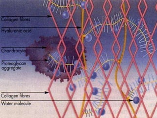
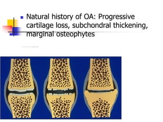
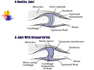
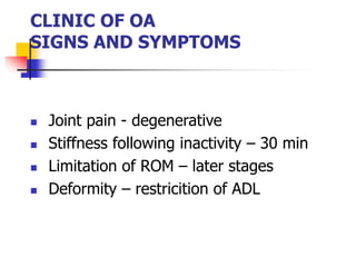
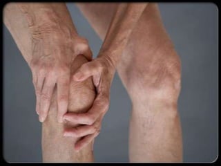
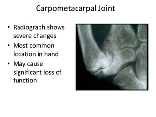
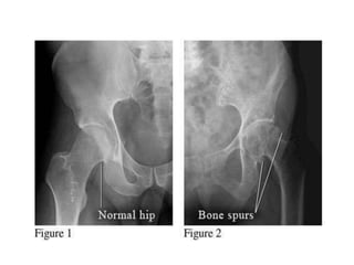
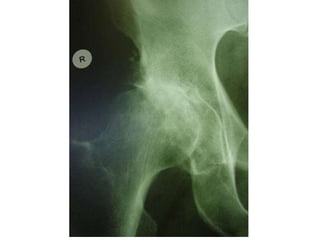
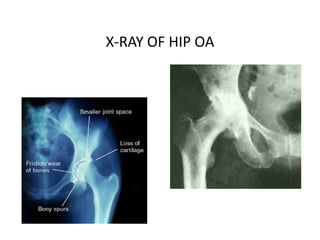
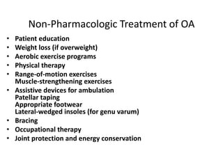
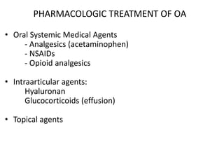
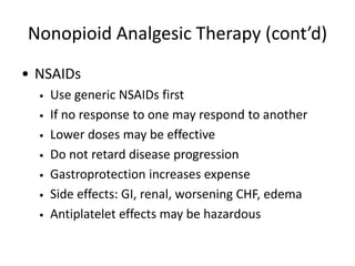
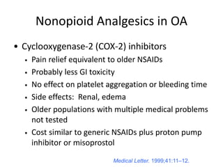
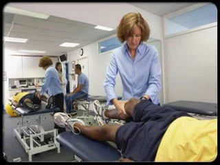
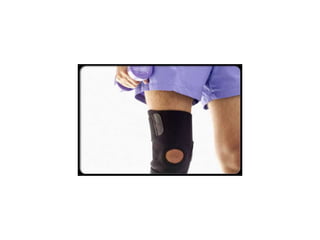
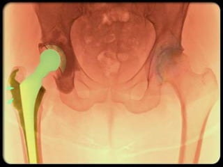
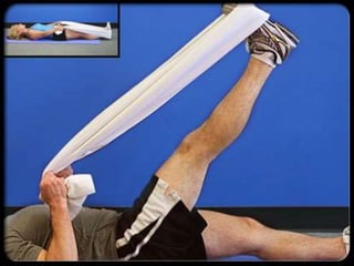
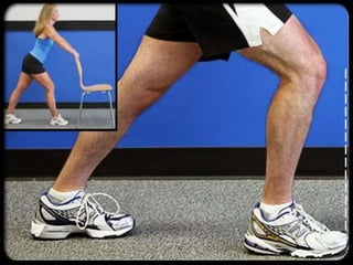
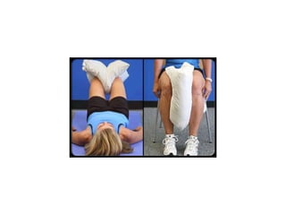
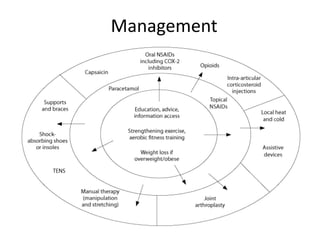
OSTEOARTHRITIS
DR. TAREK NASRALLAH
ASS. PROF.
RHEUMATOLOGY
AL AZHAR UNIVESITY
Osteoarthritis (OA): is a degenerative joint
disease characterized by the breakdown of the
joint's cartilage. It called degenerative joint
disease.
the most common form of arthritis.
EPIDEMIOLOGY
Osteoarthritis(OA) is the most common
joint disease
OA of the knee joint is found in 70% of
the population over 60 years of age
Radiological evidence of OA can be
found in over 90 % of the population
LIMITED FUNCTION
OA may cause functional loss of daily
living Activites
Most important cause of disability in
old age
Major indication for joint replacement
surgery
CLASSIFICATION OF OA
Primary OA Secondary OA
Etiology is unknown Etiology is known
AGE
Primary OA > 40 years
Direct correlation
Aging process
RISK FACTORS FOR PRIMARY OA
Age
Sex
Obesity
Genetics
Trauma (daily)
SECONDARY OSTOARTHRITIS
Trauma
Previous joint disorders;
Congenital hip dislocation
Infection: Septic arthritis, Brucella, Tb
Inflammatory: RA, AS
Metabolic: Gout
Hematologic: Hemophilia
Endocrine: DM
ETIOLOGY OF OA
Cartilage properties
Biomechanical problem
PATHOLOGY OF OA
 Fibrillation
 Eburnation
 Osteophytes
 Subcondral cysts
QuickTime™ and a
Photo CD Decompressor
are needed to use this picture
 Natural history of OA: Progressive
cartilage loss, subchondral thickening,
marginal osteophytes
CLINIC OF OA
SIGNS AND SYMPTOMS
 Joint pain - degenerative
 Stiffness following inactivity – 30 min
 Limitation of ROM – later stages
 Deformity – restricition of ADL
OA OF KNEE JOINT
 More common in obese females
 over 50 years of age
 Joint stiffness (<30 minutes)
 Mechanical pain
 Physical examination findings: Crepitus
 Pain on pressure
 Painful ROM and functional limitation
 Limitation of ROM in later stages of OA (first
extension)
 Laboratory analysis within normal limits
Morphology of Primary OA
Primary Generalized OA
LABORATORY FINDINGS OF OA
• There are no pathognomonic laboratory
findings for OA
• Laboratory analysis is performed for
differential diagnosis
RADIOLOGIC FINDINGS OF OA
• Narrowing of joint space
(due to loss of cartilage)
• Osteophytes
• Subchondral (paraarticular) sclerosis
• Bone cysts
Distal and Proximal Interphalangeal
Joints
• Radiograph shows
severe changes
• Most common
location in hand
• May cause
significant loss of
function
Carpometacarpal Joint
X-RAY OF HIP OA
Peripheral Joints
• Hands
• Feet
• X-ray shows
osteophytes,
subchondral
sclerosis, and
complete loss of joint
space
• Patients often
present with deep
groin pain that
radiates into the
medial thigh
Hip Joint
Secondary OA: Diabetic Neuropathy
• MTPs 2 to 5 involved
in addition to the 1st
bilaterally
• Destructive changes
on x-ray far in excess
of those seen in
primary OA
• Midfoot involvement
also common
Underlying Disease Associations of
OA and CPPD Disease (pseudogout)
• Hemochromatosis
• Hyperparathyroidism
• Hypothyroidism
• Hypophosphatasia
• Hypomagnesemia
• Neuropathic joints
• Trauma
• Aging, hereditary
TREATMENT OF OA
• Symptomatic treatment
• Structure modifying treatment
• Surgical treatment
STRUCTURE MODIFYING TREATMENT
• Hyaluronic acid injection (HA)
• Glycose amino glycans (GAG)
PRIMARY PREVENTION OF OA ??
• Regular exercises
• Weight control
• Prevention of trauma
AIMS OF OA TREATMENT
• Pain relief
• Preservation and restoration of joint
function
• Education
Non-Pharmacologic Treatment of OA
• Patient education
• Weight loss (if overweight)
• Aerobic exercise programs
• Physical therapy
• Range-of-motion exercises
Muscle-strengthening exercises
• Assistive devices for ambulation
Patellar taping
Appropriate footwear
Lateral-wedged insoles (for genu varum)
• Bracing
• Occupational therapy
• Joint protection and energy conservation
PHARMACOLOGIC TREATMENT OF OA
• Oral Systemic Medical Agents
- Analgesics (acetaminophen)
- NSAIDs
- Opioid analgesics
• Intraarticular agents:
Hyaluronan
Glucocorticoids (effusion)
• Topical agents
Nonopioid Analgesic Therapy
• First-line—Acetaminophen
• Pain relief comparable to NSAIDs, less toxicity
• Beware of toxicity from use of multiple
acetaminophen-containing products
• Maximum safe dose = 4 grams/day
Nonopioid Analgesic Therapy (cont’d)
• NSAIDs
• Use generic NSAIDs first
• If no response to one may respond to another
• Lower doses may be effective
• Do not retard disease progression
• Gastroprotection increases expense
• Side effects: GI, renal, worsening CHF, edema
• Antiplatelet effects may be hazardous
Nonopioid Analgesics in OA
• Cyclooxygenase-2 (COX-2) inhibitors
• Pain relief equivalent to older NSAIDs
• Probably less GI toxicity
• No effect on platelet aggregation or bleeding time
• Side effects: Renal, edema
• Older populations with multiple medical problems
not tested
• Cost similar to generic NSAIDs plus proton pump
inhibitor or misoprostol
Medical Letter. 1999;41:11–12.
Nonopioid Analgesics in OA (cont’d)
• Tramadol
• Affects opioid and serotonin pathways
• Nonulcerogenic
• May be added to NSAIDs, acetaminophen
• Side effects: Nausea, vomiting, lowered seizure
threshold, rash, constipation, drowsiness,
dizziness
Opioid Analgesics for OA
• Codeine, oxycodone
• Anticipate and prevent constipation
• Long-acting oxycodone may have fewer CNS side
effects
• Propoxyphene
• Morphine and fentanyl patches for severe
pain interfering with daily activity and sleep
Topical Agents for Analgesia in OA
• Local cold or heat: Hot packs, hydrotherapy
• Capsaicin-containing topicals
• Use well supported by evidence
• Use daily for up to 2 weeks before benefit
• Compliance poor without full instruction
• Avoid contact with eyes
• Liniments = methyl salicylates
• Temporary benefit
OA: Intra-articular Therapy
• Intra-articular steroids
• Good pain relief
• Most often used in
knees, up to q 3 mo
• With frequent injections,
risk infection, worsening
diabetes, or CHF
• Joint lavage
• Significant symptomatic
benefit demonstrated
• Hyaluronate injections*
• Symptomatic relief
• Improved function
• Expensive
• Require series of
injections
• No evidence of long-
term benefit
• Limited to knees
* Altman, et al. J Rheumatol. 1998;25:2203.
OA: Unconventional Therapies
• Polysulfated glycosaminoglycans—
nutriceuticals
• Glucosamine +/- chondroitin sulfate: Symptomatic
benefit, no known side effects, long-term
controlled trials pending
• Tetracyclines as protease/cytokine inhibitors
• Under study
• Have disease-modifying potential
HAND OA - RESTING SPLINT
INDICATIONS OF SURGICAL
INTERVENTION
• Severe joint pain,
resistant to conservative treatment
methods
• Limitation of daily living activities
• Deformity, angular deviations, instability
INVASIVE METHODS
• Joint lavage
• Arthroscopy
• Cartilage grefting- genetic engineering
• Surgery
Osteotomy
Joint replacement
Osteoarthritis- !THINK! Function
Management
Osteoarthritis

Osteoarthritis

  • 1.
    OSTEOARTHRITIS DR. TAREK NASRALLAH ASS.PROF. RHEUMATOLOGY AL AZHAR UNIVESITY
  • 2.
    Osteoarthritis (OA): isa degenerative joint disease characterized by the breakdown of the joint's cartilage. It called degenerative joint disease. the most common form of arthritis.
  • 3.
    EPIDEMIOLOGY Osteoarthritis(OA) is themost common joint disease OA of the knee joint is found in 70% of the population over 60 years of age Radiological evidence of OA can be found in over 90 % of the population
  • 4.
    LIMITED FUNCTION OA maycause functional loss of daily living Activites Most important cause of disability in old age Major indication for joint replacement surgery
  • 5.
    CLASSIFICATION OF OA PrimaryOA Secondary OA Etiology is unknown Etiology is known
  • 6.
    AGE Primary OA >40 years Direct correlation Aging process
  • 7.
    RISK FACTORS FORPRIMARY OA Age Sex Obesity Genetics Trauma (daily)
  • 8.
    SECONDARY OSTOARTHRITIS Trauma Previous jointdisorders; Congenital hip dislocation Infection: Septic arthritis, Brucella, Tb Inflammatory: RA, AS Metabolic: Gout Hematologic: Hemophilia Endocrine: DM
  • 9.
    ETIOLOGY OF OA Cartilageproperties Biomechanical problem
  • 11.
    PATHOLOGY OF OA Fibrillation  Eburnation  Osteophytes  Subcondral cysts
  • 12.
    QuickTime™ and a PhotoCD Decompressor are needed to use this picture  Natural history of OA: Progressive cartilage loss, subchondral thickening, marginal osteophytes
  • 14.
    CLINIC OF OA SIGNSAND SYMPTOMS  Joint pain - degenerative  Stiffness following inactivity – 30 min  Limitation of ROM – later stages  Deformity – restricition of ADL
  • 15.
    OA OF KNEEJOINT  More common in obese females  over 50 years of age  Joint stiffness (<30 minutes)  Mechanical pain  Physical examination findings: Crepitus  Pain on pressure  Painful ROM and functional limitation  Limitation of ROM in later stages of OA (first extension)  Laboratory analysis within normal limits
  • 16.
  • 17.
  • 22.
    LABORATORY FINDINGS OFOA • There are no pathognomonic laboratory findings for OA • Laboratory analysis is performed for differential diagnosis
  • 23.
    RADIOLOGIC FINDINGS OFOA • Narrowing of joint space (due to loss of cartilage) • Osteophytes • Subchondral (paraarticular) sclerosis • Bone cysts
  • 26.
    Distal and ProximalInterphalangeal Joints
  • 27.
    • Radiograph shows severechanges • Most common location in hand • May cause significant loss of function Carpometacarpal Joint
  • 30.
  • 31.
  • 32.
    • X-ray shows osteophytes, subchondral sclerosis,and complete loss of joint space • Patients often present with deep groin pain that radiates into the medial thigh Hip Joint
  • 33.
    Secondary OA: DiabeticNeuropathy • MTPs 2 to 5 involved in addition to the 1st bilaterally • Destructive changes on x-ray far in excess of those seen in primary OA • Midfoot involvement also common
  • 34.
    Underlying Disease Associationsof OA and CPPD Disease (pseudogout) • Hemochromatosis • Hyperparathyroidism • Hypothyroidism • Hypophosphatasia • Hypomagnesemia • Neuropathic joints • Trauma • Aging, hereditary
  • 35.
    TREATMENT OF OA •Symptomatic treatment • Structure modifying treatment • Surgical treatment
  • 36.
    STRUCTURE MODIFYING TREATMENT •Hyaluronic acid injection (HA) • Glycose amino glycans (GAG)
  • 37.
    PRIMARY PREVENTION OFOA ?? • Regular exercises • Weight control • Prevention of trauma
  • 38.
    AIMS OF OATREATMENT • Pain relief • Preservation and restoration of joint function • Education
  • 39.
    Non-Pharmacologic Treatment ofOA • Patient education • Weight loss (if overweight) • Aerobic exercise programs • Physical therapy • Range-of-motion exercises Muscle-strengthening exercises • Assistive devices for ambulation Patellar taping Appropriate footwear Lateral-wedged insoles (for genu varum) • Bracing • Occupational therapy • Joint protection and energy conservation
  • 40.
    PHARMACOLOGIC TREATMENT OFOA • Oral Systemic Medical Agents - Analgesics (acetaminophen) - NSAIDs - Opioid analgesics • Intraarticular agents: Hyaluronan Glucocorticoids (effusion) • Topical agents
  • 41.
    Nonopioid Analgesic Therapy •First-line—Acetaminophen • Pain relief comparable to NSAIDs, less toxicity • Beware of toxicity from use of multiple acetaminophen-containing products • Maximum safe dose = 4 grams/day
  • 42.
    Nonopioid Analgesic Therapy(cont’d) • NSAIDs • Use generic NSAIDs first • If no response to one may respond to another • Lower doses may be effective • Do not retard disease progression • Gastroprotection increases expense • Side effects: GI, renal, worsening CHF, edema • Antiplatelet effects may be hazardous
  • 43.
    Nonopioid Analgesics inOA • Cyclooxygenase-2 (COX-2) inhibitors • Pain relief equivalent to older NSAIDs • Probably less GI toxicity • No effect on platelet aggregation or bleeding time • Side effects: Renal, edema • Older populations with multiple medical problems not tested • Cost similar to generic NSAIDs plus proton pump inhibitor or misoprostol Medical Letter. 1999;41:11–12.
  • 44.
    Medical Letter. 1999;41:11–12. NonopioidAnalgesics in OA (cont’d) • Tramadol • Affects opioid and serotonin pathways • Nonulcerogenic • May be added to NSAIDs, acetaminophen • Side effects: Nausea, vomiting, lowered seizure threshold, rash, constipation, drowsiness, dizziness
  • 45.
    Opioid Analgesics forOA • Codeine, oxycodone • Anticipate and prevent constipation • Long-acting oxycodone may have fewer CNS side effects • Propoxyphene • Morphine and fentanyl patches for severe pain interfering with daily activity and sleep
  • 46.
    Topical Agents forAnalgesia in OA • Local cold or heat: Hot packs, hydrotherapy • Capsaicin-containing topicals • Use well supported by evidence • Use daily for up to 2 weeks before benefit • Compliance poor without full instruction • Avoid contact with eyes • Liniments = methyl salicylates • Temporary benefit
  • 47.
    OA: Intra-articular Therapy •Intra-articular steroids • Good pain relief • Most often used in knees, up to q 3 mo • With frequent injections, risk infection, worsening diabetes, or CHF • Joint lavage • Significant symptomatic benefit demonstrated • Hyaluronate injections* • Symptomatic relief • Improved function • Expensive • Require series of injections • No evidence of long- term benefit • Limited to knees * Altman, et al. J Rheumatol. 1998;25:2203.
  • 48.
    OA: Unconventional Therapies •Polysulfated glycosaminoglycans— nutriceuticals • Glucosamine +/- chondroitin sulfate: Symptomatic benefit, no known side effects, long-term controlled trials pending • Tetracyclines as protease/cytokine inhibitors • Under study • Have disease-modifying potential
  • 50.
    HAND OA -RESTING SPLINT
  • 54.
    INDICATIONS OF SURGICAL INTERVENTION •Severe joint pain, resistant to conservative treatment methods • Limitation of daily living activities • Deformity, angular deviations, instability
  • 55.
    INVASIVE METHODS • Jointlavage • Arthroscopy • Cartilage grefting- genetic engineering • Surgery Osteotomy Joint replacement
  • 60.
  • 61.