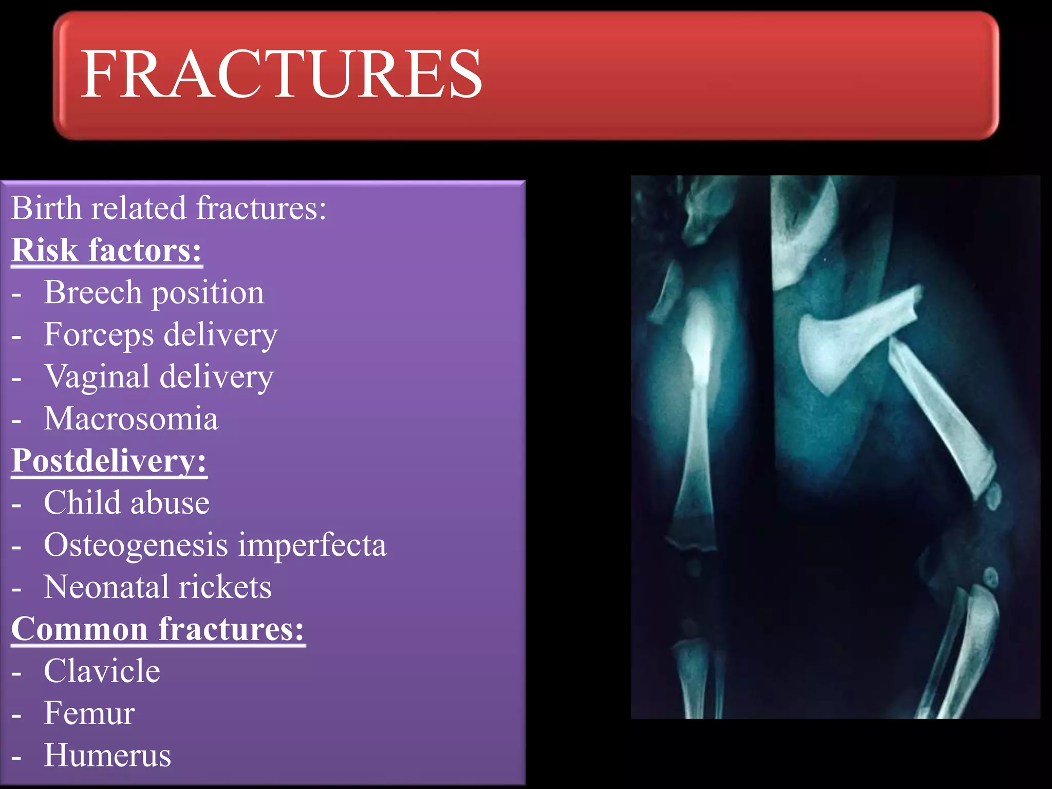
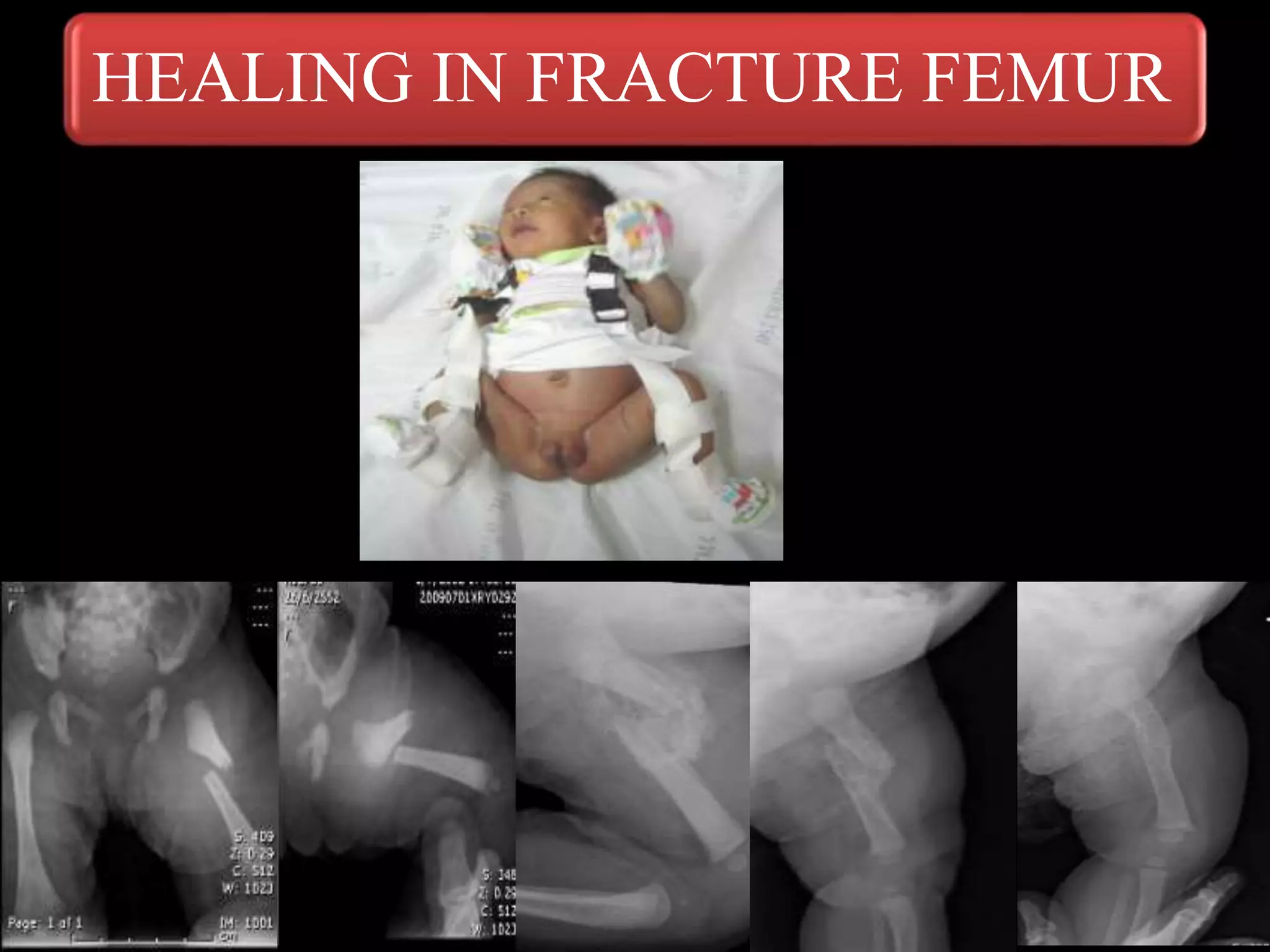
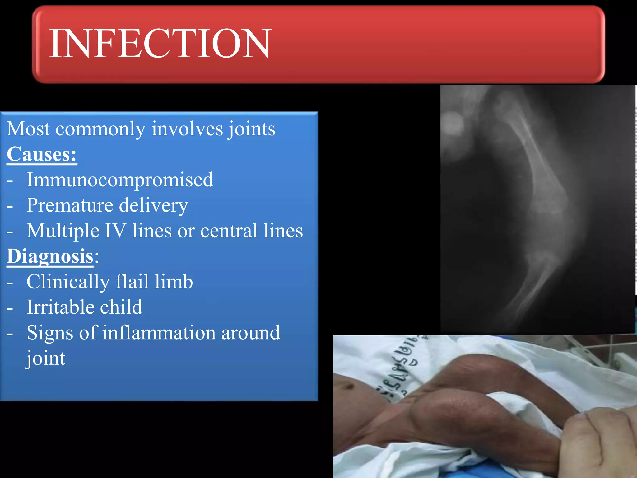
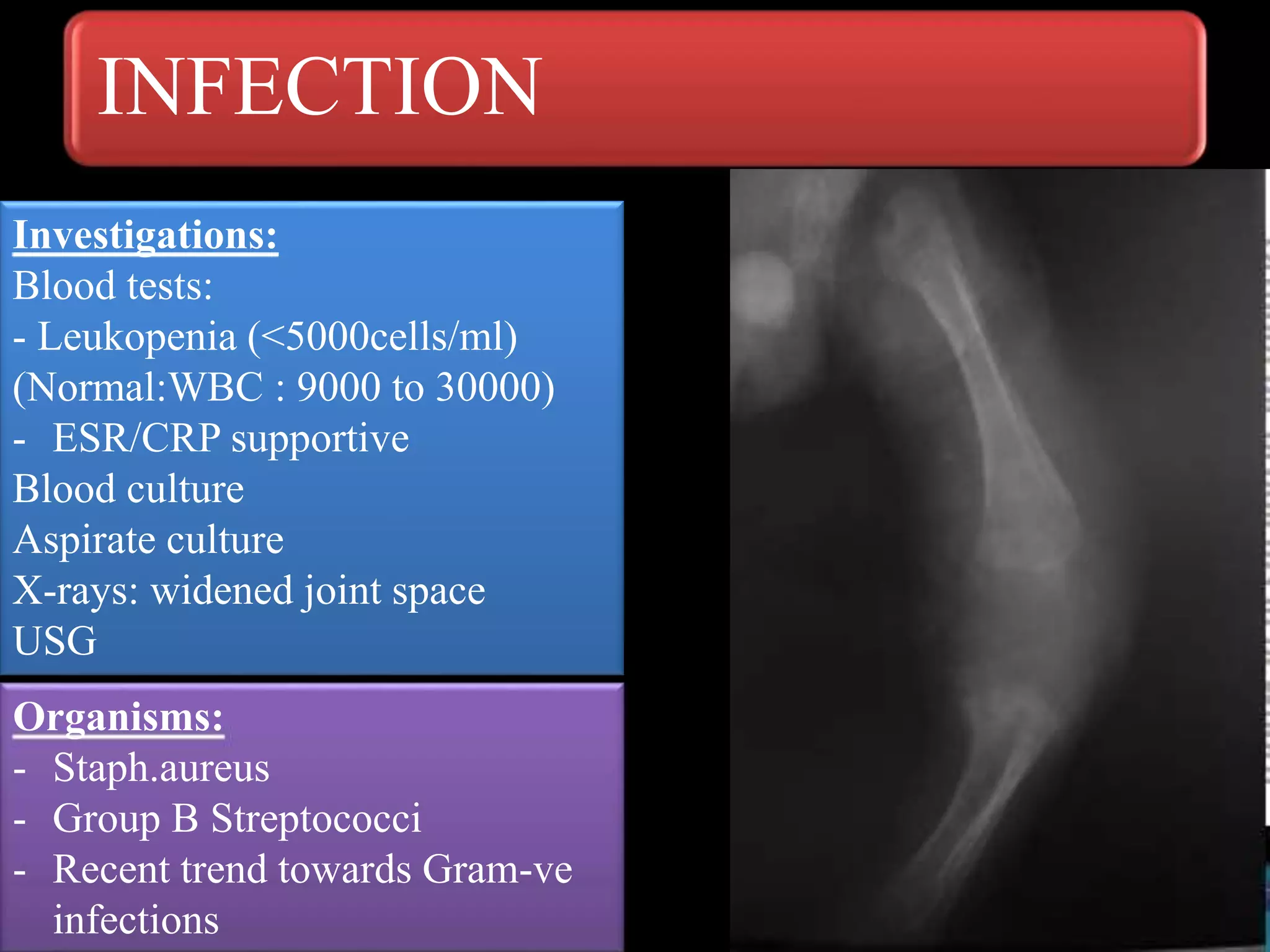
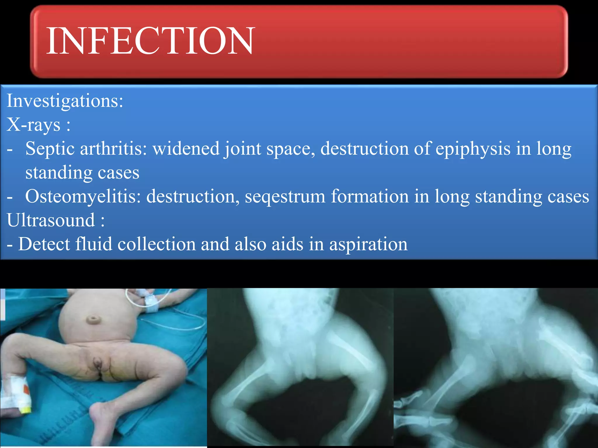
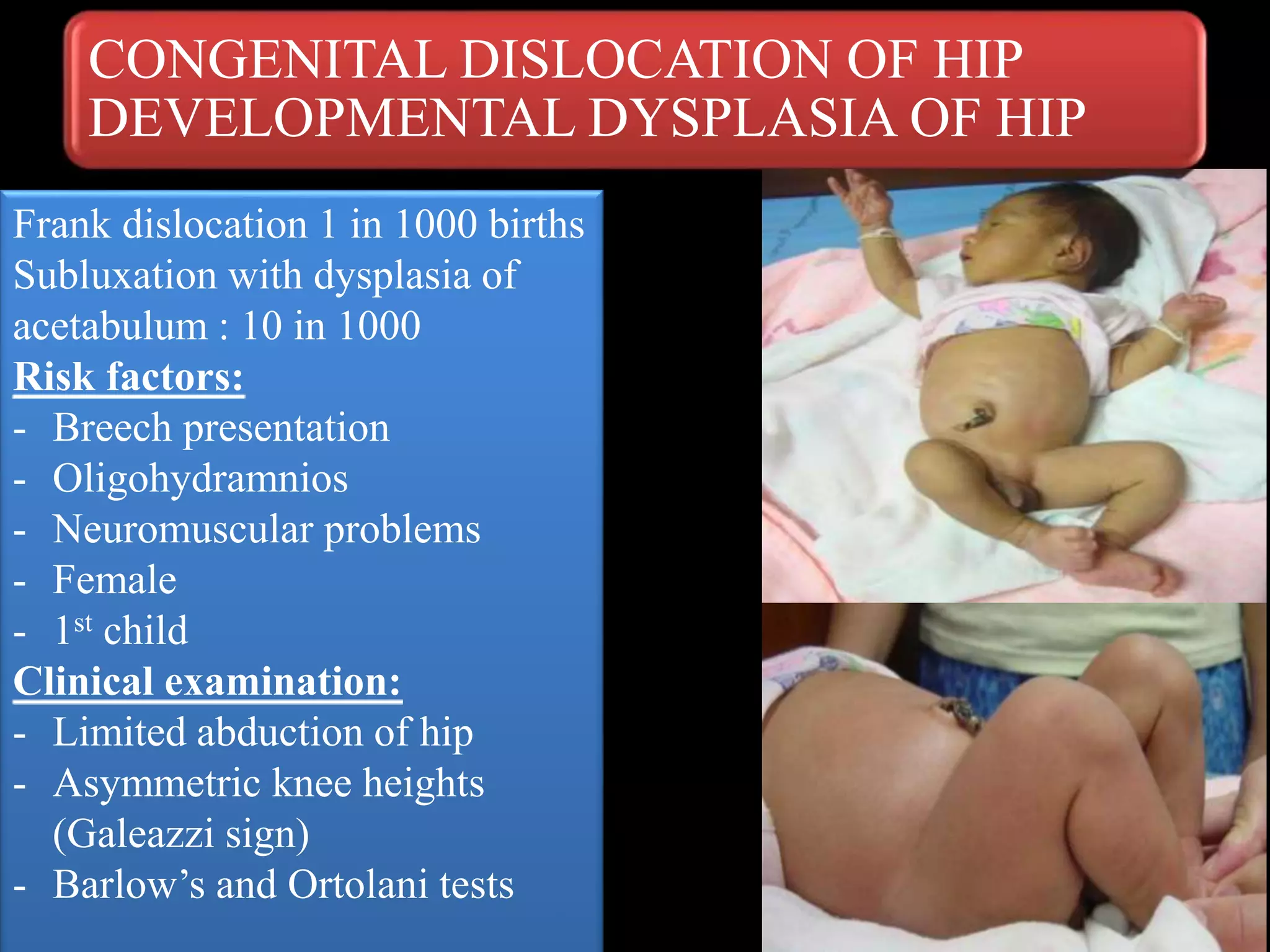
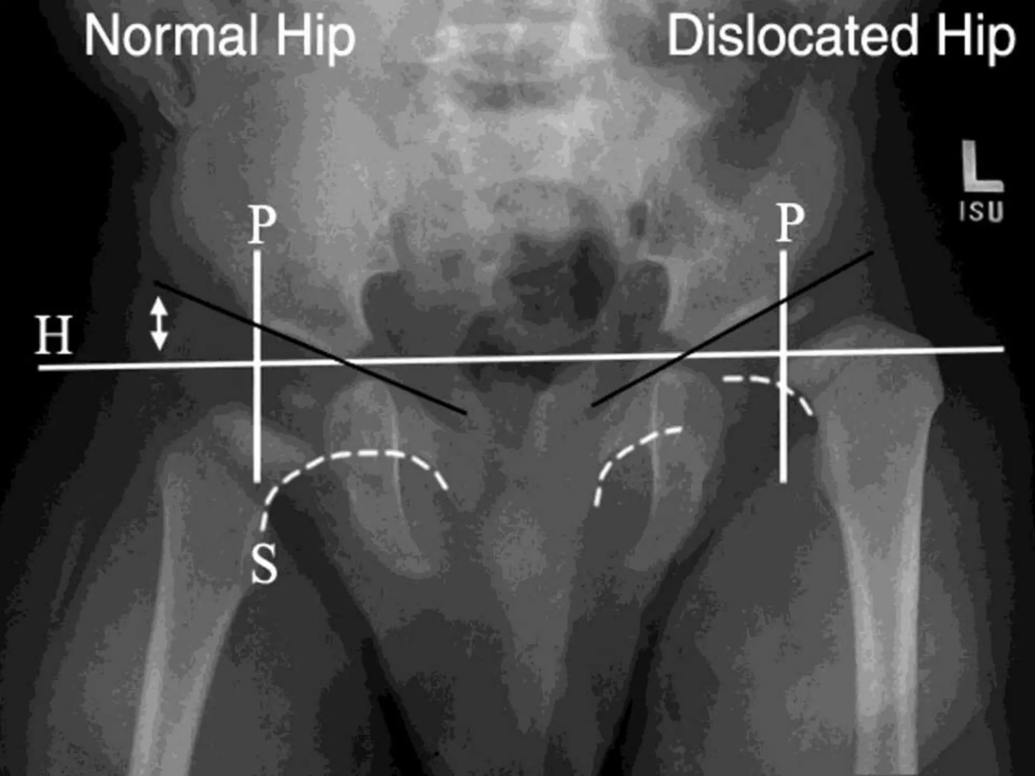
ORTHOPEDIC PROBLEMS IN
NEWBORN
Dr.A.Mohan Krishna
Consultant Orthopedic surgeon
Apollo Hospitals
Dr. A.MOHAN KRISHNA
M.S.ORTHO, MCh ORTH(U.K)
Consultant Orthopaedic surgeon ,
Trauma, Arthroscopy,
Arthroplasty Surgeon
Apollo hospitals, Hyderabad
9247258989
9441184590
www.drmohankrishna.com
FLAIL EXTREMITY
Orthopedic consultation in
NICU after delivery:
Failure to move the limb or
limbs
- Fractures
- Infection
- Brachial plexus palsy in
upper limb
- Pseudoparalysis : fracture or
infection
FRACTURES
Birth related fractures:
Risk factors:
- Breech position
- Forceps delivery
- Vaginal delivery
- Macrosomia
Postdelivery:
- Child abuse
- Osteogenesis imperfecta
- Neonatal rickets
Common fractures:
- Clavicle
- Femur
- Humerus
FRACTURES
Diagnosis:
- Flail extremity after delivery
- Deformity in case of long bone fractures
- Irritable during feeds
Radiographs: by the time radiographs are
ordered callus formation is seen in neonates
Management:
Fractures heal rapidly in neonates and
outcome is good if proper splinting is done
- Neonatal fractures require
immobilization for 2 weeks
- Femur fractures : pavilik harness, or
spica cast
- Humerus fractures : sling or splint
immobilization
HEALING IN FRACTURE FEMUR
INFECTION
Most commonly involves joints
Causes:
- Immunocompromised
- Premature delivery
- Multiple IV lines or central lines
Diagnosis:
- Clinically flail limb
- Irritable child
- Signs of inflammation around
joint
INFECTION
Investigations:
Blood tests:
- Leukopenia (<5000cells/ml)
(Normal:WBC : 9000 to 30000)
- ESR/CRP supportive
Blood culture
Aspirate culture
X-rays: widened joint space
USG
Organisms:
- Staph.aureus
- Group B Streptococci
- Recent trend towards Gram-ve
infections
INFECTION
Investigations:
X-rays :
- Septic arthritis: widened joint space, destruction of epiphysis in long
standing cases
- Osteomyelitis: destruction, seqestrum formation in long standing cases
Ultrasound :
- Detect fluid collection and also aids in aspiration
BRACHIAL PLEXUS PALSY
Cause for flail limb
- Incidence 0.13 to 3.6 per 1000
live births
Causes / Risk factors:
- Forceps delivery
- Shoulder dystocia
- Fracture clavicle, Humerus
- Prolonged vaginal delivery
- High birth weight
BRACHIAL PLEXUS PALSY
Diagnosis: Clinical
- Flail upper limb
Upper brachial plexus palsy:
(C5-C6) Erb’s palsy : Waiter’s tip
deformity
Internal rotation of shoulder
Extension of elbow
Forearm pronation and wrist
flexion
Total plexus palsy:
- Completely flaccid limb
- Horner’s syndrome-
Ptosis,Miosis,enopthalmos
- Ipsilateral diaphragmatic palsy
Investigations:
- X-rays to rule out fractures
- MRI to asses the level of injury
CONGENITAL DISLOCATION OF HIP
DEVELOPMENTAL DYSPLASIA OF HIP
Frank dislocation 1 in 1000 births
Subluxation with dysplasia of
acetabulum : 10 in 1000
Risk factors:
- Breech presentation
- Oligohydramnios
- Neuromuscular problems
- Female
- 1st child
Clinical examination:
- Limited abduction of hip
- Asymmetric knee heights
(Galeazzi sign)
- Barlow’s and Ortolani tests
CONGENITAL DISLOCATION OF HIP
DEVELOPMENTAL DYSPLASIA OF HIP
Barlow’s testOrtolani test
X-RAY- IN CDH/DDH
High resolution Ultrasound
(HRUS):
- Preferred modality in newborn:
- Help to identify mild subluxation
and acetabular dysplasia.
- Progression of treatment
Ultrasound
Femoral head
Abductors
Ilium
HRUS (ULTRASOUND)
Treatment:
- Pavlik harness
- Adductor tenotomy
- Hip Spica
- Open reduction and hip
Spica
- Corrective osteotomies in
later child hood
CONGENITAL DISLOCATION OF HIP
DEVELOPMENTAL DYSPLASIA OF HIP
TREATMENT ALGORITHM OF CDH/DDH
CONGENITAL KNEE DISLOCATION
Knee is hyperextended at birth
Causes:
- Contracture of quadriceps.
- Associated deformities- club foot,
arthrogryposis,myelodysplasia
Diagnosis:
Clinical:
- Knee is hyperextended
- Childs foot can touch the face.
- In complete dislocations knee
cannot be flexed
Investigations:
- X-rays: position of tibia in relation
to femur and grading of dislocation
CONGENITAL KNEE DISLOCATION
Management:
Non-operative treatment: gentle
reduction and serial casting in
progressive flexion
-Concomitant DDH Knee
dislocation should be reduced first
and child later put on Pavlik
harness.
Surgery:
- Not responding to conservative
management
- 6 months of age
- Open reduction and quadriceps
lengthening
CTEV (CLUB FOOT)
CONGENITAL TALIPEE EQUINO VARUS
1 to 2 in 1000 births
Deformities: Cavus,
Forefoot Adductus,Hindfoot Varus
Equinus
Etiology:
- Intrauterine Mechanical factors
- Germ plasm defect
- Neuromuscular
- Fibrosis of Medial soft-tissue
structures
Types:
Postural
Congenital
Syndromic or rigid
CTEV (CLUB FOOT)
CONGENITAL TALIPEE EQUINO VARUS
Investigations:
X-rays –Foot and ankle : Any bony
deformities Medico-legal
Hips and spine – to rule out spinal
malformations and DDH
Treatment:
- Ponseti method of serial POP
castings in out-patient setting
(Every 15 days)
- Percutaneous Achillis tendon
lengthening
- Foot abduction braces
Surgery:
Posteromedial soft tissue release at 9
months of age
CTEV (CLUB FOOT)
CONGENITAL TALIPEE EQUINO VARUS
CALCANEOVALGUS FOOT
Hind foot externally rotates and dorsiflexes.
Clinically: dorsum of foot comes in contact
with anterior tibia.
Treatment:
Gentle stretching of foot into plantar flexion
and inversion can be helpful.
Most deformities resolve by age of 3 to 6
months.
METATARSUS ADDUCTUS
- Medial deviation Metatarsotarsal joint
- Mild degree: resolve with growth
- Severe degree: Serial plasters in
corrected position.
CONGENITAL MUSCULAR TORTICOLLIS
Deformity of neck caused by contracture of sternocleidomastoid
Cause: Intrauterine compartment syndrome causing fibrosis of
sternocleidomastoid.
Risk factors: Breech position
Associated with DDH, metatarsus adductus
Clinical features:
- Head tilted towards involved muscle.
- Chin pointing towards opposite shoulder.
- Palpable mass in sternocleidomastoid muscle.
CONGENITAL MUSCULAR TORTICOLLIS
Investigations:
- X-rays: rule out klippel feil
syndrome, cervical spine
problems
Treatment:
- Gentle stretching initially up to 6
months.
- Surgery: 5 years of age- release
of Sternocleidomastoid muscle
POLYDACTYLY
SYNDACTYLY
Orthopedic problems in newborn

Orthopedic problems in newborn

  • 1.
    ORTHOPEDIC PROBLEMS IN NEWBORN Dr.A.MohanKrishna Consultant Orthopedic surgeon Apollo Hospitals
  • 2.
    Dr. A.MOHAN KRISHNA M.S.ORTHO,MCh ORTH(U.K) Consultant Orthopaedic surgeon , Trauma, Arthroscopy, Arthroplasty Surgeon Apollo hospitals, Hyderabad 9247258989 9441184590 www.drmohankrishna.com
  • 3.
    FLAIL EXTREMITY Orthopedic consultationin NICU after delivery: Failure to move the limb or limbs - Fractures - Infection - Brachial plexus palsy in upper limb - Pseudoparalysis : fracture or infection
  • 4.
    FRACTURES Birth related fractures: Riskfactors: - Breech position - Forceps delivery - Vaginal delivery - Macrosomia Postdelivery: - Child abuse - Osteogenesis imperfecta - Neonatal rickets Common fractures: - Clavicle - Femur - Humerus
  • 5.
    FRACTURES Diagnosis: - Flail extremityafter delivery - Deformity in case of long bone fractures - Irritable during feeds Radiographs: by the time radiographs are ordered callus formation is seen in neonates Management: Fractures heal rapidly in neonates and outcome is good if proper splinting is done - Neonatal fractures require immobilization for 2 weeks - Femur fractures : pavilik harness, or spica cast - Humerus fractures : sling or splint immobilization
  • 6.
  • 7.
    INFECTION Most commonly involvesjoints Causes: - Immunocompromised - Premature delivery - Multiple IV lines or central lines Diagnosis: - Clinically flail limb - Irritable child - Signs of inflammation around joint
  • 8.
    INFECTION Investigations: Blood tests: - Leukopenia(<5000cells/ml) (Normal:WBC : 9000 to 30000) - ESR/CRP supportive Blood culture Aspirate culture X-rays: widened joint space USG Organisms: - Staph.aureus - Group B Streptococci - Recent trend towards Gram-ve infections
  • 9.
    INFECTION Investigations: X-rays : - Septicarthritis: widened joint space, destruction of epiphysis in long standing cases - Osteomyelitis: destruction, seqestrum formation in long standing cases Ultrasound : - Detect fluid collection and also aids in aspiration
  • 10.
    BRACHIAL PLEXUS PALSY Causefor flail limb - Incidence 0.13 to 3.6 per 1000 live births Causes / Risk factors: - Forceps delivery - Shoulder dystocia - Fracture clavicle, Humerus - Prolonged vaginal delivery - High birth weight
  • 11.
    BRACHIAL PLEXUS PALSY Diagnosis:Clinical - Flail upper limb Upper brachial plexus palsy: (C5-C6) Erb’s palsy : Waiter’s tip deformity Internal rotation of shoulder Extension of elbow Forearm pronation and wrist flexion Total plexus palsy: - Completely flaccid limb - Horner’s syndrome- Ptosis,Miosis,enopthalmos - Ipsilateral diaphragmatic palsy Investigations: - X-rays to rule out fractures - MRI to asses the level of injury
  • 12.
    CONGENITAL DISLOCATION OFHIP DEVELOPMENTAL DYSPLASIA OF HIP Frank dislocation 1 in 1000 births Subluxation with dysplasia of acetabulum : 10 in 1000 Risk factors: - Breech presentation - Oligohydramnios - Neuromuscular problems - Female - 1st child Clinical examination: - Limited abduction of hip - Asymmetric knee heights (Galeazzi sign) - Barlow’s and Ortolani tests
  • 13.
    CONGENITAL DISLOCATION OFHIP DEVELOPMENTAL DYSPLASIA OF HIP Barlow’s testOrtolani test
  • 15.
  • 16.
    High resolution Ultrasound (HRUS): -Preferred modality in newborn: - Help to identify mild subluxation and acetabular dysplasia. - Progression of treatment
  • 17.
  • 18.
    Treatment: - Pavlik harness -Adductor tenotomy - Hip Spica - Open reduction and hip Spica - Corrective osteotomies in later child hood CONGENITAL DISLOCATION OF HIP DEVELOPMENTAL DYSPLASIA OF HIP
  • 19.
  • 20.
    CONGENITAL KNEE DISLOCATION Kneeis hyperextended at birth Causes: - Contracture of quadriceps. - Associated deformities- club foot, arthrogryposis,myelodysplasia Diagnosis: Clinical: - Knee is hyperextended - Childs foot can touch the face. - In complete dislocations knee cannot be flexed Investigations: - X-rays: position of tibia in relation to femur and grading of dislocation
  • 21.
    CONGENITAL KNEE DISLOCATION Management: Non-operativetreatment: gentle reduction and serial casting in progressive flexion -Concomitant DDH Knee dislocation should be reduced first and child later put on Pavlik harness. Surgery: - Not responding to conservative management - 6 months of age - Open reduction and quadriceps lengthening
  • 22.
    CTEV (CLUB FOOT) CONGENITALTALIPEE EQUINO VARUS 1 to 2 in 1000 births Deformities: Cavus, Forefoot Adductus,Hindfoot Varus Equinus Etiology: - Intrauterine Mechanical factors - Germ plasm defect - Neuromuscular - Fibrosis of Medial soft-tissue structures Types: Postural Congenital Syndromic or rigid
  • 23.
    CTEV (CLUB FOOT) CONGENITALTALIPEE EQUINO VARUS Investigations: X-rays –Foot and ankle : Any bony deformities Medico-legal Hips and spine – to rule out spinal malformations and DDH Treatment: - Ponseti method of serial POP castings in out-patient setting (Every 15 days) - Percutaneous Achillis tendon lengthening - Foot abduction braces
  • 24.
    Surgery: Posteromedial soft tissuerelease at 9 months of age CTEV (CLUB FOOT) CONGENITAL TALIPEE EQUINO VARUS
  • 25.
    CALCANEOVALGUS FOOT Hind footexternally rotates and dorsiflexes. Clinically: dorsum of foot comes in contact with anterior tibia. Treatment: Gentle stretching of foot into plantar flexion and inversion can be helpful. Most deformities resolve by age of 3 to 6 months.
  • 26.
    METATARSUS ADDUCTUS - Medialdeviation Metatarsotarsal joint - Mild degree: resolve with growth - Severe degree: Serial plasters in corrected position.
  • 27.
    CONGENITAL MUSCULAR TORTICOLLIS Deformityof neck caused by contracture of sternocleidomastoid Cause: Intrauterine compartment syndrome causing fibrosis of sternocleidomastoid. Risk factors: Breech position Associated with DDH, metatarsus adductus Clinical features: - Head tilted towards involved muscle. - Chin pointing towards opposite shoulder. - Palpable mass in sternocleidomastoid muscle.
  • 28.
    CONGENITAL MUSCULAR TORTICOLLIS Investigations: -X-rays: rule out klippel feil syndrome, cervical spine problems Treatment: - Gentle stretching initially up to 6 months. - Surgery: 5 years of age- release of Sternocleidomastoid muscle
  • 29.
  • 30.