
Recommended
More Related Content
What's hot
What's hot (20)
Similar to ORGANS OF THE IMMUNE SYSTEM by pranzly.ppt
Similar to ORGANS OF THE IMMUNE SYSTEM by pranzly.ppt (20)
More from Pranzly Rajput
More from Pranzly Rajput (15)
Recently uploaded
Recently uploaded (20)
ORGANS OF THE IMMUNE SYSTEM by pranzly.ppt
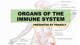
- 1. ORGANS OF THE IMMUNE SYSTEM PRESENTED BY PRANZLY
- 2. TYPES PRIMARY LYMPHOID ORGANS • Bone marrow • Thymus where maturation of lymphocytes takes place SECONDARY LYMPHOID ORGANS • Spleen • Lymph nodes • Mucosal associated lymphoid tissues (MALT) • gut-associated lymphoid tissue (GALT) which trap antigen and provide sites for mature lymphocytes to interact with that antigen
- 3. PRIMARY LYMPHOID ORGANS • Immature lymphocytes generated in hematopoiesis mature and become committed to a particular antigenic specificity within the primary lymphoid organs • Only after a lymphocytes has matured within a primary lymphoid organ is the cell immunocompetent (capable of mounting an immune response). • T cells arise in the thymus, and in many mammals— humans • B cells originate in bone marrow
- 4. BONE MARROW • supports self-renewal and differentiation of hematopoietic stem cells (HSCs) into mature blood cells. • bone marrow is the site of B-cell origin and development • the long bones (femur, humerus), hip bones (ileum), and sternum tend to be the most active • contains several cell types that coordinate HSC development, including 1. OSTEOBLASTS, versatile cells that both generate bone and control the differentiation sites of hematopoiesis of HSCs, 2. ENDOTHELIAL CELLS that line the blood vessels and also regulate HSC differentiation, 3. RETICULAR CELLS that send processes connecting cells to bone and blood vessels, and, unexpectedly, 4. SYMPATHETIC NEURONS, which can control the release of hematopoietic cells from the bone marrow
- 5. microenvironments within the bone marrow ENDOSTEAL NICHE the area directly surrounding the bone and in contact with bone- producing osteoblasts) . appears to be occupied by quiescent HSCs in close association with osteoblasts that regulate stem cell proliferation VASCULAR NICHE the area directly surrounding the blood vessels and in contact with endothelial cells •appears to be occupied by HSCs that have been mobilized to leave the endosteal niche to either differentiate or circulate
- 6. Thymus • the site of T-cell development and maturation. • It is a flat, bilobed organ situated above the heart. • Each lobe is surrounded by a capsule and is divided into lobules, which are separated from each other by strands of connective tissue called trabeculae. Lobules consists of two compartments CORTEX the outer compartment, is densely packed with immature T cells, called thymocytes MEDULLA the inner compartment, is sparsely populated with thymocytes
- 7. composed of epithelial cells, dendritic cells, and macrophages, which make up the framework of the organ and contribute to the growth and maturation of thymocytes. in the outer cortex, called nurse cells, have long membrane extensions that surround as many as 50 thymocytes, forming large multicellular complexes FUNCTION- to generate and select a repertoire of T cells that will protect the body from infection.
- 8. SECONDARY LYMPHOID ORGANS • Lymph nodes and the spleen are the most highly organized of the secondary lymphoid organs and are compartmentalized from the rest of the body by a fibrous capsule. • lymphoid tissue is organized into structures called lymphoid follicles, • Until it is activated by antigen, a lymphoid follicle— called a primary follicle—comprises a network of follicular dendritic cells and small resting B cells. • After an antigenic challenge, a primary follicle becomes a larger secondary follicle—a ring of concentrically packed B lymphocytes surrounding a center (the germinal center)
- 9. SPLEEN • It is a large, ovoid secondary lymphoid organ situated high in the left abdominal cavity. • the spleen specializes in filtering blood and trapping blood-borne antigens; thus, it can respond to systemic infections. • Functions- in iron metabolism, thrombocyte storage, haematopoiesis) • compartments 1. the red pulp and white pulp, which are separated by a specialized region called the marginal zone
- 11. • THE SPLENIC RED PULP consists of a network of sinusoids populated by red blood cells, macrophages, and some lymphocytes. It is the site where old and defective red blood cells are destroyed and removed • THE SPLENIC WHITE PULP surrounds the branches of the splenic artery, and consists of the periarteriolar lymphoid sheath (PALS) populated by T lymphocytes as well as B-cell follicles. The marginal zone, which borders the white pulp, is populated by unique and specialized macrophages and B cells, which are the first line of defense against certain blood-borne pathogen
- 12. LYMPH NODES divided into three roughly concentric regions THE CORTEX The outermost layer,, contains lymphocytes (mostly B cells), macro- phages, and follicular dendritic cells arranged in primary follicles THE PARACORTEX which is populated largely by T lymphocytes and also contains interdigitating dendritic cells THE MEDULLA The innermost layer, is more sparsely populated with lymphoid-lineage cells; of those present, many are plasma cells actively secreting antibody molecules
- 14. WHEN A FOREIGN ANTIGEN GAINS ENTRANCE TO THE TISSUES, IT IS PICKED UP BY THE LYMPHATIC SYSTEM (WHICH DRAINS ALL THE TISSUES OF THE BODY) AND IS CARRIED TO VARIOUS ORGANIZED LYMPHOID TISSUES SUCH AS LYMPH NODES ANTIGEN-PRESENTING CELLS THAT ENGULF AND PROCESS THE ANTIGEN ALSO CAN GAIN ACCESS TO LYMPH AS LYMPH PASSES FROM THE TISSUES TO LYMPHATIC VESSELS, IT BECOMES PROGRESSIVELY ENRICHED IN SPECIFIC LEUKOCYTES, INCLUDING LYMPHOCYTES, DENDRITIC CELLS, AND MACROPHAGES. WHERE THE LYMPHOCYTES CAN INTERACT WITH THE TRAPPED ANTIGEN AND UNDERGO ACTIVATION ALL IMMUNE CELLS THAT TRAFFIC THROUGH TISSUES, BLOOD, AND LYMPH NODES ARE GUIDED BY SMALL MOLECULES KNOWN AS CHEMOKINES. THESE PROTEINS ARE SECRETED BY STROMAL CELLS, ANTIGENPRESENTING CELLS, LYMPHOCYTES, AND GRANULOCYTES, AND FORM GRADIENTS THAT ACT AS ATTRACTANTS AND GUIDES FOR OTHER IMMUNE CELLS
- 15. LYMPHOID TISSUE 1. Mucosa-associated lymphoid tissue (MALT). 2. Bronchus-associated lymphoid tissue (BALT) 3. Nasal-associated lymphoid tissue (NALT), 4. Gut-associated lymphoid tissue (GALT) 5. Cutaneous-Associated Lymphoid Tissue
- 16. MUCOSA-ASSOCIATED LYMPHOID TISSUE (MALT) • MALT INCLUDES 1. TONSILS 2. PEYER’S PATCHES (IN THE SMALL INTESTINE), 3. THE APPENDIX, 4. AS WELL AS NUMEROUS LYMPHOID FOLLICLES WITHIN THE LAMINA PROPRIA OF THE INTESTINES AND IN THE MUCOUS MEMBRANES LINING THE UPPER AIRWAYS, BRONCHI, AND GENITOURINARY TRACT
- 17. TONSILS • THE TONSILS ARE FOUND IN THREE LOCATIONS: 1. LINGUAL AT THE BASE OF THE TONGUE; 2. PALATINE AT THE SIDES OF THE BACK OF THE MOUTH; 3. AND PHARYNGEAL (ADENOIDS) IN THE ROOF OF THE NASOPHARYNX ALL THREE TONSIL GROUPS ARE NODULAR STRUCTURES CONSISTING OF A MESHWORK OF RETICULAR CELLS AND FIBERS INTERSPERSED WITH LYMPHOCYTES, MACROPHAGES, GRANULOCYTES, AND MAST CELLS.
- 18. PEYER’S PATCHES • Peyer’s patches, nodules of 30 to 40 lymphoid follicles, extend into the muscle layers that are just below the lamina propria.
- 19. GUT-ASSOCIATED LYMPHOID TISSUE (GALT) • The outer mucosal epithelial layer contains intraepithelial lymphocytes (IELs), many of which are T cells. • The lamina propria, which lies under the epithelial layer, contains large numbers of B cells, plasma cells, activated T cells, and macrophages in loose clusters • In the digestive tract, specialized M cells transport antigen across the epithelium, they are flattened epithelial cells lacking the microvilli that characterize the rest of the mucosal epithelium. • M cells have a deep invagination, or pocket, in the basolateral plasma membrane; this pocket is filled with a cluster of B cells, T cells, and macrophages
- 20. M cells are located in so-called inductive sites—small regions of a mucous membrane that lie over organized lymphoid follicles. Antigens transported across the mucous membrane by M cells can activate B cells within these lymphoid follicles. The activated B cells differentiate into plasma cells, which leave the follicles and secrete the IgA class of antibodies.
- 21. Cutaneous-Associated Lymphoid Tissue (CALT) • The epidermal (outer) layer of the skin is composed largely of specialized epithelial cells called keratinocytes. • These cells secrete a number of cytokines that may function to induce a local inflammatory reaction. • These cells express high levels of class II MHC molecules and function as potent activators of naive TH cells • The epidermis also contains so- called intraepidermal lymphocytes.