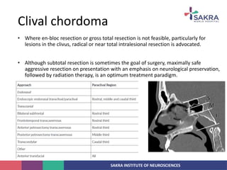
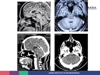
SAKRA INSTITUTE OF NEUROSCIENCES
Chordoma
Dr Abhishek Rai
DNB Neurosurgery
SAKRA INSTITUTE OF NEUROSCIENCES
• Chordoma is a rare malignant tumor of the skull base and axial skeleton, with
an incidence of less than 0.1/100,000 per year.
• They are thought to arise from remnants of the embryonal notochord and were
first described by Virchow in 1857.
• Müller was first to suggest in 1858 that these tumours may be of notochordal
origin.
• In humans, most notochordal remnants disappear during the first years of life.
• Notochordal tissue remains along the axial skeleton, which explains the
locations where chordoma occurs: the bulk arises at the sacrum or clivus and
the remainder occur at varying levels of the mobile spine.
Introduction
SAKRA INSTITUTE OF NEUROSCIENCES
• Rare, locally aggressive primary malignant
neoplasm with a phenotype that
recapitulates the notochord.
• Etiology
• Skull base chordomas probably arise from
cranial end of primitive notochord
remnants.
• Signal transducer and activation of
transcription (STAT) proteins regulate key
cellular fates, including proliferation and
apoptosis. STAT 3 is activated in chordoma
Introduction
SAKRA INSTITUTE OF NEUROSCIENCES
• 3 major histologic forms:
1. Conventional (classic)
2. Chondroid
3. Dededifferentiated
• Conventional chordoma consist of physaliphorous cells that contain mucin and
glycogen granules, giving characterstic “bubbly” appearance to its cytoplasm.
• Chondroid chordoma have stromal elements that resemble hyaline cartilage
with neoplastic cells nested within lacunae.
• Dedifferentiated chordoma represent <5% of chordoma and typically occurs in
sacrococcygeal region.
Pathology
SAKRA INSTITUTE OF NEUROSCIENCES
• Chordomas were fi rst characterised microscopically by Virchow in 1857.
• Described unique, intracellular, bubble-like vacuoles that he referred to as
physaliferous, a term now synonymous with their histopathology.
• Virchow hypothesised that chordomas were derived from cartilage; however, more
contemporary evidence suggests that they are derived from undiff erentiated
notochordal remnants that reside within the vertebral bodies and throughout the
axial skeleton.
• Ribbert first introduced the term chordoma in the 1890s,9 in view of the notochord
hypothesis.
• Examination of human embryos and fetuses and cell-fate-tracking experiments in
mice showed that notochordal cell nests topographically correspond and distribute
to the sites of occurrence of chordoma.
Pathogenesis
SAKRA INSTITUTE OF NEUROSCIENCES
• Perhaps the most compelling evidence of the notochordal hypothesis was the
discovery of a gene duplication in the transcription factor T gene (brachyury) in
familial chordoma.
• High-resolution array comparative genomic hybridisation showed unique
duplications in the 6q27 region in tumour samples from patients with familial
chordoma.
• This duplicated region contained only the brachyury gene, which was known to be
uniquely overexpressed in almost all sporadic chordomas compared with other bone
or cartilaginous lesions.
• Although it is still unclear role of brachyury in the pathogenesis of chordomas,
identification of the duplication and the remarkable overexpression seen in samples
suggests that it might be a crucial molecular driver in the initiation and propagation
of this cancer.
Pathogenesis
SAKRA INSTITUTE OF NEUROSCIENCES
• In summary, three fundamental observations support the notochordal hypothesis:
1. The site of notochord vestiges corresponds closely to the distribution of chordomas;
2. There is a morphological similarity between these remnants and the histopathology
exhibited by chordomas;
3. Both share a similar immunophenotype
Pathogenesis
SAKRA INSTITUTE OF NEUROSCIENCES
• Chordomas are indolent and slow growing, therefore they are often clinically silent
until the late stages of disease.
• The clinical manifestations vary and depend on location. Skull-base chordomas often
grow in the clivus and present with cranial-nerve palsies.
• Depending on their size and involvement of the sella, endocrinopathy can also occur.
• Other rare presentations include epistaxis and intracranial haemorrhage.
• Chordomas of the mobile spine and sacrum can present with localised deep pain or
radiculopathies related to the spinal level at which they occur.
• Diagnosis is further complicated by the fact that lytic sacral lesions might be
overlooked on plain radiographs, and CT and MRI studies often do not extend below
the S2 level
Clinical presentation
SAKRA INSTITUTE OF NEUROSCIENCES
• Studies show that neurological deficit is more often observed in chordomas of the
mobile spine than in chordomas in the sacrococcygeal region.
• Most sacrococcygeal chordomas involve the fourth and fifth sacral vertebrae, and
although large tumours can protrude anteriorly into the pelvis, invasion into pelvic
structures is often limited by presacral fascia.
• Cervical chordomas can present with airway obstruction or dysphagia, and might
even present as an oropharyngeal mass
Clinical presentation
SAKRA INSTITUTE OF NEUROSCIENCES
• NCCT:
• Relatively well circumscribed, moderately hyperdense midline or paramedian clival
mass with permative lytic bony changes.
• Intratumoral calcification generally represent sequestration from destroyed bone.
Imaging
SAKRA INSTITUTE OF NEUROSCIENCES
• Chordomas of the mobile spine and sacrum:
• In the 1970s, Stener and Gunterberg first introduced the idea of wide en-bloc
surgical resection for the treatment of sacral tumours. Since then, en-bloc excision
has remained a central tenant in the surgical management of sacral chordoma.
• Kaiser and colleagues showed that local recurrence was almost two-times higher in
patients who received en-bloc resection where the tumour capsule was entered at
the time of surgery than in those who had complete en-bloc excision of the tumour
without violation of the capsule.
• One landmark study on the surgical management of chordomas showed a
remarkable difference in recurrence between patients who underwent radical
resection and those who had subtotal excision of sacral chordomas; the time from
surgery to local recurrence was 2¡27 years compared with 8 months, respectively.
Surgical management
SAKRA INSTITUTE OF NEUROSCIENCES
• The surgical approach for many of these tumours depends on the extent of the
lesion. For example, en-bloc resection of chordomas below the sacroiliac joint
mainly involves a posterior–transperineal exposure, which can be achieved without
elaborate reconstructive efforts.
• Chordomas that are caudal to the sacroiliac joint are more technically challenging,
requiring both anterior and posterior exposures or a combined anterior–posterior
approach.
• Sacrectomies that spare the S2 nerve root are associated with up to a 50% chance of
normal bladder and bowel control, a percentage that can be improved if an S3 nerve
root is also preserved.
• Preservation of the bilateral S2 nerve root with the unilateral S3 nerve root is
associated with normal bladder and bowel function, whereas sacrifi ce of any of the
S2 nerve roots typically leads to at least loss of voluntary control.
Surgical management
SAKRA INSTITUTE OF NEUROSCIENCES
Clival chordoma
SAKRA INSTITUTE OF NEUROSCIENCES
• Skull-base chordomas have a broad surgical approach strategy that is based on the
location of the tumour and the surgeon’s preference.
• Transphenoidal, trans maxillary, transnasal, high anterior cervical retro pharyngeal,
and transoral approaches have been well documented in the literature, as have
endoscopic techniques.
Clival chordoma
SAKRA INSTITUTE OF NEUROSCIENCES
• Where en-bloc resection or gross total resection is not feasible, particularly for
lesions in the clivus, radical or near total intralesional resection is advocated.
• Although subtotal resection is sometimes the goal of surgery, maximally safe
aggressive resection on presentation with an emphasis on neurological preservation,
followed by radiation therapy, is an optimum treatment paradigm.
Clival chordoma
SAKRA INSTITUTE OF NEUROSCIENCES
• The challenge of preventing cerebrospinal fluid leakage after violation of the
anterior skull-base dura is being addressed with novel, local fl ap repair techniques.
• This approach is part of the armamentarium of skull-base surgeons, complementing
traditional surgical access techniques such as transoral and transmaxillary
approaches.
• A series instituting this management strategy along with universal adjuvant
radiotherapy reported excellent outcomes.
• 11 patients underwent removal of skull-base chordomas, with resection being
subtotal or partial in seven. Transient neurological deterioration (cranial-nerve defi
cits) occurred in seven patients, all of whom returned to neurological baseline.
Clival chordoma
SAKRA INSTITUTE OF NEUROSCIENCES
• Park and colleagues showed that treatment with proton or photon-beam radiation
in combination with surgery resulted in higher local control in patients with primary
sacral chordoma than in those with recurrence, underscoring the importance of
radiation early in the treatment of the disease.
• Unfortunately, the availability of hadron-based therapy is limited because of the
associated construction and operational expenses.
• Alternatives include intensity-modulated radiation therapy (IMRT) and stereotactic
delivery techniques.
Radiation therapy
SAKRA INSTITUTE OF NEUROSCIENCES
• Anthracycline, cisplatin, alkylating agents, and camptothecin analogues have been
reported to affect chordomas.
• Studies have identified a common gene duplication of the transcriptional regulator,
brachyury, in patients with familial and sporadic chordoma.
• Tyrosine kinases and transcriptional regulators have been shown to be
overexpressed in chordoma.
• Studies targeting these kinase domains and their downstream effectors could
provide translational therapies and will improve our understanding and treatment of
patients with chordoma.
Medical therapy
SAKRA INSTITUTE OF NEUROSCIENCES
Because of their propensity to grow in a destructive and invasive way, radical
resection is challenging—even for the experienced surgeon—and is often not
possible without causing morbidity or even mortality.
Additionally, these tumours are fairly resistant to radiation therapy and systemic
therapy.
Introduction

Chordoma

  • 1.
    SAKRA INSTITUTE OFNEUROSCIENCES Chordoma Dr Abhishek Rai DNB Neurosurgery
  • 2.
    SAKRA INSTITUTE OFNEUROSCIENCES • Chordoma is a rare malignant tumor of the skull base and axial skeleton, with an incidence of less than 0.1/100,000 per year. • They are thought to arise from remnants of the embryonal notochord and were first described by Virchow in 1857. • Müller was first to suggest in 1858 that these tumours may be of notochordal origin. • In humans, most notochordal remnants disappear during the first years of life. • Notochordal tissue remains along the axial skeleton, which explains the locations where chordoma occurs: the bulk arises at the sacrum or clivus and the remainder occur at varying levels of the mobile spine. Introduction
  • 3.
    SAKRA INSTITUTE OFNEUROSCIENCES • Rare, locally aggressive primary malignant neoplasm with a phenotype that recapitulates the notochord. • Etiology • Skull base chordomas probably arise from cranial end of primitive notochord remnants. • Signal transducer and activation of transcription (STAT) proteins regulate key cellular fates, including proliferation and apoptosis. STAT 3 is activated in chordoma Introduction
  • 4.
    SAKRA INSTITUTE OFNEUROSCIENCES • 3 major histologic forms: 1. Conventional (classic) 2. Chondroid 3. Dededifferentiated • Conventional chordoma consist of physaliphorous cells that contain mucin and glycogen granules, giving characterstic “bubbly” appearance to its cytoplasm. • Chondroid chordoma have stromal elements that resemble hyaline cartilage with neoplastic cells nested within lacunae. • Dedifferentiated chordoma represent <5% of chordoma and typically occurs in sacrococcygeal region. Pathology
  • 5.
    SAKRA INSTITUTE OFNEUROSCIENCES
  • 6.
    SAKRA INSTITUTE OFNEUROSCIENCES • Chordomas were fi rst characterised microscopically by Virchow in 1857. • Described unique, intracellular, bubble-like vacuoles that he referred to as physaliferous, a term now synonymous with their histopathology. • Virchow hypothesised that chordomas were derived from cartilage; however, more contemporary evidence suggests that they are derived from undiff erentiated notochordal remnants that reside within the vertebral bodies and throughout the axial skeleton. • Ribbert first introduced the term chordoma in the 1890s,9 in view of the notochord hypothesis. • Examination of human embryos and fetuses and cell-fate-tracking experiments in mice showed that notochordal cell nests topographically correspond and distribute to the sites of occurrence of chordoma. Pathogenesis
  • 7.
    SAKRA INSTITUTE OFNEUROSCIENCES • Perhaps the most compelling evidence of the notochordal hypothesis was the discovery of a gene duplication in the transcription factor T gene (brachyury) in familial chordoma. • High-resolution array comparative genomic hybridisation showed unique duplications in the 6q27 region in tumour samples from patients with familial chordoma. • This duplicated region contained only the brachyury gene, which was known to be uniquely overexpressed in almost all sporadic chordomas compared with other bone or cartilaginous lesions. • Although it is still unclear role of brachyury in the pathogenesis of chordomas, identification of the duplication and the remarkable overexpression seen in samples suggests that it might be a crucial molecular driver in the initiation and propagation of this cancer. Pathogenesis
  • 8.
    SAKRA INSTITUTE OFNEUROSCIENCES • In summary, three fundamental observations support the notochordal hypothesis: 1. The site of notochord vestiges corresponds closely to the distribution of chordomas; 2. There is a morphological similarity between these remnants and the histopathology exhibited by chordomas; 3. Both share a similar immunophenotype Pathogenesis
  • 9.
    SAKRA INSTITUTE OFNEUROSCIENCES • Chordomas are indolent and slow growing, therefore they are often clinically silent until the late stages of disease. • The clinical manifestations vary and depend on location. Skull-base chordomas often grow in the clivus and present with cranial-nerve palsies. • Depending on their size and involvement of the sella, endocrinopathy can also occur. • Other rare presentations include epistaxis and intracranial haemorrhage. • Chordomas of the mobile spine and sacrum can present with localised deep pain or radiculopathies related to the spinal level at which they occur. • Diagnosis is further complicated by the fact that lytic sacral lesions might be overlooked on plain radiographs, and CT and MRI studies often do not extend below the S2 level Clinical presentation
  • 10.
    SAKRA INSTITUTE OFNEUROSCIENCES • Studies show that neurological deficit is more often observed in chordomas of the mobile spine than in chordomas in the sacrococcygeal region. • Most sacrococcygeal chordomas involve the fourth and fifth sacral vertebrae, and although large tumours can protrude anteriorly into the pelvis, invasion into pelvic structures is often limited by presacral fascia. • Cervical chordomas can present with airway obstruction or dysphagia, and might even present as an oropharyngeal mass Clinical presentation
  • 11.
    SAKRA INSTITUTE OFNEUROSCIENCES • NCCT: • Relatively well circumscribed, moderately hyperdense midline or paramedian clival mass with permative lytic bony changes. • Intratumoral calcification generally represent sequestration from destroyed bone. Imaging
  • 12.
    SAKRA INSTITUTE OFNEUROSCIENCES
  • 13.
    SAKRA INSTITUTE OFNEUROSCIENCES
  • 14.
    SAKRA INSTITUTE OFNEUROSCIENCES • Chordomas of the mobile spine and sacrum: • In the 1970s, Stener and Gunterberg first introduced the idea of wide en-bloc surgical resection for the treatment of sacral tumours. Since then, en-bloc excision has remained a central tenant in the surgical management of sacral chordoma. • Kaiser and colleagues showed that local recurrence was almost two-times higher in patients who received en-bloc resection where the tumour capsule was entered at the time of surgery than in those who had complete en-bloc excision of the tumour without violation of the capsule. • One landmark study on the surgical management of chordomas showed a remarkable difference in recurrence between patients who underwent radical resection and those who had subtotal excision of sacral chordomas; the time from surgery to local recurrence was 2·27 years compared with 8 months, respectively. Surgical management
  • 15.
    SAKRA INSTITUTE OFNEUROSCIENCES • The surgical approach for many of these tumours depends on the extent of the lesion. For example, en-bloc resection of chordomas below the sacroiliac joint mainly involves a posterior–transperineal exposure, which can be achieved without elaborate reconstructive efforts. • Chordomas that are caudal to the sacroiliac joint are more technically challenging, requiring both anterior and posterior exposures or a combined anterior–posterior approach. • Sacrectomies that spare the S2 nerve root are associated with up to a 50% chance of normal bladder and bowel control, a percentage that can be improved if an S3 nerve root is also preserved. • Preservation of the bilateral S2 nerve root with the unilateral S3 nerve root is associated with normal bladder and bowel function, whereas sacrifi ce of any of the S2 nerve roots typically leads to at least loss of voluntary control. Surgical management
  • 16.
    SAKRA INSTITUTE OFNEUROSCIENCES Clival chordoma
  • 17.
    SAKRA INSTITUTE OFNEUROSCIENCES • Skull-base chordomas have a broad surgical approach strategy that is based on the location of the tumour and the surgeon’s preference. • Transphenoidal, trans maxillary, transnasal, high anterior cervical retro pharyngeal, and transoral approaches have been well documented in the literature, as have endoscopic techniques. Clival chordoma
  • 18.
    SAKRA INSTITUTE OFNEUROSCIENCES • Where en-bloc resection or gross total resection is not feasible, particularly for lesions in the clivus, radical or near total intralesional resection is advocated. • Although subtotal resection is sometimes the goal of surgery, maximally safe aggressive resection on presentation with an emphasis on neurological preservation, followed by radiation therapy, is an optimum treatment paradigm. Clival chordoma
  • 19.
    SAKRA INSTITUTE OFNEUROSCIENCES • The challenge of preventing cerebrospinal fluid leakage after violation of the anterior skull-base dura is being addressed with novel, local fl ap repair techniques. • This approach is part of the armamentarium of skull-base surgeons, complementing traditional surgical access techniques such as transoral and transmaxillary approaches. • A series instituting this management strategy along with universal adjuvant radiotherapy reported excellent outcomes. • 11 patients underwent removal of skull-base chordomas, with resection being subtotal or partial in seven. Transient neurological deterioration (cranial-nerve defi cits) occurred in seven patients, all of whom returned to neurological baseline. Clival chordoma
  • 20.
    SAKRA INSTITUTE OFNEUROSCIENCES • Park and colleagues showed that treatment with proton or photon-beam radiation in combination with surgery resulted in higher local control in patients with primary sacral chordoma than in those with recurrence, underscoring the importance of radiation early in the treatment of the disease. • Unfortunately, the availability of hadron-based therapy is limited because of the associated construction and operational expenses. • Alternatives include intensity-modulated radiation therapy (IMRT) and stereotactic delivery techniques. Radiation therapy
  • 21.
    SAKRA INSTITUTE OFNEUROSCIENCES • Anthracycline, cisplatin, alkylating agents, and camptothecin analogues have been reported to affect chordomas. • Studies have identified a common gene duplication of the transcriptional regulator, brachyury, in patients with familial and sporadic chordoma. • Tyrosine kinases and transcriptional regulators have been shown to be overexpressed in chordoma. • Studies targeting these kinase domains and their downstream effectors could provide translational therapies and will improve our understanding and treatment of patients with chordoma. Medical therapy
  • 22.
    SAKRA INSTITUTE OFNEUROSCIENCES
  • 23.
    SAKRA INSTITUTE OFNEUROSCIENCES
  • 24.
    SAKRA INSTITUTE OFNEUROSCIENCES
  • 25.
    SAKRA INSTITUTE OFNEUROSCIENCES Because of their propensity to grow in a destructive and invasive way, radical resection is challenging—even for the experienced surgeon—and is often not possible without causing morbidity or even mortality. Additionally, these tumours are fairly resistant to radiation therapy and systemic therapy. Introduction

Editor's Notes

  • #3 Virchow initially who mistakenly thought the tissue was softened cartilage with hydropic degeneration of cells and hence coined the term ecchondrosis physaliphora
  • #4 Virchow initially who mistakenly thought the tissue was softened cartilage with hydropic degeneration of cells and hence coined the term ecchondrosis physaliphora
  • #6 Intraoperatively obtained chordoma tissue with physaliferous phenotype; haematoxylin and eosin (H&E) stained, frozen tissue smear (A,B,C). Intraoperatively obtained chordoma tissue with physaliferous phenotype; H&E stained, formalin-fixed tissue (D,E,F). Chordoma tissue is positive for S-100β in A, for cytokeratin AE1/AE3 in B, and for brachyury in C; immunohistochemistry with diaminobenzidine chromogen.
  • #8 An important transcription factor in notochord development, brachyury is expressed in normal, undiff erentiated embryonic notochord in the axial skeleton
  • #10 Unfortunately, the non-specifi c nature of these symptoms and insidious onset of pain often delays the diagnosis until late in the disease course, such that bowel or bladder function can be compromised
  • #11 Unfortunately, the non-specifi c nature of these symptoms and insidious onset of pain often delays the diagnosis until late in the disease course, such that bowel or bladder function can be compromised
  • #14 Sagittal graphic shows expansile destructive, lobulated clival mass with athumb of tumor
  • #15 This study provided the fi rst compelling evidence that contamination of the surgical wound via cell seeding is responsible for recurrence, and therefore advocated complete excision of the tumour during initial surgery over any decompression
  • #17  the upper red horizontal line represents the sellar floor plane; the lower red horizontal line represents the sphenoid sinus floor plane.
  • #20 When local invasiveness into surrounding skull-base neurovascular structures is present, one management philosophy is to minimise neurological dysfunction, even at the price of postoperative residual tumour
  • #25 Sagittal graphic shows expansile destructive, lobulated clival mass with a thumb of tumor