Download as PPSX, PPTX


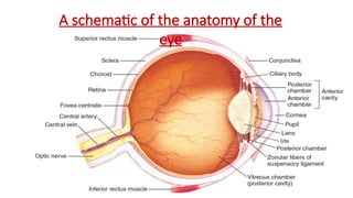
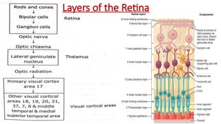
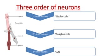
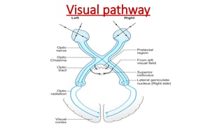
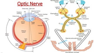
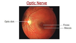
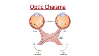
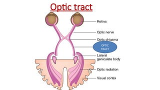
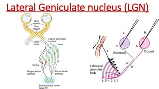
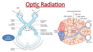
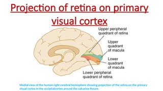


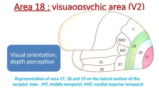
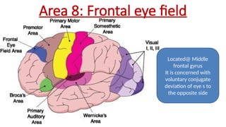
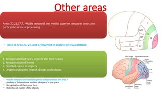
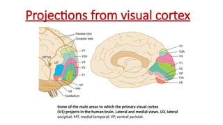
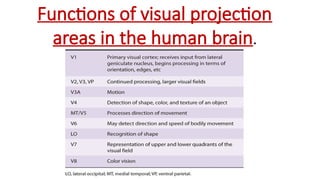

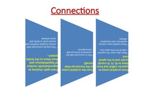
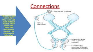


The document covers the visual pathways in the human brain, detailing the structure and function of the retina, optic nerve, optic chiasm, and visual centers in the occipital cortex. It explains the roles of various brain areas in visual processing, including recognition of objects, motion detection, and coordination of eye movements. Additionally, it addresses optic pathway lesions and their effects on vision, highlighting terms such as anopia and hemianopia.


