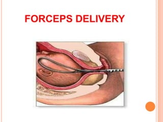
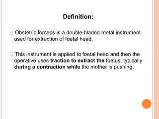
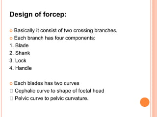
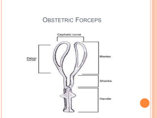
4. Dilatation and evacuation
• Dilatation of the cervix and evacuation of the products
of conception from uterine cavity
One stage operation
– Dilatation of cervix and evacuation of uterus done in
the same sitting
Two stage operation
– First phase : slow method
– Second phase : rapid method
5. One stage operation
Steps ;
• Dilate the cervix to desired extent
• Products are removed by ovum forceps
• IV methergine 0.2mg to be given
• Uterus is massaged bimanually with both external and
internal hand
• Vagina and perineum is toileted, with sterile vulval pad
Placed
Indication
Incomplete abortion
Inevitable abortion
MTP
Hydatidiform mole
6. Two stage operation
1. First phase
– Introduction of laminaria tent (MgSO4,sponge)
2. Second phase
– Further dilatation of cervix with metal dilators followed by
evacuation
Patient is brought back to OT after 12 hours
Conducted under IV diazepam/GA sedation
Indication
Induction of 1st trimester abortion
Missed abortion
Hydatidiform mole
7.
8. Complication
– Excessive hemorrhage : due to incomplete
evacuation or atonic uterus
– Injury : cervical laceration, uterine perforation
– Shock
– Sepsis
– Hematometra
– Continuation of pregnancy (failure)
10. Definition:
Obstetric forceps is a double-bladed metal instrument
used for extraction of foetal head.
This instrument is applied to foetal head and then the
operative uses traction to extract the foetus, typically
during a contraction while the mother is pushing.
11. Design of forcep:
Basically it consist of two crossing branches.
Each branch has four components:
1. Blade
2. Shank
3. Lock
4. Handle
Each blades has two curves
Cephalic curve to shape of foetal head
Pelvic curve to pelvic curvature.
13. TYPES OF FORCEP APPLICATION
CEPHALIC APPLICATION:
The forceps is applied on the sides of the foetal head in
the mentovertical diameter so, injury of the fetal face, eyes
and facial nerve is avoided.
PELVIC APPLICATION:
The forceps is applied along the maternal pelvic wall
irrespective to the position of the head. It is easier for
application but carries a great risk of foetal injuries.
CEPHALO-PELVIC APPLICATION:
It is the ideal and possible application when the occiput is
directly anterior or in mento-anterior diameter position.
14.
15. INDICATIONS OF FORCEP DELIVERY
Prolonged 2nd stage
It is the prolongation for more than 1 hour in
primigravida or 30 mins in multipara.
This may be due to:
Poor voluntary bearing down
Large fetus
Rigid perineum
Malposition: persistent occipito-posterior and
deep transverse arrest.
16. MATERNAL INDICATIONS
Maternal distress
Pulse greater than 100 beats per min
Temperature greater than 38 C
Sign of dehydration
Maternal diseases as:
Heart disease
Pulmonary TB
Pre-eclampsia and eclampsia
18. PRE-REQUISITIES FOR FORCEPS APPLICATION
Anesthesia: general ,epidural, spinal, pudental block.
Adequate pelvic outlet.
Aseptic measures
Bladder and bowel evacuation
Contractions of the uterus should be present.
Dilatation of the cervix should be fully.
Engaged head.
19. MANAGEMENT:
Re-assessment: the forcep is removed and the patient
is re-examined to detect the cause and correct it if
possible.
Caesarean section: it is indicated in uncorrectable
causes as CPD and contracted outlet.
Exploration of the birth canal: for any injuries.
24. INTRODUCTION
“Instrumental device designed to assist delivery by
applying traction to a suction cup attached to the
fetal scalp”
• Any condition threatened to mother or foetus that is
likely to be relieved by delivery
• Fatus of at least 34 weeks
25. Instrumentation
Components:
• a suction cup with four sizes(30mm,40mm,50mm,
60mm)
– Metal cup
– Soft cup
– Silastic cup
– Rigid plastic cup
• vacuum pump,
• traction tubing
27. Maternal indication
1. Maternal distress
2. Prolonged second stage of labor
( Nulliparous: >3hrs
Parous: >2hrs )
3. Maternal medical disorders such as heart disease,
hypertensive disorders and moderate to severe anemia.
4. Previous caesarean section or genital prolapse repair.
5. Intra partum infection, certain neurological
conditions.
28. Fetal indication
1. Prolapse of umbilical cord
2. Premature separation of placenta
3. Fetal distress
4. Occipito-posterior position
29. Contraindication
• Operator inexperience
• Inability to assess foetal position
• Suspicion of cephalo-pelvic disproportion
• Fetal coagulopathy
• Preterm babies (<34 weeks) due to risk of foetal
intraventricular hemorrhage
• Macrosomia (≥4 kg)
• Soft tissues obstruction in the pelvis
• Breach presentation and face presentation
30. Technique
• The woman's bladder should be empty (via voiding or
catheterization).
• The patient is placed in the lithotomy position.
• Vaginal examination to check pelvic capacity, cervical
dilatation, présentation, position, station and degree of
flexion of head and that the membranes are ruptured
• Determination of flexion point
31.
32. Continue….
• Proper cup placement over flexion point
• Exclude maternal soft tissue entrapment by palpation
• Vacuum creation by increasing the suction in
increments of 0.2 kg/cm2 every 2 mins until 0.8 kg/cm2
• A check is made using the fingers round the cup
to ensure that no cervical or vaginal tissue is
trapped inside the cup
33. • The pressure is gradually raised at the rate of
0.1kg/cm2 per minute until the effective vacuum
of 0.8kg/cm2 is achieved in about 10 minutes
time
• The scalp is sucked into the cup and an artificial
caput succedaneum is produced, which
disappears within few hours.
• Instrument handle is grasped, and initiation of
traction
34. Continue….
• Traction is initiated by using a two-handed
technique, i.e the fingers of one hand are placed
against the suction cup, while the other hand grasps
the handle of the instrument
• Traction must be at right angle to the cup
35. • Traction should be synchronous with the uterine
contractions; released in between the contractions.
• Once head is extracted, vacuum pressure is relieved; cup
is removed; vaginal delivery followed
• The total time from the application until delivery should not
exceed 20 minutes
• If >20 minutes, the risk of fetal scalp trauma and
intracranial damage increases
36. Fetal Complications
• Scalp laceration and bruising
• Subgalial hematoma, Cephalohematoma
• Intracranial haemorrhage, intraventricular and cerebral
hemorrhages
• Retinal and sub-conjunctival hemorrhages
• Neonatal jaundice
• Clavicular fracture, Shoulder dystocia
• Hypoxia, particularly when extraction has taken a long
time and has been difficult
• Fetal death
37. Maternal Complications
• Soft tissues injuries such as cervical tears, vaginal
tears, perineal lacerations and tears, extension of
episiotomy, vaginal wall and perineal hematomas.
• Traumatic postpartum hemorrhages
• Infection
• Genital prolapse
38. Management
• To assess the effect on the mother and the fetus
• To start a Ringer’s solution drip and to arrange for
blood transfusion, if required
• To exclude rupture of the uterus
• To assess if procedure is to be abandoned and consider
delivery by caesarean section
• Laparotomy should be done in a case with rupture of
uterus.
• To administer parenteral antibiotic
40. INTRODUCTION
An operative procedure that is carried out under
anaesthesia whereby the foetus, placenta and
membranes are delivered through an incision in
abdominal wall and the uterus
Usually carried out after viability has been reached i.e.
24-48 weeks of gestation onwards.
The first operation performed on a women is referred to
as a primary caesarean section.
When operation is performed in subsequent
pregnancies, it is called repeat caesarean section.(C/S)
41. INCIDENCE:
The incidence of caesarean is steadily raising.
Factors responsible are increased safety of operation
due to improved anaesthesia, availability of blood
transfusion and antibiotics.
Increased awareness of foetal well being and
identification of risk factors have caused reduction of
difficult operation or manipulative vaginal deliveries.
42. Indication for Caesarean section
. Absolute:
Advanced carcinoma of cervix
Cervical or broad of contracted pelvis.
Severe degree of contracted pelvis.
. Relatives:
Cephalopelvic disproportion
Previous uterine scar
Fetal distress.
Malpresentations
Antepartum hemorrhage
Elderly primigravidae
Chronic hypertension
Diabetes
Pelvis atresia
45. Time of operation:
A. Elective caesarean section:
The term elective indicates that the decision to
deliver the baby by caesarean has been made
during the pregnancy and before the onset of labor.
It means pre-planning for doing caesarean section.
Indication:
CPD
Placenta previa
Bad obstetric history
46. B. Emergency caesarean delivery
When the operation is performed due to unforeseen
complication arising either during pregnancy or labour
without wasting time following the decision.
Indication:
Cord prolapse
Uterine rupture
Eclampsia
Prolonged first stage of labour
Abnormal uterine contraction
Placenta previa diagnosed in labor.
47. Types of operation:
1. Lower segment caesarean section:
Is lesser muscular than the upper segment of the uterus.
Transverse incision is made in the lower segment this
heals faster and sucessfully than an incision in the upper
segment of the uterus.
There is less muscle and more fibrous tissue in lower
segment which reduces the risk of rupture in a
subsequent pregnancy.
48. 2. Classical caesarean section:
In this baby is extracted through an incision made in
upper segment of uterus.
Is rarely performed.
Operation is done only under forced circumstances, such
as:
carcinoma of cervix
Big fibroid on lower segment
lower segment is difficult or risky
example: placenta previa
49. Nursing Management
A. Pre-operative management:
Patient should be physically prepared i.e. abdomen, back
,private parts and upper part or thigh are shaved and
cleaned.
Prepare mother psychologically by providing assurance
and explaining the indication, procedure and need of
caesarean section.
Administration of IV infusion of 50% dextrose to avoid
hypotension following spinal anaesthesia, the infusion line
is maintained patent by an intra venous cannula.
Blood grouped and cross matched for emergency
requirement.
50. Bladder should be empty by inserting foleys catheter. This
may be done before and after induction of
anesthesia.
Mother should be in NPO for about 8 hours.
Patient should be in clean gown, valuable ornament
should be taken off and all make up should be
removed.
If elective caesarean section then Ranitidine 150mg
should be given orally in the night before and repeated
one hour before surgery to prevent
51. B. Post operative care :
1. Immediate care (4-6 hours):
• In the immediate recovery period,the blood pressure is
recorded in every 2 hourly.
• The wound must be inspected half hourly to detect any
blood loss.
• The lochia are inspected and drainage should be small
initially Following general anaesthesia, the women is
nursed in left lateral or recovery position until she is full
conscious.
• Analgesic is given as prescribed.
52. 2. First 24 hours:
IV fluids are continued, blood transfusion is helpful in anemia
mothers.
Parental antibiotic is usually given for 1st 48 hour, analgesics is
the form of pethidine 75-100mg are given as needed.
Ambulation is encouraged following day of surgery and baby is
given to mother.
After 24 hours:
TPR are usually checked every 4 hourly
Orally feeding is started with clear liquid and then advanced to
normal diet and IV fluid are continued for about 48 hours.
Catheter may be removed on following day when the women is
able to get up to the toilet. She should be helped to get out of bed.
The mother must be encouraged to take rest and provide care to
the baby and should breast feed the baby.
53. Complication
Mother:
partum hemorrhage related to uterine atony and
rarely blood coagulation disorders.
Shocks related to blood loss.
Anesthesia hazards
Sepsis, secondary PPH.
Thrombosis
Lung infection post.
56. Craniotomy
• Operation to make a perforation on the fetal head,
evacuated the contents followed by extraction of the uterus
• Indications
– Cephalic presentation producing obstructed labor with dead fetus
– Hydrocephalus even in living fetus
– Interlocking head of twin
Condition to be fulfilled
√cervix fully dilated
√ baby must be dead
C/I
Severely contracted pelvis
Rupture of uterus
57.
58. Decapitation
• Head is severed from the trunk, delivery is completed
with extraction of trunk and that decapitated head per
vagina
Indication
Neglected shoulder presentation with dead fetus where
neck is easy accesible
Interlocking head of twins
59. Evisceration
• Removal of thoracic and abdominal contents piecemeal
through an opening at the most accessible site
• Together with spondylectomy
Indication
Neglected shoulder presentation (dead fetus)
Fetal malformations
61. Cleidotomy
• Reduction in the bulk of the shoulder girdle by
division of one or both the clavicles
• Clavicle are divided by embryotomy scissor/long
straight scissor
Indication
Only in dead fetus with shoulder dystocia
63. Postoperative care for destructive
operation
• Exploration of utero-vaginal canal
• Self retaining Foley’s catheter to be put inside
following craniotomy
• Dextrose saline drip – to be continued
• Ceftriaxone IV 1g infusion