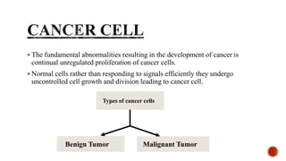
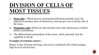
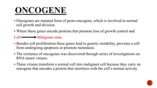
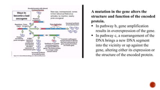
ONCOGENES
Sana Sidhik.K.V.
MSC Biotechnology
 The fundamental abnormalities resulting in the development of cancer is
continual unregulated proliferation of cancer cells.
 Normal cells rather than responding to signals efficiently they undergo
uncontrolled cell growth and division leading to cancer cell.
Types of cancer cells
Benign Tumor Malignant Tumor
 There are two types of genetic alterations that develop into cancer.
I. Germ-line mutation:- Those that we inherit from our parents; this type
won’t contribute much to disease.
II. Somatic mutation:-Those that occur during their own life time; but this
type can cause greatest impact.
 The development of malignant tumor(tumorigenesis) is multistep process
characterised by a progression of permanent genetic alterations in a single line
of cell.
 Malignant cells won’t undergo the process of apoptosis hence the cell becomes
less responsive to normal regulatory machinery.
I. Stem cells :- Which possess unlimited proliferation potential, have the
capacity to produce more of themselves, and can give rise to all the cells of
the tissue.
II. Progenitor cells:-Which are derived from stem cells and possess a limited
ability to proliferate.
III. The differentiated end products of the tissue, which generally lack the
capability to divide.
Examples of common cancer:-
Breast, Colon, Prostate and lungs, also arise in epithelial cells which undergo
high level of cell division.
Cancer Gene
Tumor Suppressor Gene Oncogene
 Oncogenes are mutated form of proto-oncogene, which is involved in normal
cell growth and division.
 Where these genes encode proteins that promote loss of growth control and
Cell Malignant state.
 Besides cell proliferation these genes lead to genetic instability, prevents a cell
from undergoing apoptosis or promote metastasis.
 The existence of oncogenes was discovered through series of investigations on
RNA tumor viruses.
 These viruses transform a normal cell into malignant cell because they carry an
oncogene that encodes a protein that interferes with the cell’s normal activity.
 Based on the studies done in 1976, they discovered oncogene called src,
carried by an RNA tumor virus called Avian sarcoma virus, was actually
present in the genome of uninfected cell.
 They are proto-oncogene that have the potential to subvert the cell’s own
activities and push the cell to malignant state.
 Proto-oncogene Oncogene by several mechanisms.
I. The gene can be mutated in a way that alerts the properties of the gene
product so that it no longer functions normally.
II. The gene can become duplicated one or more times, resulting in gene
amplification and excess production of the encoded protein.
III. A chromosome rearrangement can occur that brings a DNA sequence form a
distinct site in the genome into close proximity of the gene, which can either
alter the expression of the gene or the nature of the gene product.
A mutation in the gene alters the
structure and function of the encoded
protein.
 In pathway b, gene amplification
results in overexpression of the gene.
 In pathway c, a rearrangement of the
DNA brings a new DNA segment
into the vicinity or up against the
gene, altering either its expression or
the structure of the encoded protein.
 Any of these gene alterations can cause a cell to become less responsive to
normal growth controls.
 Oncogenes act dominantly that is a single copy of oncogene can cause cell to
express the altered phenotype.
 Development of human malignancy requires more than a single genetic
alteration.
 There are two types of genes responsible for tumor formation; Oncogene and
Tumor Supressor Gene.
 Most tumors contain alterations in both tumor suppressor genes and oncogenes
by the loss of a tumor-suppressor function with in a cell must be accompanied
by the conversion of proto-oncogene in to oncogene before the cell become
fully malignant.
 Different oncogenes become activated in different types of tumors, which reflects the
variation in the signaling pathways that operate in diverse cell types.
 The oncogene mutated mostly in human is RAS, which encodes a GTP- binding protein that
functions as an on-off switch for a number of key signalling pathways controlling cell
proliferation and metabolism.
 Oncogenic RAS mutants typically encode a protein whose GTPase activity cannot be
stimulated, which leaves the molecule in an active GTP- bound form, sending continuous
proliferation signals along the pathway.
 Cellular gene for platelet- derived growth factor(PDGF), a protein present in human blood.
Cultured cells that are transformed with this virus secrete large amount of PDGF into the
medium, which causes the cell to proliferate in an uncontrolled fashion.
 Overexpression of PDGF has been implicated in the development of brain tumors(gliomas).
 Oncogenic virus, avian erythroblastosis virus, was found to carry an oncogene (erbB) that
encodes an EGF receptor .
 One might expect that the altered receptor would be unable to signal a cell to divide, but
just the reverse is true.
 This altered version of the receptor stimulates the cell constitutively, that is, regardless of
whether the growth factor is present in the medium.
 This is the reason why cultured cells that carry the altered gene proliferate in an
uncontrolled manner.
There are oncogenes that code for variety of factors they are:-
1. Oncogenes that encode cytoplasmic protein Kinase
 protein kinases function as oncogenes by generating signals that lead to
inappropriate cell proliferation or survival.
 Raf, for example, is a serine‐threonine protein kinase that heads the MAP kinase
cascade, the primary growth‐controlling signaling pathway in cells.
 It is evident that Raf is well positioned to cause great damage within a cell should
its enzymatic activity become altered as the result of mutation.
 As with the growth factor receptors and Ras, mutations that turn Raf into an
enzyme that remains in the “on” position are most likely to convert the
proto‐oncogene into an oncogene and contribute to the cell’s loss of growth
control.
 Raf is most closely linked to melanoma, where BRAF mutations play a causative
role in the development of approximately 70 percent of these cancers.
 Another group of cytoplasmic kinases that are often deregulated in cancer are the
cyclin‐dependent kinases, especially Cdk4 and Cdk6.
2. Oncogene that encode transcription factors
 A number of oncogenes encode proteins that act as transcription factors. The
progression of cells through the cell cycle requires the timely activation
(repression) of a large variety of genes whose products contribute in various ways
to cell growth and division.
 Therefore, that alterations in the proteins that control the expression of these genes
could seriously disturb a cell’s normal growth patterns.
 The best studied oncogene whose product acts as a transcription factor is MYC.
 Myc regulates the expression of a huge number of proteins and noncoding RNAs
(rRNAs, tRNAs, and miRNAs) involved in cell growth and proliferation.
 When MYC expression is selectively blocked, the progression of the cell through
G1 is blocked.
 The MYC gene is one of the proto‐oncogenes most commonly altered in human
cancers, often being amplified within the genome or rearranged as the result of a
chromosome translocation.
 These chromosomal changes are thought to remove the MYC gene from its
normal regulatory influences and increase its level of expression in the cell,
producing an excess of the Myc protein.
 One of the most common types of cancer among populations in Africa, called
Burkitt’s lymphoma, results from the translocation of a MYC gene to a position
adjacent to an antibody gene.
3. Oncogene that encode proteins that affect the epigenetic state of chromatin
Two of the most important factors in determining the epigenetic state of
chromatin are :
(1) whether particular sites in the DNA of gene promoters are methylated or not.
(2) The particular modifications present in the tails of certain core histones within
the nucleosomes of these same gene promoters.
 DNA methylation tends to silence genes whereas histone modifications may
either activate or repress gene transcription.
 Recent studies have indicated that a number of oncogenes encode proteins that
affect DNA methylation or histone modifications.
 These include DNA methyltransferases, histone acetylases and deacetylases,
histone methyltransferases and demethylases, and proteins present within
chromatin remodeling complexes.
 Mutations in any of these classes of genes can promote tumorigenesis by
increasing or decreasing transcription of genes involved in the various
signaling and regulatory pathways that affect cell proliferation, survival,
migration, and other functions.
 Example, acute myeloid leukemia is characterized by recurrent mutations in
DNMT3A, a gene whose product is involved in maintaining DNA methylation
patterns during DNA replication.
 A reduction in the level of DNA methylation could lead to increased movement
of transposable elements, which would cause genetic instability, as well as
increased transcription of certain proto‐oncogenes.
 Conversely, an increase in the level of DNA methylation of the promoter regions
of tumor suppressor genes is known to silence the expression of genes that exert a
key inhibitory influence on tumorigenesis.
4. Oncogene that encode metabolic enzymes
 Tumor cells depend much more on glycolysis than do normal cells.
 This is only one of a number of major differences in metabolism between normal
and tumor cells.
 Another difference, which was one of the surprising discoveries to emerge from
genome sequencing studies, was the repeated presence of mutations in the TCA
cycle enzyme isocitrate dehydrogenase (IDH1 and IDH2) in the tumor cells of
patients with glioblastoma (brain cancer) and acute myeloid leukemia (AML).
 These mutations cause the enzyme to lose its normal activity of converting
isocitrate to α‐ketoglutarate and instead convert the substrate to an abnormal
metabolite called 2‐hydroxyglutarate (2‐HG), which accumulates to high levels in
the tumor.
 The elevated levels of 2‐HG have an impact on a number of processes including
histone demethylation and DNA methylation. It is proposed that the disruption of
these epigenetic processes would likely result in the aberrant regulation of gene
expression within tumor cells.
5. Oncogenes that encode products that affect apoptosis
 Apoptosis is one of the body’s key mechanisms to rid itself of tumor cells at an
early stage in their progression toward malignancy. Consequently, any alteration
that diminishes a cell’s ability to self‐ destruct would be expected to increase the
likelihood of that cell giving rise to a tumor.
 The oncogene most closely linked to apoptosis is BCL‐2, which encodes a
membrane‐bound protein that inhibits apoptosis .
 The role of BCL‐2 in apoptosis is most clearly revealed in the phenotypes of
knockout mice that are lacking a BCL‐2 gene. Once formed, the lymphoid tissues of
these mice undergo dramatic regression as the result of widespread apoptosis.
 Like MYC, the product of the BCL‐2 gene becomes oncogenic when it is
expressed at higher‐than‐normal levels, as can occur when the gene is translocated
to an abnormal site on the chromosome.
 Certain human lymphoid cancers (called follicular B‐cell lymphomas) are
correlated with the translocation of the BCL‐2 gene next to a gene that codes for
the heavy chain of antibody molecules.
 It is suggested that overexpression of the BCL‐2 gene leads to the suppression of
apoptosis in lymphoid tissues, allowing abnormal cells to proliferate to form
lymphoid tumors.
 The BCL‐2 gene may also play a role in reducing the effectiveness of
chemotherapy by keeping tumor cells alive and proliferating despite damage by the
drug treatment.
1. Karp Cellbiology
2. www.mayoclinic.org
3. ScienceDirect.com
THANK YOU

Oncogene.pptx

  • 1.
  • 2.
     The fundamentalabnormalities resulting in the development of cancer is continual unregulated proliferation of cancer cells.  Normal cells rather than responding to signals efficiently they undergo uncontrolled cell growth and division leading to cancer cell. Types of cancer cells Benign Tumor Malignant Tumor
  • 3.
     There aretwo types of genetic alterations that develop into cancer. I. Germ-line mutation:- Those that we inherit from our parents; this type won’t contribute much to disease. II. Somatic mutation:-Those that occur during their own life time; but this type can cause greatest impact.  The development of malignant tumor(tumorigenesis) is multistep process characterised by a progression of permanent genetic alterations in a single line of cell.  Malignant cells won’t undergo the process of apoptosis hence the cell becomes less responsive to normal regulatory machinery.
  • 4.
    I. Stem cells:- Which possess unlimited proliferation potential, have the capacity to produce more of themselves, and can give rise to all the cells of the tissue. II. Progenitor cells:-Which are derived from stem cells and possess a limited ability to proliferate. III. The differentiated end products of the tissue, which generally lack the capability to divide. Examples of common cancer:- Breast, Colon, Prostate and lungs, also arise in epithelial cells which undergo high level of cell division.
  • 5.
  • 6.
     Oncogenes aremutated form of proto-oncogene, which is involved in normal cell growth and division.  Where these genes encode proteins that promote loss of growth control and Cell Malignant state.  Besides cell proliferation these genes lead to genetic instability, prevents a cell from undergoing apoptosis or promote metastasis.  The existence of oncogenes was discovered through series of investigations on RNA tumor viruses.  These viruses transform a normal cell into malignant cell because they carry an oncogene that encodes a protein that interferes with the cell’s normal activity.
  • 7.
     Based onthe studies done in 1976, they discovered oncogene called src, carried by an RNA tumor virus called Avian sarcoma virus, was actually present in the genome of uninfected cell.  They are proto-oncogene that have the potential to subvert the cell’s own activities and push the cell to malignant state.  Proto-oncogene Oncogene by several mechanisms. I. The gene can be mutated in a way that alerts the properties of the gene product so that it no longer functions normally. II. The gene can become duplicated one or more times, resulting in gene amplification and excess production of the encoded protein. III. A chromosome rearrangement can occur that brings a DNA sequence form a distinct site in the genome into close proximity of the gene, which can either alter the expression of the gene or the nature of the gene product.
  • 8.
    A mutation inthe gene alters the structure and function of the encoded protein.  In pathway b, gene amplification results in overexpression of the gene.  In pathway c, a rearrangement of the DNA brings a new DNA segment into the vicinity or up against the gene, altering either its expression or the structure of the encoded protein.
  • 9.
     Any ofthese gene alterations can cause a cell to become less responsive to normal growth controls.  Oncogenes act dominantly that is a single copy of oncogene can cause cell to express the altered phenotype.  Development of human malignancy requires more than a single genetic alteration.  There are two types of genes responsible for tumor formation; Oncogene and Tumor Supressor Gene.  Most tumors contain alterations in both tumor suppressor genes and oncogenes by the loss of a tumor-suppressor function with in a cell must be accompanied by the conversion of proto-oncogene in to oncogene before the cell become fully malignant.
  • 10.
     Different oncogenesbecome activated in different types of tumors, which reflects the variation in the signaling pathways that operate in diverse cell types.  The oncogene mutated mostly in human is RAS, which encodes a GTP- binding protein that functions as an on-off switch for a number of key signalling pathways controlling cell proliferation and metabolism.  Oncogenic RAS mutants typically encode a protein whose GTPase activity cannot be stimulated, which leaves the molecule in an active GTP- bound form, sending continuous proliferation signals along the pathway.  Cellular gene for platelet- derived growth factor(PDGF), a protein present in human blood. Cultured cells that are transformed with this virus secrete large amount of PDGF into the medium, which causes the cell to proliferate in an uncontrolled fashion.  Overexpression of PDGF has been implicated in the development of brain tumors(gliomas).
  • 11.
     Oncogenic virus,avian erythroblastosis virus, was found to carry an oncogene (erbB) that encodes an EGF receptor .  One might expect that the altered receptor would be unable to signal a cell to divide, but just the reverse is true.  This altered version of the receptor stimulates the cell constitutively, that is, regardless of whether the growth factor is present in the medium.  This is the reason why cultured cells that carry the altered gene proliferate in an uncontrolled manner. There are oncogenes that code for variety of factors they are:-
  • 12.
    1. Oncogenes thatencode cytoplasmic protein Kinase  protein kinases function as oncogenes by generating signals that lead to inappropriate cell proliferation or survival.  Raf, for example, is a serine‐threonine protein kinase that heads the MAP kinase cascade, the primary growth‐controlling signaling pathway in cells.  It is evident that Raf is well positioned to cause great damage within a cell should its enzymatic activity become altered as the result of mutation.  As with the growth factor receptors and Ras, mutations that turn Raf into an enzyme that remains in the “on” position are most likely to convert the proto‐oncogene into an oncogene and contribute to the cell’s loss of growth control.  Raf is most closely linked to melanoma, where BRAF mutations play a causative role in the development of approximately 70 percent of these cancers.  Another group of cytoplasmic kinases that are often deregulated in cancer are the cyclin‐dependent kinases, especially Cdk4 and Cdk6.
  • 13.
    2. Oncogene thatencode transcription factors  A number of oncogenes encode proteins that act as transcription factors. The progression of cells through the cell cycle requires the timely activation (repression) of a large variety of genes whose products contribute in various ways to cell growth and division.  Therefore, that alterations in the proteins that control the expression of these genes could seriously disturb a cell’s normal growth patterns.  The best studied oncogene whose product acts as a transcription factor is MYC.  Myc regulates the expression of a huge number of proteins and noncoding RNAs (rRNAs, tRNAs, and miRNAs) involved in cell growth and proliferation.  When MYC expression is selectively blocked, the progression of the cell through G1 is blocked.
  • 14.
     The MYCgene is one of the proto‐oncogenes most commonly altered in human cancers, often being amplified within the genome or rearranged as the result of a chromosome translocation.  These chromosomal changes are thought to remove the MYC gene from its normal regulatory influences and increase its level of expression in the cell, producing an excess of the Myc protein.  One of the most common types of cancer among populations in Africa, called Burkitt’s lymphoma, results from the translocation of a MYC gene to a position adjacent to an antibody gene. 3. Oncogene that encode proteins that affect the epigenetic state of chromatin Two of the most important factors in determining the epigenetic state of chromatin are : (1) whether particular sites in the DNA of gene promoters are methylated or not. (2) The particular modifications present in the tails of certain core histones within the nucleosomes of these same gene promoters.
  • 15.
     DNA methylationtends to silence genes whereas histone modifications may either activate or repress gene transcription.  Recent studies have indicated that a number of oncogenes encode proteins that affect DNA methylation or histone modifications.  These include DNA methyltransferases, histone acetylases and deacetylases, histone methyltransferases and demethylases, and proteins present within chromatin remodeling complexes.  Mutations in any of these classes of genes can promote tumorigenesis by increasing or decreasing transcription of genes involved in the various signaling and regulatory pathways that affect cell proliferation, survival, migration, and other functions.  Example, acute myeloid leukemia is characterized by recurrent mutations in DNMT3A, a gene whose product is involved in maintaining DNA methylation patterns during DNA replication.  A reduction in the level of DNA methylation could lead to increased movement of transposable elements, which would cause genetic instability, as well as increased transcription of certain proto‐oncogenes.
  • 16.
     Conversely, anincrease in the level of DNA methylation of the promoter regions of tumor suppressor genes is known to silence the expression of genes that exert a key inhibitory influence on tumorigenesis. 4. Oncogene that encode metabolic enzymes  Tumor cells depend much more on glycolysis than do normal cells.  This is only one of a number of major differences in metabolism between normal and tumor cells.  Another difference, which was one of the surprising discoveries to emerge from genome sequencing studies, was the repeated presence of mutations in the TCA cycle enzyme isocitrate dehydrogenase (IDH1 and IDH2) in the tumor cells of patients with glioblastoma (brain cancer) and acute myeloid leukemia (AML).  These mutations cause the enzyme to lose its normal activity of converting isocitrate to α‐ketoglutarate and instead convert the substrate to an abnormal metabolite called 2‐hydroxyglutarate (2‐HG), which accumulates to high levels in the tumor.
  • 17.
     The elevatedlevels of 2‐HG have an impact on a number of processes including histone demethylation and DNA methylation. It is proposed that the disruption of these epigenetic processes would likely result in the aberrant regulation of gene expression within tumor cells. 5. Oncogenes that encode products that affect apoptosis  Apoptosis is one of the body’s key mechanisms to rid itself of tumor cells at an early stage in their progression toward malignancy. Consequently, any alteration that diminishes a cell’s ability to self‐ destruct would be expected to increase the likelihood of that cell giving rise to a tumor.  The oncogene most closely linked to apoptosis is BCL‐2, which encodes a membrane‐bound protein that inhibits apoptosis .  The role of BCL‐2 in apoptosis is most clearly revealed in the phenotypes of knockout mice that are lacking a BCL‐2 gene. Once formed, the lymphoid tissues of these mice undergo dramatic regression as the result of widespread apoptosis.
  • 18.
     Like MYC,the product of the BCL‐2 gene becomes oncogenic when it is expressed at higher‐than‐normal levels, as can occur when the gene is translocated to an abnormal site on the chromosome.  Certain human lymphoid cancers (called follicular B‐cell lymphomas) are correlated with the translocation of the BCL‐2 gene next to a gene that codes for the heavy chain of antibody molecules.  It is suggested that overexpression of the BCL‐2 gene leads to the suppression of apoptosis in lymphoid tissues, allowing abnormal cells to proliferate to form lymphoid tumors.  The BCL‐2 gene may also play a role in reducing the effectiveness of chemotherapy by keeping tumor cells alive and proliferating despite damage by the drug treatment.
  • 19.
    1. Karp Cellbiology 2.www.mayoclinic.org 3. ScienceDirect.com
  • 20.

Editor's Notes

  • #14 MYC – MASTER REGULATOR OF CELL CYCLE ENTRY AND PROLIFERATIVE METABOLISM.