The document discusses chemoreceptors, which include taste receptors and olfactory receptors. Taste and smell rely on chemical receptors being stimulated by certain molecules. Humans can taste sweet, sour, bitter, salty, and umami; taste and smell are directly related because they use the same types of receptors. Olfactory receptors are located in the nose, while taste receptors are located in the tongue and oral cavity. Both system detect chemicals and transmit signals to the brain.
The vertebrate brain
The vertebrate brain is the main part of the central nervous system. The brain and the spinal cord make up the central nervous system,
In most of the vertebrates the brain is at the front, in the head. It is protected by the skull and close to the main sense organs.
Brains are extremely complex and the part of human and animal body. The brain controls the other organs of the body, either by activating muscles or by causing secretion of chemicals such as hormones and neurotransmitters.
Muscular action allows rapid and coordinated responses to changes in the environment.
The brain of an adult human weights about 1300–1400 grams .
In vertebrates, the spinal cord by itself can cause reflex responses as well as simple movement such as swimming or walking. However, sophisticated control of behaviour requires a centralized brain.
The structure of all vertebrate brains is basically the same.
At the same time, during the course of evolution, the vertebrate brain has undergone changes, and become more effective.
In so-called 'lower' animals, most or all of the brain structure is inherited, and therefore their behaviour is mostly instinctive.
In mammals, and especially in man, the brain is developed further during life by learning. This has the benefit of helping them fit better into their environment. The capacity to learn is seen best in the cerebral cortex.
Three principles
The brain and nervous system is essentially a system which makes connections. It has input from sense organs and output to muscles. It is connected in several ways with the endocrine system, which makes hormones, and the digestive system and sex system. Hormones work slowly, so those changes are gradual.
The brain is a kind of department store. It has, all inter-connected, departments which do different things. They all help each other gather senses.
Much of what the body does is not conscious. Basically, much of the body runs on automatic (breathing, heart beat, hungry, hair growth) adjusted by the autonomic nervous system. The brain, too, does much of its work without a person noticing it. The unconscious mind refers to the brain activities which are hardly ever noticed.
Origin of the Lateral Line System
Lateral line is a canal along the side of a fish containing pores that open into tubes supplied with sense organs sensitive to low vibrations.
Robert H. Denison explained the origin of the lateral line system. He explained that early vertebrates had a pore-canal system in the dermis which functioned as a primitive sensory system in detecting water movement.
Through the evidences from fossils, embryology and comparative anatomy, Denison (1966) established that the inner ear is closely related to the lateral line system. He found a distinct relationship between the pore canal system and the lateral line in Osteotraci.
The inner ear and the lateral line are developed from ectodermal thickenings, called dorso-lateral placodes. These have a number of similarities, including receptors with sensory hairs, and are both innervated by fibers in the acoustico-lateral area of the brain.
The pore canal system is present and developed in Osteostraci (ostracoderm).
It is also present in Heterostraci which is another group of ostracoderms and includes early vertebrates such as lungfishes and crossopterygians.
As its presence is extensive, it is reasonable to suggest that the pore canal system was a primitive character in early vertebrates .
In transverse sections also , it is very difficult to differentiate the pore canal system from a lateral line canal.
Structure of the Lateral Line System
Epidermal structures called neuromasts form the peripheral area of the lateral line.
Neuromasts consist of two types of cells, hair cells and supporting cells.
Hair cells have an epidermal origin and each hair cell has one high kynocyle (5-10 μm) and 30 to 150 short stereocilia (2-3 μm).
The number of hair cells in each neuromast depends on its size, and they can range from dozens to thousands.
Hair cells can be oriented in two opposite directions with each hair cell surrounded by supporting cells.
At the basal part of each hair cell, there are synaptic contacts with afferent and efferent nerve fibers. Afferent fibers, transmit signals to the neural centres of the lateral line and expand at the neuromast base. The regulation of hair cells is achieved by the action of efferent fibers.
Stereocilia and kinocilium of hair cells are immersed into a cupula and are located above the surface of the sensory epithelium.
The cupula is created by a gel-like media, which is secreted by non-receptor cells of the neuromast.
INTRODUCTION
The term urogenital refers to something that has both urinary and genital origins. The word urogenital is used because the urinary and reproductive systems in males merge.
These are grouped together because of their proximity to each other, their common embryological origin and the use of common pathways (ex. urethra).
Kidneys and urinary ducts form the urinary system.
The Urinary system performs two important homeostatic processes like excretion and osmoregulation. This system is intimately associated both anatomically, and in terms of embryonic origin with the genital system.
The genital system includes the gonads which generate gametes and the genital ducts that serve as passages for the gametes.
Though functionally different the two organ systems the urinary and the genital system are treated together as the urino- genital system, since both develop from the same segmental blocks of trunk mesoderm or adjacent tissues and share many of the ducts.
Thus although the two systems have nothing common functionally they are closely associated in their use of common ducts and are studied under the broad heading of urinogenital system.
The function of the excretory system is crucial in considering the possible environment of the ‘vertebrate life ’. Several main functions can be attributed to all vertebrate excretory systems:
Excretion of nitrogenous waste products.
Maintaining homeostasis with regard to ions (i.e. salt balance).
Regaining valuable substances (glucose, salts, amino acids, etc.)
Maintaining a physiological osmotic value (i.e. water balance).
The excretory system is formed by a series of paired, segmental nephrons that begin with a nephrostome opening into the coelomic cavity.
A pair of glomeruli per segment, supplied by branches from the aorta, projects into the coelomic cavity close to these nephrostomes.
At a later stage of development, the glomerulus/nephrostome area becomes separated from the rest of the coelomic cavity by an epithelial fold.
The nephrons connect to a duct that is formed by caudal growth of the most anterior nephric tubules. These paired urinary ducts open near the anal region.
Introduction
Ostracoderms (shell-skinned) are of several groups of extinct, primitive, jawless fishes that were covered in an armour of bony plates.
They appeared in the Cambrian, about 510 million years ago, and became extinct towards the end of the Devonian, about 377 million years ago. They were quite abundant during the upper Silurian and Devonian periods. Most of fossils of Ostracodermi were preserved in the bottom sediments of freshwater streams.
However, the opinion is sharply divided as to whether their habitat was freshwater or marine.
The first fossil fishes that were discovered were ostracoderms.
The Swiss anatomist Louis Agassiz received some fossils of bony armored fish from Scotland in the 1830s.
The ostracoderms resembled the present day cyclostomes (lampreys and hagfishes) in many respects and together with them constitute a special group of jawless vertebrates, the Agnatha.
Characteristics: They use gills exclusively for respiration but not for feeding . Earlier chordates with gills used them for both respiration and feeding. Ostracoderms had separate pharyngeal gill pouches along the side of the head, which were permanently open with no protective operculum. mostly small to medium-sized fishes, protected by a heavy, bony dermal (derived from skin) armor. bottom-dwellers; filter-feeders or grazers. no paired fins, but many with stabilizing paired flaps on either side of head.
(1) Ostracoderms were the first vertebrates.
(2) They were popularly called armoured fishes.
(4) They lived in freshwater.
(5) They were bottom dwellers.
(6) Their body was fish-like and did not exceed 30 cm in size.
(7) Paired fins were absent.
(8) Median and caudal fins were present.
(9) The caudal fin was of heterocercal type.
(10) The head and thorax were covered by heavy armour of bones. It protected ostracoderms from the giant scorpion like arthropods, eurypterids.
(11) Bony skull was well developed.
(12) Mouth was mostly present on the ventral side.
(13) They were having large number of gill slits.
(14) The nervous system had 10 pairs of cranial nerves.
(15) The head had a pair of lateral eyes, and a median pineal eye.
(16) They were filter feeders, feeding like a vacuum cleaner.
(17) The endoskeleton was either bony or cartilaginous.
The integumentary system comprises the skin and its appendages. Skin + derivatives= Integument.
It aims to protect the body from various kinds of damage, such as loss of water or damages from outside.
The integumentary system in chordates includes hair, scales, feathers, hooves, and nails.
It may serve to water proof, and protect the deeper tissues.
Excrete wastes, and regulate body temperature.
It is the attachment site for sensory receptors to detect pain, sensation, pressure, and temperature.
Sense organs are the specialized organs composed of sensory neurons, which help us to perceive and respond to our surroundings. There are five sense organs – eyes, ears, nose, tongue, and skin.
External receptors (exteroceptors): sense organs for touch, smell, taste, sight and hearing.
Internal receptors (interocepyors): these sense organs found in the body which detect the temperature, pain, hunger, thirst, fatigue and muscle position.
Evolutionary change in heart of vertebrates
Heart is situated ventral to the oseophagus in the pericardial section of the coelom.
Heart is a highly muscular pumping organ that pumps blood into arteries and sucks it back through the veins.
In vertebrates it has undergone transformation by twisting from a straight tube to a complex multi-chambered organ.
. There has been an increase in the number of chambers in heart during evolution of vertebrates.
The heart is covered by a transparent protective covering, called pericardium. It is a single layer in fish.
Within pericardium there is a pericardial fluid, protects the heart from the external injury.
The evolution of the heart is based on the separation of oxygenated blood from deoxygenated blood for efficient oxygen transport.
The main function of gills is respiration...In gills, there are many hair like projections called gill filaments..in gill filaments, there are number of lamella, from transfer of gases and water occur..
INTRODUCTION
The jaw (Upper and lower) is any opposable articulated structure at the entrance of the mouth.
It is typically used for grasping and manipulating food.
Jaw suspension means the fusion of upper jaw and lower jaw or skull for efficient biting.
There are different ways in which these attachments are attained depending upon the modifications in visceral arches in vertebrates.
In most vertebrates, the jaws are bony or cartilaginous and oppose vertically.
The vertebrate jaw is derived from the most anterior two pharyngeal arches supporting the gills, and usually bears numerous teeth.
The vertebrate jaw probably originally evolved in the Silurian period and appeared in the Placoderm fish which further diversified in the Devonian.
It is believed that the hyoid system suspends the jaw from the brain case of the skull, permitting great mobility of the jaws.
The original selective advantage offered by the jaw may not be related to feeding, but rather to increased respiration efficiency.
The jaws were used in the buccal pump (observable in modern fish and amphibians) that pumps water across the gills of fish or air into the lungs in the case of amphibians.
Over evolutionary time the more familiar use of jaws (to humans), in feeding, was selected for and became a very important function in vertebrates. Many teleost fish have substantially modified jaws for suction feeding and jaw protrusion, resulting in highly complex jaws with dozens of bones involved.
Jaw Suspension or Suspensoria:
The method by which the upper and lower jaws are suspended or attached from the chondrocranium is known as jaw suspension or suspensorium.
Amongst the visceral arches, the first (mandibular) arch consists of
= a dorsal palato pterygoquadrate bar forming the upper jaw,
= and ventral Meckel’s cartilage forms the lower jaw.
The second (hyoid) arch consists of = a dorsal hyomandibular supporting and suspending the jaws with the cranium, and a ventral hyoid.
The remaining visceral arches support the gills and are, hence, called branchial arches. Thus, splanchnocranium forms the jaws and suspends them with the chondrocranium.
Iczn(The International Commission on Zoological Nomenclature )Al Nahian Avro
The International Commission on Zoological Nomenclature (ICZN) acts as adviser and arbiter for the zoological community by generating and disseminating information on the correct use of the scientific names of animals. The ICZN is responsible for producing the International Code of Zoological Nomenclature - a set of rules for the naming of animals and the resolution of nomenclatural problems.
The basic fundamental plan of the aortic arches is similar in different vertebrates during embryonic stages.
But in adult the condition of the arrangement is changed either being lost or modified considerably.
The number of aortic arches is gradually reduced as the scale of evolution of vertebrates is ascended.
The embryonic aortic arches were basically six pairs.
But with progressive evolution , there has been consequent reduction in numbers of aortic arches.
In the basic pattern the major arterial channels consists of
A ventral aorta emerging from the heart and passing forward beneath the pharynx
A dorsal aorta paired above the pharynx and passing caudal above the digestive tract.
Six pairs of aortic arches connecting ventral aorta to with the dorsal aorta.
1st aortic arch= Mandibular aortic arch
2nd Aortic arch= hyoid aortic arch
3rd ,4th ,5th and 6th aortic arches in case of aquatic animal , known as branchial aortic arches.
The primitive blueprint for the heart and circulatory system emerged with the arrival of the third mesodermal germ layer in bilaterians. Since then, hearts in animals have evolved from a single layered tube to a multiple chambered heart in due course of time.
Central nervous system: The central nervous system consists of the brain and spinal cord. The brain plays a central role in the control of most bodily functions, including awareness, movements, sensations, thoughts, speech, and memory. Some reflex movements can occur via spinal cord pathways without the participation of brain structures. The spinal cord is connected to a section of the brain called the brainstem and runs through the spinal canal.
Peripheral Nervous System: Nerve fibers that exit the brainstem and spinal cord become part of the peripheral nervous system. Cranial nerves exit the brainstem and function as peripheral nervous system mediators of many functions, including eye movements, facial strength and sensation, hearing, and taste.
The autonomic nervous system: The autonomic nervous system is a control system that acts largely unconsciously and regulates bodily functions, such as the heart rate, digestion, respiratory rate, pupillary response, urination, and sexual arousal. This system is the primary mechanism in control of the fight-or-flight response.
The autonomic nervous system comprises two antagonistic sets of nerves, the sympathetic and parasympathetic nervous systems. The hypothalamus is the key brain site for central control of the autonomic nervous system, and the paraventricular nucleus is the key hypothalamic site for this control.
Divisions of Nervous System:
The vertebrate nervous system has three divisions:
(i) A central nervous system comprising the brain and spinal cord. Its function is to receive the stimulus from the receptors and transmit its response to the effectors. Thus, it coordinates all the functions of the body.
(ii) A peripheral nervous system consisting of cranial and spinal nerves arising from the brain and spinal cord respectively. It forms a connecting link between the receptors, central nervous system (CNS) and effectors.
(iii) An autonomic nervous system made of two ganglionated sympathetic nerves, ganglia in the head and viscera, and their connecting nerves. The autonomic nervous system is often regarded as a part of the peripheral nervous system because the two are connected. But all the three divisions of the nervous system are connected intimately both structurally and functionally.
Water pollution is a threat to aquatic organisms, including fish, and is a global concern for aquatic biodiversity and ecosystem integrity. This study evaluates the effect of waterborne pollutants on Gangetic Mystus (Mystus cavasius) collected from Chambal River at Nagda, Ujjain. Mystus appeared to be a useful biomarker to assess the impact of toxicity of water pollution.
Zooplankton are the animal component of the plankton community. They are heterotrophic, meaning they can't make their own food and must eat other organisms. In particular, they eat phytoplankton, which are generally smaller than zooplankton.11 species of zooplankton were found in the Shivna River. The most abundant species were copepods Oithona similis, Paracalanus sp., and Calanus sinicus.The species composition of zooplankton varies by season. The highest number of species were found in winter, followed by autumn, summer, and spring. The highest abundance of zooplankton was found in summer, and the lowest in post-monsoon.
This presentation explores how climate change alters the pursuit of economic development: the transformation of poor economies and their people into prosperous ones.
This is hardly the first attempt to reconcile the climate agenda with that of economic development. The United Nations’ Sustainable Development Goals are significant for defining a dual agenda where development targets for people and planet sit alongside each other in a unifying framework.1 Much commentary focuses on the compatibility of the two agendas. A radical and specious view pits progress on climate change and economic development as strict substitutes and calls for no less than the unravelling of economic development to save the planet.2 Cooler heads point instead to their complementarity: the critical role of economic development in supporting adaptation and the recognition that investments in the green transition will propel economies rather than sacrifice living standards.3
In contrast, this essay takes as its starting point that the goals and salience of economic development are immutable. The question posed here is how the quest for economic development changes in a world gripped by a changing climate. The essay argues that climate change will force three major changes: a reappraisal of the causes of and prospects for development, the rebirth of the economics of transition, and a reformulation of the problem development is trying to solve. In a final section, it asks what these changes could mean for international security and for the community of national and global actors who set policy and strategy in this field.
Climate change refers to long-term shifts in temperatures and weather patterns. Such shifts can be natural, due to changes in the sun’s activity or large volcanic eruptions. But since the 1800s, human activities have been the main driver of climate change, primarily due to the burning of fossil fuels like coal, oil and gas.
Burning fossil fuels generates greenhouse gas emissions that act like a blanket wrapped around the Earth, trapping the sun’s heat and raising temperatures.
The main greenhouse gases that are causing climate change include carbon dioxide and methane. These come from using gasoline for driving a car or coal for heating a building, for example. Clearing land and cutting down forests can also release carbon dioxide. Agriculture, oil and gas operations are major sources of methane emissions. Energy, industry, transport, buildings, agriculture and land use are among the main sectors causing greenhouse gases.
In India, bacteria that cause common infections, such as urinary tract and bloodstream infections, are becoming resistant to nearly all antibiotics. This resistance is due to a combination of factors: uncontrolled access to antibiotics, gaps in infection prevention and control (IPC) practices, and high rates of communicable diseases. Antibiotic resistance, or AR, is a serious problem throughout the country, and threatens to reduce the usefulness of antibiotics both in India and around the world.
Because of this emerging threat, India is committed to slowing the spread of AR. Two institutions within India’s Ministry of Health – the Indian Council of Medical Research and National Centre for Disease Control – each developed national networks of public and private hospitals to measure AR trends, prevent healthcare-associated infections (HAIs), and enhance appropriate use of antibiotics. The All India Institute of Medical Sciences is coordinating HAI measurement and prevention efforts in both networks. In addition, efforts in the state of Tamil Nadu focus on building district-level IPC capacity to prevent HAIs, focusing on maternal and neonatal patients.
The Indian Governamnet is is working closely with partners at the national and state level to:
Detect AR pathogens, including novel strains, by developing lab networks and lab expertise.
Use standardized surveillance to monitor and track AR infections in healthcare to learn how often these infections occur and to help develop strategies to prevent them.
Implement focused IPC activities and training.
Optimize use and reduce misuse of critical antibiotics through antibiotic stewardship programs.
Physiological and histopathological effects of Bisphenol A .Bisphenol A is less soluble in water. For that reason, dimethyl sulfoxide (DMSO) was used as a medium to obtain proper distribution in the test solution (Chen, J., et al, 2015). Working solution of commercial grade Bisphenol A (97% pure) was prepared by dilution of stock solution double distilled water immediately prior to experimental use. Serial dilutions of the stock solution were prepared using previously aerated, copper free and stored tap water. The water was continuously aerated. This was prepared by dissolving BPA (50mg) in 100ml of DMSO and the desired concentrations of BPA in tap water were prepared by adding appropriate volumes of this stock solution into test aquarium. A static non-renewable bioassay was conducted in triplicate for each concentration with four animals in each tub. No water exchange was done and the fishes were not fed during the period of the experiment. Percentage mortality was recorded at 12, 24, 48, 72 and 96 h interval. Control group was subjected to acetone at the maximum acetone volume used in the dilution of the dose concentrations. The range of LC50 for H.fossilis (mean wt. 36.78 g) under given conditions was determined to lie between 5 and 10 mg/L for BPA. Hence, for the definitive test, concentrations such as 2, 4, 6, 8, 10, 12, and 14 mg/L of BPA concentration were selected. The test was conducted in triplicate for each concentration with 10 fishes in each tank. At the end of 96 h, the fishes that had survived were anesthetized with clove oil at 100 mg/L, sampled for blood, and processed for hematological analysis. The data obtained from the experiment was processed by probit analysis using a Microsoft Excel computer program.
When pollutants are discharged from a specific location such as a drain pipe carrying industrial effluents discharged directly into a water body it represents point source pollution.
In contrast, non-point sources include discharge of pollutants from diffused sources or from a larger area such as runoff from agricultural fields, grazing lands, construction sites, abandoned mines and pits, etc.
Targets of Sustainable Development Goal 3
WHO Framework Convention on Tobacco Control; support research, development and universal access to affordable vaccines and medicines; increase health financing and support health workforce in developing countries
INTRODUCTION
Toxicology is the science of the poisons. It also studies the nature, effects, detection, assessment and treatment of their effects on biological material.
Toxicology is a multidisciplinary science. The ultimate objective of the combined research is to determine how an organism is affected by exposure to an agent.
This includes an understanding of:
How the agent moves and interact with living cells and tissues of the organism;
What parts of the organism are affected by its presence and health outcomes of this exposure.
Evaluation of the toxicity of substances whose biological effects may not have been well characterized.
The influence of chemical toxicity is mainly
determined by the dosage, duration of exposure,
route of exposure, species, age, sex, and environment.
The goal of toxicology is to contribute to the
general knowledge and harmful actions of
chemical substances.
2. to study their mechanisms of action,
3. and to estimate their possible risks to humans
HISTORY
Dioscorides, a Greek physician in the court of the Roman emperor Nero, made the first attempt to classify plants according to their toxic and therapeutic effect. Poisonous plants and animals were recognized and their extracts used for hunting or in warfare.
In 1500 BC people used hemlock, opium, arrow poisons, and certain metals to poison enemies or for state executions.
Theophrastus Phillipus Auroleus Bombastus von Hohenheim (1493–1541) (also referred to as Paracelsus, a Roman physician from the first century) is considered "the father" of toxicology.
He stated that "All things are poisonous and nothing is without poison; only the dose makes a thing not poisonous.“
Mathieu Orfila (1813) is considered the modern father of toxicology.
In 1850, Jean Stas became the first person to successfully isolate plant poisons from human tissue.
Hippolyte Visart de Bocarmé used nicotine to kill his brother-in-law. He extracted nicotine from tobacco leaves.
The 20th and 21st Centuries have marked by great advancements in the level of understanding of toxicology. DNA and various biochemicals that maintain body functions have been discovered. Our level of knowledge of toxic effects on organs and cells has expanded to the molecular level.
Introduction
Gnathostomata are the jawed vertebrates. (gnathos= "jaw" + (stoma)="mouth".
It comprises roughly 60,000 species. (99% of all living vertebrates).
Living gnathostomes have teeth, and paired appendages.
A horizontal semicircular canal is present in the inner ear.
Myelin sheaths is present on the neurons.
Adaptive immune system uses V(D) J recombination ( it is the mechanism of somatic recombination that occurs only in developing lymphocytes during the early stages of T and B cell maturation. VDJ recombination is the process by which T cells and B cells randomly assemble different gene segments – known as variable (V), diversity (D) and joining (J) genes – in order to generate unique receptors (known as antigen receptors) that can collectively recognize many different types of molecule. While Agnatha (petromyzon and hagfish) use genetic recombination in the variable lymphocyte receptor gene.
It is now assumed that Gnathostomata evolved from ancestors that already possessed a pair of both pectoral and pelvic fins.
In addition to this, some placoderms were shown to have a third pair of paired appendages, that had been modified to claspers in males and basal plates in females—a pattern not seen in any other vertebrate group.
It is believed that the jaws evolved from anterior gill support arches that had acquired a new role, being modified to pump water over the gills by opening and closing the mouth more effectively – the buccal pump mechanism.
Presence of Calcified, bony skull and vertebra are the characteristic features of Gnathostomata (fishes, amphibians, reptiles, birds and mammals).
Pelvic fins are situated just in front of the anus.
Interventrals and basiventrals present in the backbone. These are the elements of the backbone which lie under the notochord, and match the basidorsals and interdorsals respectively.
Gill arches which lie internally to the gills and branchial blood vessels, contrary to the gill arches of all jawless craniates, which are external to the gills and blood vessels.
A horizontal semicircular canal in the inner ear.
Paired nasal sacs which are independent from the hypophysial tube.
There are numerous other characteristics of the soft anatomy and physiology (e.g. myelinated nerve fibres, sperms passing through urinary ducts, etc.), which are unique to the gnathostomes among extant craniates, but cannot by observed in fossils.
Introduction:
Adaptation to environment is one of the basic characteristics of the living organisms. Living organisms are plastic and posses the inherent properties to respond to a particular environment.
It is a facet of evolution and involve structural diversities amongst living organisms that are heritable. Organisms exhibit numerous structural and functional adaptations that help them to survive as species and to overcome the tremendous competition in nature.
All classes of vertebrates have their representatives leading to partial or total aquatic life.
Water is a homogenous medium for animals.
As a medium, it is heavy in concentration than air.
Stable gaseous and osmotic concentration in a specific region.
Temperature fluctuation is minimum for a particular region.
Water bodies generally have very rich food resources.
Characters of an Aquatic Animal:
An aquatic animal should have the ability to swim to overcome the resistance of the surrounding medium.
Therefore, it should have a streamlined body with an organ or ability to float.
The animal should also have to overcome the problem of osmoregulation.
There are two types of animals living in the present day water, which have undergone aquatic adaptation.
According to their origin, they are primary and secondary aquatic animals.
Adaptations to water habitat are of two types:
Primary aquatic adaptations which includes primitive gill-breathing vertebrates (fishes); Those animals, whose ancestors and themselves are living in the water from the very beginning of their evolution, are called primary aquatic animals. In other words, primary aquatic animals never had a terrestrial ancestry. They exhibit perfect aquatic adaptations. All fishes are primary aquatic animals.
Secondary aquatic adaptations which are acquired as in reptiles, birds and mammals. Those animals whose ancestors were lung breathing land animals, migrated to the water for some reason and ultimately got adapted to live in aquatic habitat, are called secondary aquatic animals. Some of them live partially while others live totally in the water. All aquatic reptiles, aves and mammals are representatives of secondary aquatic animals. Amphibians are in a transitional form between primary and secondary aquatic life.
Sensory adaptations like, electroreception for electrolocation and electro communication, olfaction (vomeronasal system), balance (spatial orientation, movement perception), vision (cornea curvature, retinal topography), and hearing (acoustics, ear anatomy) under the underwater sound reception mechanisms in various aquatic amniotes are well developed.
Abstract: Soil contamination with heavy metals is a serious global concern due to their toxicity and bioaccumulation property. The present investigation was aimed to assess heavy metal contamination of agricultural soil around the polluted zone of the Chambal River at Nagda, Ujjain (M.P, India). Soil samples were collected at three sites S1, S2, and S3 alongside of Chambal River in December 2019 and analyzed for heavy metals like Cr, Ni, Cd, Pb, and Zn by atomic absorption spectrophotometer (AAS) methods. The Igeo results revealed that the study area has fallen in the category of uncontaminated and moderately contaminated with Cd and Pb in all study stations. Essential compositions were evaluated through the estimation of geochemical accumulation indices to find out the heavy metal contamination of soil. Significant enrichment of the soil with Cd, Zn, Cu, Ni, and Pb was observed in all study stations. The S1 station exhibited the highest concentrations of heavy metals in soil. The present outcome is useful for mitigating the impact of metallic pollution on environmental health and required strategies to prevent such effects.
Keywords: Chambal River, Geo-Accumulation Index, Heavy Metals, Industrial Pollution, Soil Quality.
The male and female reproductive systems develop initially embryonically "indifferent", it is the product of the Y chromosome SRY gene that makes the "difference".
♂ - Male ♀ - Female
The reproductive organs are developed from the intermediate mesoderm.
The permanent organs of the adult are preceded by a set of structures which are purely embryonic, and which with the exception of the ducts disappear almost entirely before the end of fetal life.
These embryonic structures are the mesonephric ducts (also known as Wolffian ducts) and the paramesonephric ducts, (also known as Müllerian ducts). The mesonephric duct remains as the duct in males which gives rise to seminal vesical, epididymes and vas deferens, and the paramesonephric duct as that of the female.
Importantly its sex chromosome dependence, late embryonic/fetal differential development, complex morphogenic changes, long time-course, hormonal sensitivity and hormonal influences make it a system prone to many different abnormalities.
Gonads:
Gonads Produce eggs and sperm cells, transport and sustain egg and sperm cells, nurture developing offspring, and produce hormones.
The gonads, ovary or testis, also develop in the intermediate mesoderm.
They originally form as swellings that lie just ventral to the anterior mesonephric kidney.
A mullarian duct also develops in the intermediate mesoderm near the mesonephric duct.
Due to fusion or failure of 1st ridge to differentiate, some vertebrates (agnathans, some female lizards & crocodilians, & most female birds) have a single testis or ovary.
Hormones cause differentiation of early gonads into either testes or ovaries.
As males develop the mesonephric duct makes connection with the testis as the primary sperm conducting duct, and the mullerian duct is lost.
Embryonic Digestive Tract:
Archenteron:
The embryonic archenteron becomes the lining of the adult digestive tract and of all its derivatives.
Splanchnic mesoderm adds layers of connective tissue and smooth muscles around the archenteron.
Ectodermal invagination of the head forms the stomodaeum leading into oral cavity, and a similar mid-ventral ectodermal invagination forms proctodaeum, which leads into the hindgut.
The stomodaeum becomes the adult buccal cavity and gives rise to teeth enamel, epithelial covering of tongue, glands, e.g., mucous, poison and salivary, etc., and Rathke’s pouch of anterior pituitary gland.
The proctodaeum forms either a small terminal part of the cloaca in lower vertebrates and rectum in mammals.
Digestive Tract of Adult:
Following outgrowths arise from the digestive tract- oral glands, Rathke’s pouch, thyroid gland, gill-clefts, tympanic cavity, thymus and other glands of gill-clefts, trachea, lungs, swim bladder, liver, pancreas, yolk sac, and urinary bladder.
Histology: The wall of the alimentary canal is made of four concentric layers.
An outermost visceral peritoneum or serous coat is made of mesothelial cells and thin layer of connective tissue. It is lacking in the oesophagus.
(ii) Below this is a muscular layer formed of smooth muscle fibres arranged in outer longitudinal and inner circular muscle fibres. Between the two layers of muscles is a network of nerve cells and nerve fibres of the autonomic nervous system, known as myenteric plexus or plexus of Auerbach.
(iii) Beneath the muscle layer is a submucosa made of connective tissue having elastic fibres, fat, blood and lymph vessels, nerve cells and fibres glands.
(iv) The innermost layer is a mucosa composed of three regions:
(a) Outer-most narrow muscularis mucosa of outer longitudinal and inner circular smooth muscle fibres.
(b) Middle thin layer of lamina propria of connective tissue, blood vessels, nerves and nodules of lymphatic tissue, and
(c) A basement membrane supporting a layer of columnar epithelial cells which are often glandular and ciliated.
Professional air quality monitoring systems provide immediate, on-site data for analysis, compliance, and decision-making.
Monitor common gases, weather parameters, particulates.
The increased availability of biomedical data, particularly in the public domain, offers the opportunity to better understand human health and to develop effective therapeutics for a wide range of unmet medical needs. However, data scientists remain stymied by the fact that data remain hard to find and to productively reuse because data and their metadata i) are wholly inaccessible, ii) are in non-standard or incompatible representations, iii) do not conform to community standards, and iv) have unclear or highly restricted terms and conditions that preclude legitimate reuse. These limitations require a rethink on data can be made machine and AI-ready - the key motivation behind the FAIR Guiding Principles. Concurrently, while recent efforts have explored the use of deep learning to fuse disparate data into predictive models for a wide range of biomedical applications, these models often fail even when the correct answer is already known, and fail to explain individual predictions in terms that data scientists can appreciate. These limitations suggest that new methods to produce practical artificial intelligence are still needed.
In this talk, I will discuss our work in (1) building an integrative knowledge infrastructure to prepare FAIR and "AI-ready" data and services along with (2) neurosymbolic AI methods to improve the quality of predictions and to generate plausible explanations. Attention is given to standards, platforms, and methods to wrangle knowledge into simple, but effective semantic and latent representations, and to make these available into standards-compliant and discoverable interfaces that can be used in model building, validation, and explanation. Our work, and those of others in the field, creates a baseline for building trustworthy and easy to deploy AI models in biomedicine.
Bio
Dr. Michel Dumontier is the Distinguished Professor of Data Science at Maastricht University, founder and executive director of the Institute of Data Science, and co-founder of the FAIR (Findable, Accessible, Interoperable and Reusable) data principles. His research explores socio-technological approaches for responsible discovery science, which includes collaborative multi-modal knowledge graphs, privacy-preserving distributed data mining, and AI methods for drug discovery and personalized medicine. His work is supported through the Dutch National Research Agenda, the Netherlands Organisation for Scientific Research, Horizon Europe, the European Open Science Cloud, the US National Institutes of Health, and a Marie-Curie Innovative Training Network. He is the editor-in-chief for the journal Data Science and is internationally recognized for his contributions in bioinformatics, biomedical informatics, and semantic technologies including ontologies and linked data.
Observation of Io’s Resurfacing via Plume Deposition Using Ground-based Adapt...Sérgio Sacani
Since volcanic activity was first discovered on Io from Voyager images in 1979, changes
on Io’s surface have been monitored from both spacecraft and ground-based telescopes.
Here, we present the highest spatial resolution images of Io ever obtained from a groundbased telescope. These images, acquired by the SHARK-VIS instrument on the Large
Binocular Telescope, show evidence of a major resurfacing event on Io’s trailing hemisphere. When compared to the most recent spacecraft images, the SHARK-VIS images
show that a plume deposit from a powerful eruption at Pillan Patera has covered part
of the long-lived Pele plume deposit. Although this type of resurfacing event may be common on Io, few have been detected due to the rarity of spacecraft visits and the previously low spatial resolution available from Earth-based telescopes. The SHARK-VIS instrument ushers in a new era of high resolution imaging of Io’s surface using adaptive
optics at visible wavelengths.
Richard's entangled aventures in wonderlandRichard Gill
Since the loophole-free Bell experiments of 2020 and the Nobel prizes in physics of 2022, critics of Bell's work have retreated to the fortress of super-determinism. Now, super-determinism is a derogatory word - it just means "determinism". Palmer, Hance and Hossenfelder argue that quantum mechanics and determinism are not incompatible, using a sophisticated mathematical construction based on a subtle thinning of allowed states and measurements in quantum mechanics, such that what is left appears to make Bell's argument fail, without altering the empirical predictions of quantum mechanics. I think however that it is a smoke screen, and the slogan "lost in math" comes to my mind. I will discuss some other recent disproofs of Bell's theorem using the language of causality based on causal graphs. Causal thinking is also central to law and justice. I will mention surprising connections to my work on serial killer nurse cases, in particular the Dutch case of Lucia de Berk and the current UK case of Lucy Letby.
A brief information about the SCOP protein database used in bioinformatics.
The Structural Classification of Proteins (SCOP) database is a comprehensive and authoritative resource for the structural and evolutionary relationships of proteins. It provides a detailed and curated classification of protein structures, grouping them into families, superfamilies, and folds based on their structural and sequence similarities.
Earliest Galaxies in the JADES Origins Field: Luminosity Function and Cosmic ...Sérgio Sacani
We characterize the earliest galaxy population in the JADES Origins Field (JOF), the deepest
imaging field observed with JWST. We make use of the ancillary Hubble optical images (5 filters
spanning 0.4−0.9µm) and novel JWST images with 14 filters spanning 0.8−5µm, including 7 mediumband filters, and reaching total exposure times of up to 46 hours per filter. We combine all our data
at > 2.3µm to construct an ultradeep image, reaching as deep as ≈ 31.4 AB mag in the stack and
30.3-31.0 AB mag (5σ, r = 0.1” circular aperture) in individual filters. We measure photometric
redshifts and use robust selection criteria to identify a sample of eight galaxy candidates at redshifts
z = 11.5 − 15. These objects show compact half-light radii of R1/2 ∼ 50 − 200pc, stellar masses of
M⋆ ∼ 107−108M⊙, and star-formation rates of SFR ∼ 0.1−1 M⊙ yr−1
. Our search finds no candidates
at 15 < z < 20, placing upper limits at these redshifts. We develop a forward modeling approach to
infer the properties of the evolving luminosity function without binning in redshift or luminosity that
marginalizes over the photometric redshift uncertainty of our candidate galaxies and incorporates the
impact of non-detections. We find a z = 12 luminosity function in good agreement with prior results,
and that the luminosity function normalization and UV luminosity density decline by a factor of ∼ 2.5
from z = 12 to z = 14. We discuss the possible implications of our results in the context of theoretical
models for evolution of the dark matter halo mass function.
Seminar of U.V. Spectroscopy by SAMIR PANDASAMIR PANDA
Spectroscopy is a branch of science dealing the study of interaction of electromagnetic radiation with matter.
Ultraviolet-visible spectroscopy refers to absorption spectroscopy or reflect spectroscopy in the UV-VIS spectral region.
Ultraviolet-visible spectroscopy is an analytical method that can measure the amount of light received by the analyte.
Richard's aventures in two entangled wonderlandsRichard Gill
Since the loophole-free Bell experiments of 2020 and the Nobel prizes in physics of 2022, critics of Bell's work have retreated to the fortress of super-determinism. Now, super-determinism is a derogatory word - it just means "determinism". Palmer, Hance and Hossenfelder argue that quantum mechanics and determinism are not incompatible, using a sophisticated mathematical construction based on a subtle thinning of allowed states and measurements in quantum mechanics, such that what is left appears to make Bell's argument fail, without altering the empirical predictions of quantum mechanics. I think however that it is a smoke screen, and the slogan "lost in math" comes to my mind. I will discuss some other recent disproofs of Bell's theorem using the language of causality based on causal graphs. Causal thinking is also central to law and justice. I will mention surprising connections to my work on serial killer nurse cases, in particular the Dutch case of Lucia de Berk and the current UK case of Lucy Letby.
Slide 1: Title Slide
Extrachromosomal Inheritance
Slide 2: Introduction to Extrachromosomal Inheritance
Definition: Extrachromosomal inheritance refers to the transmission of genetic material that is not found within the nucleus.
Key Components: Involves genes located in mitochondria, chloroplasts, and plasmids.
Slide 3: Mitochondrial Inheritance
Mitochondria: Organelles responsible for energy production.
Mitochondrial DNA (mtDNA): Circular DNA molecule found in mitochondria.
Inheritance Pattern: Maternally inherited, meaning it is passed from mothers to all their offspring.
Diseases: Examples include Leber’s hereditary optic neuropathy (LHON) and mitochondrial myopathy.
Slide 4: Chloroplast Inheritance
Chloroplasts: Organelles responsible for photosynthesis in plants.
Chloroplast DNA (cpDNA): Circular DNA molecule found in chloroplasts.
Inheritance Pattern: Often maternally inherited in most plants, but can vary in some species.
Examples: Variegation in plants, where leaf color patterns are determined by chloroplast DNA.
Slide 5: Plasmid Inheritance
Plasmids: Small, circular DNA molecules found in bacteria and some eukaryotes.
Features: Can carry antibiotic resistance genes and can be transferred between cells through processes like conjugation.
Significance: Important in biotechnology for gene cloning and genetic engineering.
Slide 6: Mechanisms of Extrachromosomal Inheritance
Non-Mendelian Patterns: Do not follow Mendel’s laws of inheritance.
Cytoplasmic Segregation: During cell division, organelles like mitochondria and chloroplasts are randomly distributed to daughter cells.
Heteroplasmy: Presence of more than one type of organellar genome within a cell, leading to variation in expression.
Slide 7: Examples of Extrachromosomal Inheritance
Four O’clock Plant (Mirabilis jalapa): Shows variegated leaves due to different cpDNA in leaf cells.
Petite Mutants in Yeast: Result from mutations in mitochondrial DNA affecting respiration.
Slide 8: Importance of Extrachromosomal Inheritance
Evolution: Provides insight into the evolution of eukaryotic cells.
Medicine: Understanding mitochondrial inheritance helps in diagnosing and treating mitochondrial diseases.
Agriculture: Chloroplast inheritance can be used in plant breeding and genetic modification.
Slide 9: Recent Research and Advances
Gene Editing: Techniques like CRISPR-Cas9 are being used to edit mitochondrial and chloroplast DNA.
Therapies: Development of mitochondrial replacement therapy (MRT) for preventing mitochondrial diseases.
Slide 10: Conclusion
Summary: Extrachromosomal inheritance involves the transmission of genetic material outside the nucleus and plays a crucial role in genetics, medicine, and biotechnology.
Future Directions: Continued research and technological advancements hold promise for new treatments and applications.
Slide 11: Questions and Discussion
Invite Audience: Open the floor for any questions or further discussion on the topic.
The ASGCT Annual Meeting was packed with exciting progress in the field advan...
Olfactory and gustatory receptors
1. Chemoreceptors
(Taste and Olfaction in Vertebrates)
Dr. P.B.Reddy
M.Sc,M.Phil,Ph.D, FIMRF,FICER,FSLSc,FISZS,FISQEM
PG DEPARTMENT OF ZOOLOGY
GOVERTNAMENT PG COLLEGE, RATLAM.M.P
reddysirr@gmail.com
2.
3. TYPES OF RECEPTORS
Detecting a taste (gustation) is fairly similar to detecting an odor (olfaction), given that
both taste and smell rely on chemical receptors being stimulated by certain molecules.
Receptors or Sense organs are present in the body to detect the environmental changes and
internal changes. All animals have sense organs for touch, smell, taste ,sight and hearing.
The senses of taste and smell are related because they use the same types of receptors and
are stimulated by molecules in solutions or air.
Key Points
Humans can taste sweet, sour, bitter, salty, and umami; umami is the savoriness of
certain foods that are commonly high in protein.
Odors come from molecules in the air that stimulate receptors in the nose; if an
organism does not have a receptor for that particular odor molecule, for that
organism, the odor has no smell.
The senses of smell and taste are directly related because they both use the same
types of receptors.
If one’s sense of smell is not functional, then the sense of taste will also not
function because of the relationship of the receptors.
Key Terms
umami: one of the five basic tastes, the savory taste of foods such as seaweed, cured
fish, aged cheeses and meats.
olfactory: concerning the sense of smell
receptor: a protein on a cell wall that binds with specific molecules so that they can
be absorbed into the cell in order to control certain functions
4. Key points
Sensory systems consist of peripheral receptor cells and integrating neurons in the
brain.
Impulses are transmitted from receptors by sensory fibres to the central nervous
system where they are interpreted as sensations or messages, which are sent to
effector organs through efferent or motor nerve fibres, for responding in an
appropriate manner.
A vertebrate has receptors or sense organs for touch, smell, taste, sight, and hearing,
which are stimulated by the environment. These sense organs are termed external
receptors or exteroceptors.
There are other sense organs found in the body, which detect temperature, pain,
hunger, thirst, fatigue, and muscle position. They are spoken of as internal receptors or
interoceptors.
Besides these two, third is proprioceptors, which are stretch receptors found in the
muscles, joints, tendons, connective tissue and skeletons. All receptors are closely
associated with the nervous system and respond to external or internal stimuli.
List of Common Senses:
1. Touch.- It includes contact, pressure, heat and cold, etc.
2. Taste. -Receive stimulus by chemicals in solution.
3. Smell.- Receive volatile chemicals and gases in air.
4. Hearing.- Receive sound vibrations.
5. Chemoreceptors
Chemoreceptors or organs of chemical sense consist of olfactory organs and organs
of taste. Both these organs are stimulated only by chemical substances or odours in
air (nostrils) and in solution (tongue).
The medium for dissolving substances for taste is water for aquatic animals and
mucus for land animals.
The olfactory organs can respond to a low concentration of the dissolved
substance, whereas organs of taste need a higher concentration of the dissolved
substance for a response.
Olfactory Organs in Vertebrates:
Odours bind to and activate olfactory receptors located on the dendrites of sensory
neurons in the nose. Olfactory organs (olfactory-receptors) are a pair of invaginations
of the ectodermal cells of the skin forming olfactory sacs on the anterior end of head.
Their external openings are called nostrils or nares.
In most fishes the olfactory organs consist of a pair of pits lined with folds or ridges
of sensory epithelium.
The cyclostomes have a single median olfactory organ. This is a blind pit in the
lampreys, but in hagfishes it opens into the pharynx.
Dipnoans resemble higher vertebrates in possessing paired nasal passages that
open by means of choanae into pharynx. The nasal passages, therefore, have both
internal and external openings. The olfactory epithelium within canals appears in the
form of folds.
6. The olfactory epithelium is located at
the top of the nasal cavity.
Unlike the receptors of the other
special senses, the olfactory receptor
membrane is found on the primary
afferent neurons (i.e., receptor=primary
afferent) .
Olfactory receptor cells are the only
neurons that are regularly replaced
throughout life (via development of
basal cells) .
Axons of olfactory receptor neurons
(ORNs) enter the CNS through the
cribriform plate to synapse in the
olfactory bulb.
Odourants are dissolved in the mucus
layer and are bound to olfactory binding
proteins .
Olfactory epithelium
7. Olfactory neurons are bipolar neurons (neurons with two processes from
the cell body).
Each neuron has a single dendrite buried in the olfactory epithelium.
Extending from this dendrite are 5 to 20 receptor-laden, hair-like cilia
that trap odorant molecules. The sensory receptors on the cilia are
proteins.
Each olfactory sensory neuron has only one type of receptor on its cilia.
The receptors are specialized to detect specific odorants, so the bipolar
neurons themselves are specialized.
When an odorant binds with a receptor that recognizes it, the sensory
neuron associated with the receptor is stimulated.
Olfactory stimulation is the only sensory information that directly
reaches the cerebral cortex, whereas other sensations are relayed through
the thalamus.
8.
9. The olfactory epithelium in amphibians is generally smooth and is
restricted to the upper part of the nasal passages .
Within each of the nasal passages, which are elongate in higher
reptiles because of the development of a secondary palate, there is a shelf
or concha which serves to increase the surface for the olfactory
epithelium.
In birds, the nasal passages have three shelves or conchae. In most
birds there are external nostrils leading into the nasal passages, but in
certain members of Pelecaniformes these are closed.
Olfactory epithelium in most birds is restricted to the surface of the
upper most or superior concha. This is correlated with the small size of
olfactory lobes and poor sense of smell. But kiwi has relatively more
developed sense organs.
In mammals, the sense of smell is highly developed in many mammals.
It is due to the development of the nasal conchae into elaborate scroll-like
structures, which greatly increase the surface available for olfactory
epithelium. In whales, the olfactory organs are essentially non-functional.
10. Organs of Jacobson or Vomero-nasal Organ:
In many tetrapoda there is a pair of vomeronasal organs which are sac-like
chambers lying below the nasal cavities but above the buccal cavity.
They have a pigmented epithelial lining like that of the olfactory organs. Each
opens by a short duct into the olfactory organ in amphibians, but in others the duct
opens into the buccal cavity.
The organ of Jacobson receives nerves from the nervus terminalis, a branch from
the olfactory nerve, and a branch from the trigeminal nerve.
The organ is believed to aid by smelling the recognition of food held in the mouth,
and in lizards and snakes it appreciates the scent introduced into it by the tip of the
tongue.
The organ of Jacobson first appears in amphibians as evaginations from the nasal
passage and is lined with olfactory epithelium. It is believed to be an aid in tasting
food. They may also be important in reproductive behaviour in case of Ensatina
(lungless plethodontid salamander) since the first act of male in courtship to nose
the females’ head and neck.
It is best developed in Sphenodon, lizards and snakes and is connected with the
roof of mouth rather the nasal canal.
It is also well formed in monotremes, marsupials, insectivores, and rodents. But in
turtles, crocodiles, birds, and many mammals such as Primates and Cetacea, it is
found only in the embryo and is absent in the adult.
11. Comparative anatomy
In the lancelet there is a ciliated pit (which later divides into two) above the anterior end of the
central nervous system, which is probably a rudiment of an unpaired olfactory organ.
In fishes there are also two lateral pits, the nostrils of which open sometimes, as in the sharks and
rays, onto the ventral surface of the snout and sometimes, as in the higher fishes, onto the dorsal
surface.
Up to this stage, the olfactory organs are mere pits, but in mudfish an opening is established from
them into the front of the roof of the mouth, and so they serve as respiratory passages and organs for
the sense of smell.
In the higher amphibians the nasal organ becomes included in the skull, and respiratory and olfactory
parts are distinguished. In this class, too, turbinal ingrowths are found, and the nasolachrymal duct
appears.
In lizards the olfactory and respiratory parts are very distinct, the latter being lined only by stratified
epithelium unconnected with the olfactory nerves. There is one true turbinal bone growing from the
outer wall, and close to this is a large nasal gland.
In crocodiles the hard palate is formed, and there is henceforward a considerable distance between
the openings of the external and internal nares. In crocodiles, also, air sinuses are first found extending
from the olfactory cavities into the skull bones.
The birds’ arrangement is very like that of the reptiles; olfactory and respiratory chambers are
present, and into the latter projects the true turbinal, though there is a pseudoturbinal in the upper or
olfactory chamber.
In mammals the olfactory chamber of the nose is variously developed: most of them are macrosmatic
and have a large area of olfactory mucous membrane; some, like seals, baleen whales, monkeys, and
humans, are microsmatic, while the toothed whales have the olfactory region practically suppressed in
the adult and are said to be anosmatic. There are generally five turbinal bones in macrosmatic mammals,
although humans have a reduced number.
12. Gustatory or Organs of Taste in Vertebrates:
Organs of taste (gustatoreceptors) consist of taste buds which perceive the sense of taste
from the dissolved substances.
Each taste bud consists of cluster of neurosensory cells and supporting cells arranged to
form a barrel-like structure embedded in stratified epithelium.
Each neurosensory cell is long and narrow with a thin taste hair at its free tip and a
sensory nerve fibre at its base.
Taste hairs project into a depression or taste pore in mammals, in others they project
above the surface, there being no taste pore.
In man, there are four fundamental sensations of taste- sweet, salty, bitter, and sour.
Taste buds are widely distributed in fishes of the mouth, pharynx and outer surface of the
head.
In some fishes they occur on the entire body surface including even the fins.
The taste buds are innervated by the V, VII, IX and X cranial nerves.
In amphibians, the taste buds are restricted to the roof of the mouth, the tongue and
mucosa that lines the jaws.
In most reptiles, the taste buds are restricted largely to the pharyngeal region.
Taste buds are lacking on the tongue of most birds, although they are found on the lining
of the mouth and pharynx.
In mammals, there are various kinds of papillae on the tongue which possess taste buds
except filiform papillae. Some taste buds of vertebrates are not for tasting but for testing
substances in the pharynx to cause reflexes which prevent solid particles from entering air
passages.
13. The senses of taste
In terrestrial vertebrates, including humans, taste receptors are confined to the oral cavity.
They are most abundant on the tongue but also occur on the palate and epiglottis and in the
upper part of the esophagus.
The taste receptor cells, with which incoming chemicals interact to produce electrical signals,
occur in groups of 50–150.
Each of these groups forms a taste bud.
On the tongue, taste buds are grouped together into taste papillae. On average, the human
tongue has 2,000–8,000 taste buds, implying that there are hundreds of thousands of receptor
cells.
However, the number of taste buds varies widely; some humans have only 500, whereas others
have as many as 20,000.
Healthy humans may have anywhere from three to several thousand taste buds per square
centimetre on the tip of the tongue, and this variability contributes to differences in the taste
sensations experienced by different people.
The taste buds are embedded in the epithelium of the tongue and make contact with the outside
environment through a taste pore. Slender processes (microvilli) extend from the outer ends of the
receptor cells through the taste pore, where the processes are covered by the mucus that lines the
oral cavity. At their inner ends the taste receptor cells synapse, or connect, with afferent sensory
neurons, nerve cells that conduct information to the brain. Each receptor cell synapses with several
afferent sensory neurons, and each afferent neuron branches to several taste papillae, where each
branch makes contact with many receptor cells. Unlike the olfactory system, in which input to the
brain involves a single nerve, the afferent sensory neurons occur in three different nerves running to
the brain—the facial nerve, the glossopharyngeal nerve, and the vagus nerve. Taste receptor cells
of vertebrates are continually renewed throughout the life of the organism.
14. The taste receptor system of terrestrial vertebrates is concerned with the detection of chemicals that are
taken into the oral cavity and are present at relatively high concentrations.
In humans, five different classes, or modalities, of taste are usually recognized: sweet, salt, sour, bitter,
and umami.
In general, animals are unable to taste proteins, but they do taste amino acids (from which proteins are
made). Some of the amino acids taste sweet to humans, whereas others taste sour, and umami taste, which
is meat like, is a response to glutamic acid and its derivatives, such as monosodium glutamate (MSG).
Sweet taste comes mainly from sugars (carbohydrates), and bitter taste derives from potentially harmful
chemicals present in food.
The constituents of inorganic salts, such as sodium chloride, potassium chloride, and calcium chloride, are
essential nutrients, but the quantities required to fulfill animal nutrient requirements are relatively small.
It is possible that the salt taste reflects an animal’s need to avoid ingesting too much salt, which would
increase the osmotic pressure in body tissues, producing adverse effects on cell metabolism.
Minor essential nutrients, such as sterols and vitamins, are not known to be tasted by animals. Except for
bitter-tasting substances, the chemicals that stimulate taste receptors are generally water soluble.
There is evidence that all taste buds exhibit sensitivity to all taste sensations.
However, in humans and some other mammals, there are certain taste papillae with receptor cells highly
sensitive to sweet taste, as well as receptors preferentially tasting salt and receptors preferentially tasting
bitter substances. The taste receptor cells of other animals can often be characterized in similar ways to
those of humans, because all animals have the same basic needs in selecting food.
In addition, some organisms have other types of receptors that permit them to distinguish between
classes of chemicals not directly related to diet and that enable them to make further distinctions within the
modalities.
15. Tastes and Odors
Both taste and odor stimuli are molecules taken in from the environment.
The primary tastes detected by humans are sweet, sour, bitter, salty, and umami.
The identification of umami as a fundamental taste occurred fairly recently.
It was identified in 1908 by Japanese scientist Kikunae Ikeda while he worked with
seaweed broth, but it was not widely accepted as a taste that could be physiologically
distinguished until many years later.
The taste of umami, also known as savoriness (pleasant), is attributable to the taste
of the amino acid L-glutamate.
In fact, monosodium glutamate, or MSG, is often used in cooking to enhance the
savory taste of certain foods. The adaptive value of being able to distinguish umami is
that savory substances tend to be high in protein.
This chemoreception in regards to taste, occurs via the presence of specialized taste
receptors within the mouth that are referred to as taste cells and are bundled together
to form taste buds. These taste buds, located in papillae which are found across the
tongue, are specific for the five modalities: salt, sweet, sour, bitter and umami. These
receptors are activated when their specific stimulus (i.e. sweet or salt molecules) is
present and signals to the brain.
In addition to the activation of the taste receptors, there are similar receptors within
the nose that coordinates with activation of the taste receptors. When you eat
something, you can tell the difference between sweet and bitter. It is the sense of smell
that is used to distinguish the difference. Although humans commonly distinguish taste
16. Uniform distribution of taste receptors (the myth of the tongue map):
Humans detect taste using receptors called taste buds.
Each of these receptors is specially adapted to determine one type of taste
sensation. Recent evidence suggests that taste receptors are uniformly
distributed across the tongue; thus, this traditional tongue map is no longer
valid.
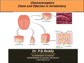
17. Taste
The taste receptors are located around the small structures known
as papillae found on the upper surface of the tongue, soft palate,
upper esophagus, the cheek, and epiglottis. These structures are
involved in detecting the five elements of taste perception: salty, sour,
bitter, sweet and umami. The primary organ of taste is the taste bud.
A taste bud is a cluster of gustatory receptors (taste cells) that are
located within the bumps on the tongue called papillae (singular:
papilla).
There are several structurally-distinct papillae.
Filiform papillae, which are located across the tongue, are tactile,
providing friction that helps the tongue move substances; they
contain no taste cells.
In contrast, fungiform papillae, which are located mainly on the
anterior two-thirds of the tongue, each contain one to eight taste
buds; they also have receptors for pressure and temperature.
The large circumvallate papillae contain up to 100 taste buds and
form a V near the posterior margin of the tongue.
18. In humans, there are five primary tastes; each taste has only one corresponding type of receptor. Thus, like
olfaction, each receptor is specific to its stimulus ( tastant ). Transduction of the five tastes happens
through different mechanisms that reflect the molecular composition of the tastant. A salty tastant
(containing NaCl) provides the sodium ions (Na+) that enter the taste neurons, exciting them directly. Sour
tastants are acids which belong to the thermoreceptor protein family. Binding of an acid or other sour-
tasting molecule triggers a change in the ion channel which increases hydrogen ion (H+) concentrations in
the taste neurons; thus, depolarizing them. Sweet, bitter, and umami tastants require a G-protein-coupled
receptor. These tastants bind to their respective receptors, thereby exciting the specialized neurons
associated with them.
Both tasting abilities and sense of smell change with age. In humans, the senses decline dramatically by
age 50 and continue to decline. A child may find a food to be too spicy, whereas an elderly person may
find the same food to be bland and unappetizing.