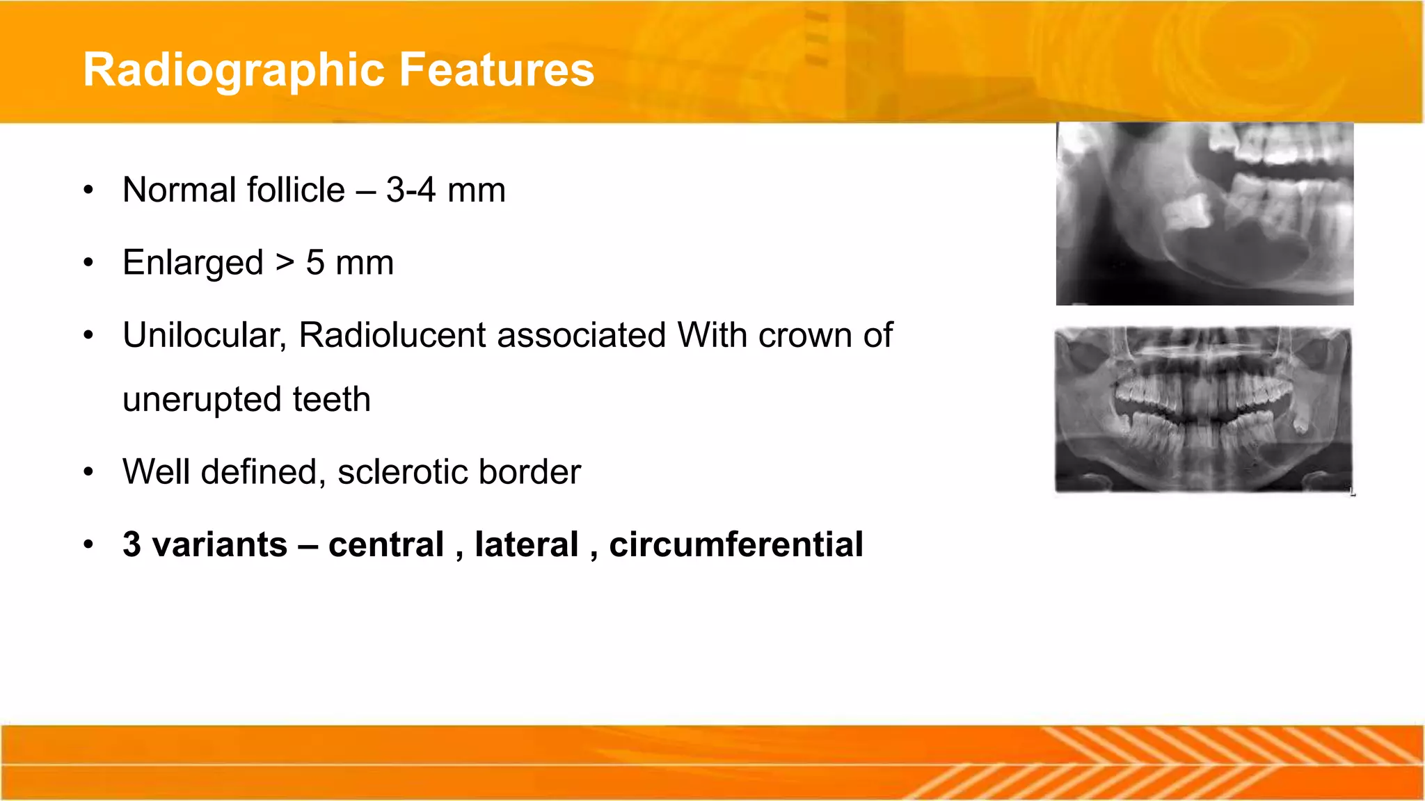
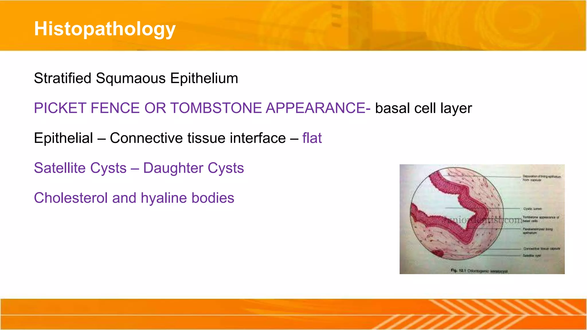
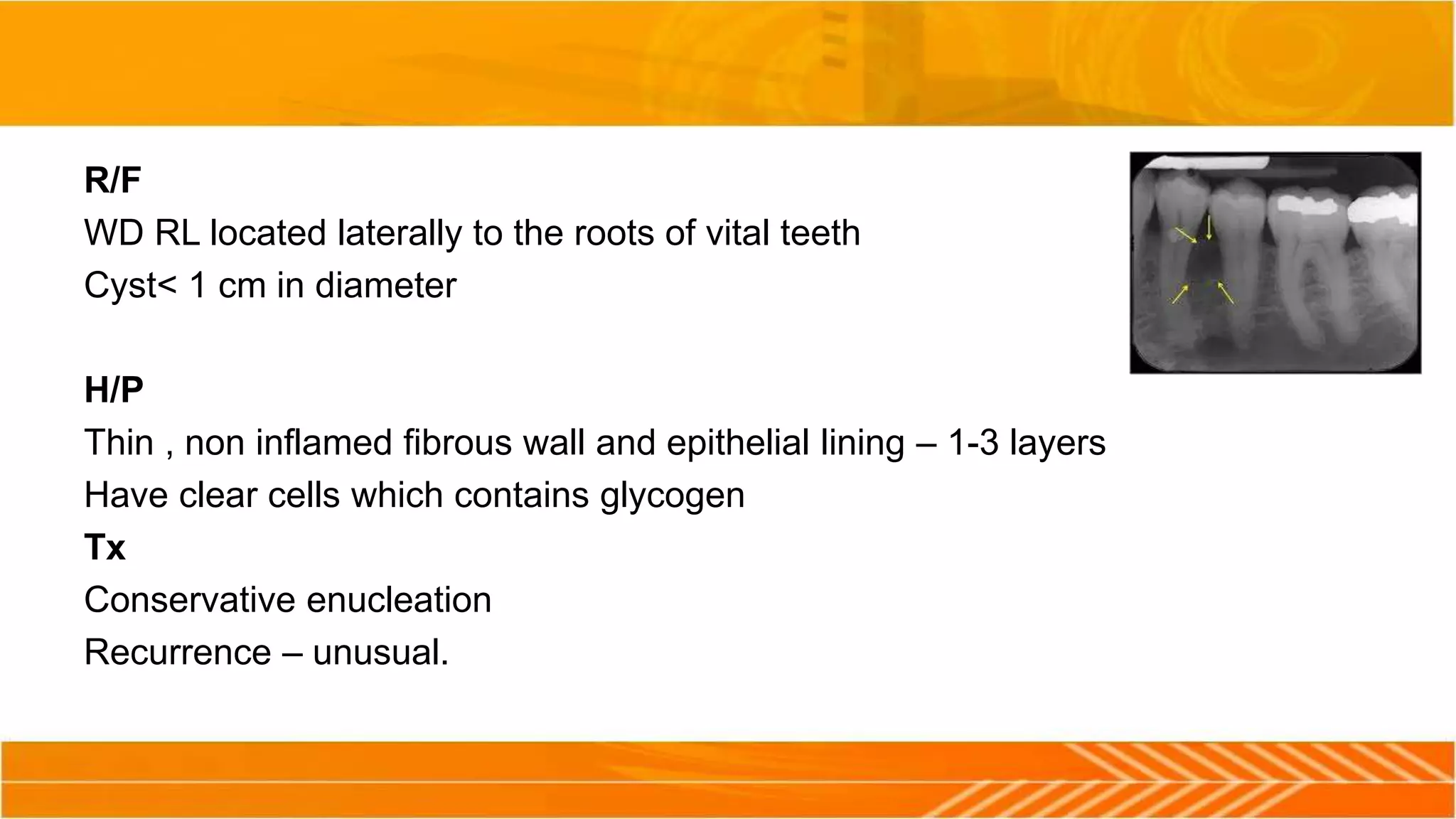
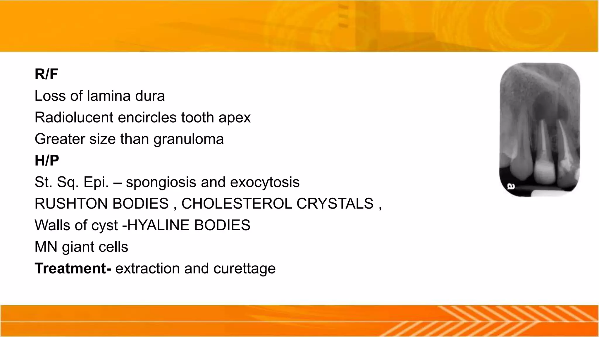
CYSTS OF THE JAW
Dr. Ashish Kr. Kushwaha
MDS
Maxillofacial Surgeon
CYST DEFINITION
 Kramer (1974)
• Cyst is a pathological cavity having fluid, semi fluid or gaseous content which is not
created by accumulation of pus, which may or may not be lined by epithelial tissue.
Types of Cyst –
1. TRUE CYSTS:
which is lined by epithelium (Dentigerous cyst, Radicular cyst etc).
2. PSEUDO CYSTS:
which is not lined by epithelium (Solitary bone cyst, Aneurismal bone cyst etc).
CYST OF JAW
EPITHELIAL
• ODONTOGENIC
• DEVELOPMENTAL
• DENTIGEROUS CYST
• ODONTOGENIC KERATOCYST
• LATERAL PERIODONTAL CYST
• CALCIFYING ODONTOGENIC CYST
• GINGIVAL CYST OF ADULT
• GINGIVAL CYST OF INFANTS
• ERUPTION CYST
• INFLAMMATORY
• PERIAPICAL CYST
• RESIDUAL CYST
• PARADENTAL CYST
• NON ODONTOGENIC
• NASOLABIAL CYST
• NASOPALATINE CYST
• MID PALATINE RAPHE CYST
NON EPITHELIAL
• ANEURSYMAL CYST
• SOLITARY BONE CYST
CYST
ASSOCIATED
WITH MAXILLARY
ANTRUM
BENIGN MUCOSAL
CYST OF MAX.
ANTRUM
POSTOPERATIVE
MAXILLARY CYST
CYST OF THE
SOFT TISSUE ,
MAXILLA , FACE
AND NECK
DERMOID CYST
EPIDERMOID
CYST
WHO - 1992
(Given by KRAMER, J.J. PINDBORG
AND MERVYN SHEAR)
Pathogenesis of Cyst
• There are 2 phases of cyst formation.
I. Cyst Initiation
II. Cyst Enlargement
I. Cyst Initiation
• Initiation of cyst formation is mostly from odontogenic epithelium.
• Following factors involved –
• Proliferation of epithelial lining
• Fluid accumulation in cyst cavity
• Bone resorption
II. Cyst Enlargement
Once cyst formation has been initiated, It continues to grow & enlarge.
Following factors are involved –
• Increase in the volume of content.
• Increase in the surface area of sac.
• Displacement of surrounding soft tissue.
• Resorption of surrounding bones.
1
2 3
4 5
Theories of Cyst Enlargement Classified By – Harris 1974
I. Mural Growth Theory
a. Peripheral Cell Division
b. Accumulation of Cellular Contents
II. Hydrostatic Enlargement
I. Secretion
II. Transudation & Exudation
III. Bone Resorbing Factors
Mural Growth Theory –
Mural growth in the form of epithelial proliferation is one of the essential process by which
the surface area of sac increases basically by peripheral cell division or by accumulation of
cellular contents.
Low grade infection stimulates cell such as cell rests of malassez to proliferate & form
arcades.
Hydrostatic Cyst Enlargement
Certain cyst s having lining which secrete mucin. Accumulation of mucus increases the cyst
volume and hence the enlargement.
Transudation and Exudation -
• Inflammatory cells release cofactors, lymphocytes release lymphokines & osteoclast
activating factors and monocytes release IL – 1.
• All these factors stimulates fibroblast to release prostaglandin.
• Prostaglandin produces a hyperosmolar cyst fluid.
• Increased hyperosmalrity draws fluid from surroundings.
• This increases hydrostatic pressure leading to cyst enlargement.
Bone Resorption -
• Growth of cyst, there is proliferation of residual developmental epithelial cells leading to
formation of solid mass of epithelial cells.
• As the mass enlarges, the epithelial cells in the center are degenerated, and thus creating a
lumen in the center.
• Intracellular products make lumen hypertonic and start transudation.
• Because of transudation, hydrostatic pressure increases.
• This hydrostatic pressure produces bone resorption, expansion of cyst and some times mild
paresthesia or pain.
Diagnosis Of Cyst
Clinical Examination –
Slow growing.
Painless.
Expansile (Buccal cortical expansion is more common).
Displaced or unerupted teeth.
Radiographic Examination –
IOPA, OCCLUSAL, OPG, PA View, Water’s View.
Typical Cyst appears radiolucent with a well defined radiopaque margin.
FNAC
• It is done under LA with 18 gauge needle.
• Aspirated fluid is studied to find out the nature of the lesion.
• Aspiration of air indicates that the needle has entered the antrum.
• If there are no contents on aspiration, it indicates a tumor.
Various Aspirates
Dentigerous Cyst Clear
Straw Colored fluid
Odontogenic Keratocyst Dirty
Creamy white viscoid
Periodontal Cyst Clear
Pale yellow straw colored
Infected Cyst Pus or brownish fluid
Solitary bone cyst Serous, sanguinerous fluid
Blood or empty cavity
Stafne’s bone cyst Empty cavity
Dermoid cyst Thick sebaceous material
Arterior / arterio-venous
malformation
Bright red color pulsatile.
Gingival cyst Clear fluid
Mucocele / ranula mucus
Contrast Studies- injection of water soluble dye to find out exact size of cyst.
Biopsy - Biopsy is the gold standard for determining the type of the cyst and to differentiate
from neoplasm.
Surgical Management
Treatment Modalties
• Marsupialization
 Partsch I
 Partsch II / Waldron operation – (Combination of marsupialization and enucleation)
• Enucleation
• Enucleation with chemical cauterization
• En-block resection
• Segmental resection
MARSUPIALIZATION
• Marsupialization, refers to creating a surgical window in the wall of the cyst and evacuation
of cystic content.
• This process decreases intra-cystic pressure & promotes shrinkage of the cyst.
Indications –
• Pt with developing tooth germs.
• Proximity to vital structures.
• Eruption of teeth – marsupialization permit the eruption of unerupted teeth.
• Large size of cyst.
• When apices of many adjacent teeth are involved within a large cyst.
Advantages –
• Simple procedure
• Preserve vital Structures
• Allows eruption of teeth
• Prevents oronasal, oroantral fistula
• Prevents pathological fracture
• Allows for endosteal bone formation
Disadvantages –
• Pathological tissues is left in situ.
• Prolonged healing time
• Discomfort to patient
• Prolonged follow up visit
• Periodic Irrigation of cavity
• Regular adjustment of plug
Partsch - I
• Local Anaesthesia / general Anaesthesia
• Aspiration of Cystic Content
• Incision
• Mucoperiosteum Flap Reflected
• Removal of bone
• Exposure of cystic cavity
• Irrigation of cystic cavity
• Suturing of remaining cystic lining with the edge of oral mucosa
• Packing with a ribbon gauze.
Marsupialization method. Circular incision includes mucosa and
periosteum.
Exposure of buccal cortical plate and removal of portion of bone with
round bur
Enlargement
of osseous
window with
rongeur
Exposure of cyst
after removal of
bone
Suturing of wound
margins with cystic
wall
Packing of cystic
cavity with
iodoform gauz
Cystic cavity after
insertion of gauze
Partsch II / Waldron operation - 1941
• This is 2 stage procedure that combines marsupialization and Enucleation.
Advantages -
• Development of thickened cystic lining, which makes enucleation easier.
• Preserve adjacent vital structures.
• Reduces morbidity.
• Promotes healing process
Disadvantages -
• Secondary surgery
Enucleation
• Enucleation involves complete removal of cystic lining & its contents.
• After enucleation, space fills with blood clot, which will eventually organize &
form normal bone.
Advantages-
• Primary closure of wound.
• Healing is rapid.
• Post operative care is reduced.
Disadvantages-
• Enucleation of large cyst will weaken the mandible, making it prone to jaw
fracture.
• Damage to adjacent vital structures.
• Pulpal necrosis.
• Unerupted teeth in a dentigerous cyst will be removed with the lesion.
Enucleation With Primary Closure
• Local / General Anaesthesia.
• Surgical Approach – Intra Oral / Extra Oral.
• Incision – According to region.
• Reflection of mucoperiosteum flap.
• Bone removed to expose the underlying cystic lesion.
• Cystic lining separated with help of a curette, periosteal elevator & mosquito
forcep.
• Suturing of flap.
Enucleation With Chemical Cauterization
• Stoelinga advocated the use of CARNOY’S SOLUTION to reduce the
recurrence of Cyst.
• Compostion of CARNOY’S SOLUTION
• Ethanol – 60 %
• Chloroform - 30 %
• Glacial Acetic Acid – 10 %
• Ferric Chloride – 1 gram
Panoramic radiograph showing an
extensive radicular lesion at the region
of teeth 22, 23, 24
Clinical photograph of case
Removal of maxillary cyst, with labial access. Incision for
creating a trapezoidal flap.
Reflection of flap and exposure of surgical field.
Removal of bone at the labial aspect respective to the lesion.
Osseous window created to expose part of the lesion.
Removal of cyst from bony cavity, using hemostat and curette.
Surgical field after removal of lesion.
Operation site after placement of sutures.
Panoramic radiograph and clinical photograph taken 2 months
after the surgical procedure.
Enucleation & Packing
• When previous infection or in infected large cysts, a primary closure would be
unsuccessful, difficulty in approximating the wound edges.
• In such cases, enucleation is performed & cystic cavity is packed as in
marsupialization.
• The wound heals with granulation tissue.
• En-Block Resection
• In this procedure, the Cyst is removed along with a rim of uninvolved bone,
while maintaining the continuity of jaw.
• Segmental Resection
• In this, continuity of the inferior border is not maintained.
• Hemimandibulectomy
• Partial mandibulectomy
• maxilectomy
Reconstruction
• Reconstruction with plate.
• Reconstruction with Graft (Iliac graft/ Costochondral Graft).
• Reconstruction with Distraction Osteogenesis.
DENTIGEROUS CYST / FOLLICULAR CYST
• Term “Dentigerous Cyst” coined by – Paget 1863.
• Most common – 20% of all cysts
PATHOGENESIS –
• Accumulation of fluid between REE and tooth crown.
• Attached at CEJ.
Clinical Features
•AGE – 1st to 3rd decade.
SITES – 38/48 > 18/28 > 13/23 > 35/45
• SEX – M : F- 3 : 2.
• Always associated with unerupted or impacted teeth.
• Painless expansion of bone. (Pain if infected).
• Egg shell cracking sound on palpation
• B/L and multiple – cleidocranial dysplasias
Radiographic Features
• Normal follicle – 3-4 mm
• Enlarged > 5 mm
• Unilocular, Radiolucent associated With crown of
unerupted teeth
• Well defined, sclerotic border
• 3 variants – central , lateral , circumferential
Cystic Content
• Thin watery straw coloured fluid .
• Protein content > 4gm / 100 ml
Differential Diagnosis
• AMELOBLASTOMA
Treatment - I/O or E/O
• Marsupialization
• Enucleation of cyst with removal of tooth.
ODONTOGENIC KERATOCYST/PRIMORDIAL CYST
• Also known as Primordial Cyst.
• Term “Primordial Cyst” was popularized by Robinson 1945.
• Term “Keratocyst” was coined by - Philipsen 1956
• Highest tendency for recurrence
• 2 types – Orthokeratinized & Parakeratinized.
• About 5 – 10 % of odontogenic cyst.
• Associated with basal cell nevoid syndrome
Pathogenesis
• Arises from odontogenic epithelium.
• Main source –
 Dental lamina or its remnants.
 Basal cell from overlying oral mucosa
 Enamel organ
Clinical Features
• Age :10 - 40 yrs
• Male prediliction
• Site : Mandible > Maxilla, Posterior > Anterior
• Grows in antero posterior direction.
• Displacement of the teeth.
• Dull / hollow sound present on percussion of teeth.
• Expansion of buccal cortical bone is more common.
• Paraesthesia may be present.
Radiographic Features
• Unilocular or Multilocular
• Well Defined,
• Radiolucent,
• Smooth Corticated margins.
• Unerupted teeth associated - 20-40 %
Histopathology
Stratified Squmaous Epithelium
PICKET FENCE OR TOMBSTONE APPEARANCE- basal cell layer
Epithelial – Connective tissue interface – flat
Satellite Cysts – Daughter Cysts
Cholesterol and hyaline bodies
Cystic Content
• Dirty White.
• Viscoid Suspension of keratin which has appearance of pus.
• Total protein content < 4 gm / 100 ml, mostly albumin.
• Smear stained & examined for Keratin cells.
Differential Diagnosis
• Ameloblastoma
• Dentigerous Cyst
Treatment of OKC
• Marsupialization – incorrect – owing to the high tendency to recur
• Bramley (1971,1974) –Outlined the Surgical Management
1. Small surgical cyst – enucleated from an intraoral approach
2. Larger cyst - from an extraoral approach
3. Large Multilocular lesions with /without cortical perforation – Resection of
the involved bone followed by primary/ secondary reconstruction.
• Stoelinga & Von Hoelst 1981- proposed a more conservative approach to
large OKC.
Enucleation
Cautrization of cystic cavity by CARNOY’S SOLUTION
CALCIFYING ODONTOGENIC CYST/GORLIN CYST
• Also known as Gorlin gold cyst / dentinogenic ghost cell tumor/ calcifying
ghost cell odontogenic cyst.
Clinical features -
• Age – 2nd – 3rd decade
• Gender – equal
• Types – Intra-osseous & Extra-osseous
• Sites – max: mand ---1:1
R/F
• Uni Lateral, Well Defined Radiolucent lesion.
• Ass. With Unerupted teeth – canine
• Size – btw 2 cm and 4 cm.
H/P
Well defined lesion – fibrous capsule
Ghost cell epi -Altered epi cell with loss of nuclei , preservation of basic cell
outline, intercellular oedema , normal/ aberrant keratinization of OE
Ass. With odontoma
Treatment-
Enucleation
Recurrence - common
LATERAL PERIODONTAL CYST
• Occurs along a lateral surface of tooth
Arise from
1 . REE
2. Remnants of dental lamina
3. Cell rests of malassez
C/F
Asymptomatic
detected during Radiographic Examination.
Age – 5th – 7th decades
Site – mandibular Premolar, Canine, Lateral Incisor
R/F
WD RL located laterally to the roots of vital teeth
Cyst< 1 cm in diameter
H/P
Thin , non inflamed fibrous wall and epithelial lining – 1-3 layers
Have clear cells which contains glycogen
Tx
Conservative enucleation
Recurrence – unusual.
RADICULAR CYST
• apical periodontal cyst , root end cyst
• Most common cyst
Patho – epi. At the apex of a nonvital tooth – stimulated by an inflammation to
form a true lined cyst ( rests of malassez)
Causes - . 1. caries 2. trauma 3. restoration
C/F
Asymptomatic
Age - At any age
Site – incisors involved , not ass. With deciduous dentition
Mobility possible – due to cyst enlargement
Non vital tooth
R/F
Loss of lamina dura
Radiolucent encircles tooth apex
Greater size than granuloma
H/P
St. Sq. Epi. – spongiosis and exocytosis
RUSHTON BODIES , CHOLESTEROL CRYSTALS ,
Walls of cyst -HYALINE BODIES
MN giant cells
Treatment- extraction and curettage
RESIDUAL CYST
• Retained Periapical cyst
• From teeth that have been removed
C/F – any tooth bearing area of max. and mand
R/F – Well defined radiolucent area, Do not expand bone
Treatment - Surgical Curettage , Enucleation
GLOBULOMAXILLARY CYST
• Patho – epi entrapped during fusion of MNP AND Max. Process
C/F
• Site- btw Max. LI and Cuspid teeth
R/F
• Well defined unilocular radiolucent area – resembling INVERTED PEAR
SHAPED Between apices of teeth
H/P
• Inflamed St. Sq. Epi are consistent with periapical cysts
Treatment -
Surgical enucleation
Tooth – non vital – Extraction / RCT , recurrence - Rare
NASOLABIAL CYST
• Non odontogenic cyst
• Developed from the remnants of nasolacrimal duct mucus cyst arising from epithelial lining of
the floor of mouth
C/F
SWELLING – upper lip obliterated labial sulcus
Unilateral painless
Cystic content – straw coloured whitish mucous fluid
H/P
Lined by pseudostratified columnar, cuboidal or ciliated epithelium
Treatment - Surgical Enucleation.
NASOPALATINE CYST
• Most common non odontogenic cyst – 1%
C/F
Swelling of the anterior palate, pain and drainage
R/F
Well defined radiolucent area in or near the midline of the anterior maxilla
btw Central Incisor teeth.
Size – 6mm to 6 cm
Treatment
Enucleation
ANY QUESTIONS???

Cyst of the jaw

  • 1.
    CYSTS OF THEJAW Dr. Ashish Kr. Kushwaha MDS Maxillofacial Surgeon
  • 2.
    CYST DEFINITION  Kramer(1974) • Cyst is a pathological cavity having fluid, semi fluid or gaseous content which is not created by accumulation of pus, which may or may not be lined by epithelial tissue.
  • 3.
    Types of Cyst– 1. TRUE CYSTS: which is lined by epithelium (Dentigerous cyst, Radicular cyst etc). 2. PSEUDO CYSTS: which is not lined by epithelium (Solitary bone cyst, Aneurismal bone cyst etc).
  • 4.
    CYST OF JAW EPITHELIAL •ODONTOGENIC • DEVELOPMENTAL • DENTIGEROUS CYST • ODONTOGENIC KERATOCYST • LATERAL PERIODONTAL CYST • CALCIFYING ODONTOGENIC CYST • GINGIVAL CYST OF ADULT • GINGIVAL CYST OF INFANTS • ERUPTION CYST • INFLAMMATORY • PERIAPICAL CYST • RESIDUAL CYST • PARADENTAL CYST • NON ODONTOGENIC • NASOLABIAL CYST • NASOPALATINE CYST • MID PALATINE RAPHE CYST NON EPITHELIAL • ANEURSYMAL CYST • SOLITARY BONE CYST CYST ASSOCIATED WITH MAXILLARY ANTRUM BENIGN MUCOSAL CYST OF MAX. ANTRUM POSTOPERATIVE MAXILLARY CYST CYST OF THE SOFT TISSUE , MAXILLA , FACE AND NECK DERMOID CYST EPIDERMOID CYST WHO - 1992 (Given by KRAMER, J.J. PINDBORG AND MERVYN SHEAR)
  • 5.
    Pathogenesis of Cyst •There are 2 phases of cyst formation. I. Cyst Initiation II. Cyst Enlargement
  • 6.
    I. Cyst Initiation •Initiation of cyst formation is mostly from odontogenic epithelium. • Following factors involved – • Proliferation of epithelial lining • Fluid accumulation in cyst cavity • Bone resorption
  • 7.
    II. Cyst Enlargement Oncecyst formation has been initiated, It continues to grow & enlarge. Following factors are involved – • Increase in the volume of content. • Increase in the surface area of sac. • Displacement of surrounding soft tissue. • Resorption of surrounding bones.
  • 8.
  • 9.
    Theories of CystEnlargement Classified By – Harris 1974 I. Mural Growth Theory a. Peripheral Cell Division b. Accumulation of Cellular Contents II. Hydrostatic Enlargement I. Secretion II. Transudation & Exudation III. Bone Resorbing Factors
  • 10.
    Mural Growth Theory– Mural growth in the form of epithelial proliferation is one of the essential process by which the surface area of sac increases basically by peripheral cell division or by accumulation of cellular contents. Low grade infection stimulates cell such as cell rests of malassez to proliferate & form arcades.
  • 11.
    Hydrostatic Cyst Enlargement Certaincyst s having lining which secrete mucin. Accumulation of mucus increases the cyst volume and hence the enlargement.
  • 12.
    Transudation and Exudation- • Inflammatory cells release cofactors, lymphocytes release lymphokines & osteoclast activating factors and monocytes release IL – 1. • All these factors stimulates fibroblast to release prostaglandin. • Prostaglandin produces a hyperosmolar cyst fluid. • Increased hyperosmalrity draws fluid from surroundings. • This increases hydrostatic pressure leading to cyst enlargement.
  • 13.
    Bone Resorption - •Growth of cyst, there is proliferation of residual developmental epithelial cells leading to formation of solid mass of epithelial cells. • As the mass enlarges, the epithelial cells in the center are degenerated, and thus creating a lumen in the center. • Intracellular products make lumen hypertonic and start transudation.
  • 14.
    • Because oftransudation, hydrostatic pressure increases. • This hydrostatic pressure produces bone resorption, expansion of cyst and some times mild paresthesia or pain.
  • 15.
    Diagnosis Of Cyst ClinicalExamination – Slow growing. Painless. Expansile (Buccal cortical expansion is more common). Displaced or unerupted teeth.
  • 16.
    Radiographic Examination – IOPA,OCCLUSAL, OPG, PA View, Water’s View. Typical Cyst appears radiolucent with a well defined radiopaque margin.
  • 17.
    FNAC • It isdone under LA with 18 gauge needle. • Aspirated fluid is studied to find out the nature of the lesion. • Aspiration of air indicates that the needle has entered the antrum. • If there are no contents on aspiration, it indicates a tumor.
  • 18.
    Various Aspirates Dentigerous CystClear Straw Colored fluid Odontogenic Keratocyst Dirty Creamy white viscoid Periodontal Cyst Clear Pale yellow straw colored Infected Cyst Pus or brownish fluid Solitary bone cyst Serous, sanguinerous fluid Blood or empty cavity Stafne’s bone cyst Empty cavity Dermoid cyst Thick sebaceous material Arterior / arterio-venous malformation Bright red color pulsatile. Gingival cyst Clear fluid Mucocele / ranula mucus
  • 19.
    Contrast Studies- injectionof water soluble dye to find out exact size of cyst. Biopsy - Biopsy is the gold standard for determining the type of the cyst and to differentiate from neoplasm.
  • 20.
  • 21.
    Treatment Modalties • Marsupialization Partsch I  Partsch II / Waldron operation – (Combination of marsupialization and enucleation) • Enucleation • Enucleation with chemical cauterization • En-block resection • Segmental resection
  • 22.
    MARSUPIALIZATION • Marsupialization, refersto creating a surgical window in the wall of the cyst and evacuation of cystic content. • This process decreases intra-cystic pressure & promotes shrinkage of the cyst.
  • 23.
    Indications – • Ptwith developing tooth germs. • Proximity to vital structures. • Eruption of teeth – marsupialization permit the eruption of unerupted teeth. • Large size of cyst. • When apices of many adjacent teeth are involved within a large cyst.
  • 24.
    Advantages – • Simpleprocedure • Preserve vital Structures • Allows eruption of teeth • Prevents oronasal, oroantral fistula • Prevents pathological fracture • Allows for endosteal bone formation
  • 25.
    Disadvantages – • Pathologicaltissues is left in situ. • Prolonged healing time • Discomfort to patient • Prolonged follow up visit • Periodic Irrigation of cavity • Regular adjustment of plug
  • 26.
    Partsch - I •Local Anaesthesia / general Anaesthesia • Aspiration of Cystic Content • Incision • Mucoperiosteum Flap Reflected • Removal of bone • Exposure of cystic cavity • Irrigation of cystic cavity • Suturing of remaining cystic lining with the edge of oral mucosa • Packing with a ribbon gauze.
  • 27.
    Marsupialization method. Circularincision includes mucosa and periosteum. Exposure of buccal cortical plate and removal of portion of bone with round bur Enlargement of osseous window with rongeur
  • 28.
    Exposure of cyst afterremoval of bone Suturing of wound margins with cystic wall
  • 29.
    Packing of cystic cavitywith iodoform gauz Cystic cavity after insertion of gauze
  • 30.
    Partsch II /Waldron operation - 1941 • This is 2 stage procedure that combines marsupialization and Enucleation. Advantages - • Development of thickened cystic lining, which makes enucleation easier. • Preserve adjacent vital structures. • Reduces morbidity. • Promotes healing process Disadvantages - • Secondary surgery
  • 31.
    Enucleation • Enucleation involvescomplete removal of cystic lining & its contents. • After enucleation, space fills with blood clot, which will eventually organize & form normal bone.
  • 32.
    Advantages- • Primary closureof wound. • Healing is rapid. • Post operative care is reduced. Disadvantages- • Enucleation of large cyst will weaken the mandible, making it prone to jaw fracture. • Damage to adjacent vital structures. • Pulpal necrosis. • Unerupted teeth in a dentigerous cyst will be removed with the lesion.
  • 33.
    Enucleation With PrimaryClosure • Local / General Anaesthesia. • Surgical Approach – Intra Oral / Extra Oral. • Incision – According to region. • Reflection of mucoperiosteum flap. • Bone removed to expose the underlying cystic lesion. • Cystic lining separated with help of a curette, periosteal elevator & mosquito forcep. • Suturing of flap.
  • 34.
    Enucleation With ChemicalCauterization • Stoelinga advocated the use of CARNOY’S SOLUTION to reduce the recurrence of Cyst. • Compostion of CARNOY’S SOLUTION • Ethanol – 60 % • Chloroform - 30 % • Glacial Acetic Acid – 10 % • Ferric Chloride – 1 gram
  • 35.
    Panoramic radiograph showingan extensive radicular lesion at the region of teeth 22, 23, 24 Clinical photograph of case
  • 36.
    Removal of maxillarycyst, with labial access. Incision for creating a trapezoidal flap. Reflection of flap and exposure of surgical field.
  • 37.
    Removal of boneat the labial aspect respective to the lesion. Osseous window created to expose part of the lesion.
  • 38.
    Removal of cystfrom bony cavity, using hemostat and curette. Surgical field after removal of lesion.
  • 39.
    Operation site afterplacement of sutures. Panoramic radiograph and clinical photograph taken 2 months after the surgical procedure.
  • 40.
    Enucleation & Packing •When previous infection or in infected large cysts, a primary closure would be unsuccessful, difficulty in approximating the wound edges. • In such cases, enucleation is performed & cystic cavity is packed as in marsupialization. • The wound heals with granulation tissue.
  • 41.
    • En-Block Resection •In this procedure, the Cyst is removed along with a rim of uninvolved bone, while maintaining the continuity of jaw. • Segmental Resection • In this, continuity of the inferior border is not maintained. • Hemimandibulectomy • Partial mandibulectomy • maxilectomy
  • 42.
    Reconstruction • Reconstruction withplate. • Reconstruction with Graft (Iliac graft/ Costochondral Graft). • Reconstruction with Distraction Osteogenesis.
  • 43.
    DENTIGEROUS CYST /FOLLICULAR CYST • Term “Dentigerous Cyst” coined by – Paget 1863. • Most common – 20% of all cysts PATHOGENESIS – • Accumulation of fluid between REE and tooth crown. • Attached at CEJ.
  • 44.
    Clinical Features •AGE –1st to 3rd decade. SITES – 38/48 > 18/28 > 13/23 > 35/45 • SEX – M : F- 3 : 2. • Always associated with unerupted or impacted teeth. • Painless expansion of bone. (Pain if infected). • Egg shell cracking sound on palpation • B/L and multiple – cleidocranial dysplasias
  • 45.
    Radiographic Features • Normalfollicle – 3-4 mm • Enlarged > 5 mm • Unilocular, Radiolucent associated With crown of unerupted teeth • Well defined, sclerotic border • 3 variants – central , lateral , circumferential
  • 46.
    Cystic Content • Thinwatery straw coloured fluid . • Protein content > 4gm / 100 ml Differential Diagnosis • AMELOBLASTOMA Treatment - I/O or E/O • Marsupialization • Enucleation of cyst with removal of tooth.
  • 47.
    ODONTOGENIC KERATOCYST/PRIMORDIAL CYST •Also known as Primordial Cyst. • Term “Primordial Cyst” was popularized by Robinson 1945. • Term “Keratocyst” was coined by - Philipsen 1956 • Highest tendency for recurrence • 2 types – Orthokeratinized & Parakeratinized. • About 5 – 10 % of odontogenic cyst. • Associated with basal cell nevoid syndrome
  • 48.
    Pathogenesis • Arises fromodontogenic epithelium. • Main source –  Dental lamina or its remnants.  Basal cell from overlying oral mucosa  Enamel organ
  • 49.
    Clinical Features • Age:10 - 40 yrs • Male prediliction • Site : Mandible > Maxilla, Posterior > Anterior • Grows in antero posterior direction. • Displacement of the teeth. • Dull / hollow sound present on percussion of teeth. • Expansion of buccal cortical bone is more common. • Paraesthesia may be present.
  • 50.
    Radiographic Features • Unilocularor Multilocular • Well Defined, • Radiolucent, • Smooth Corticated margins. • Unerupted teeth associated - 20-40 %
  • 51.
    Histopathology Stratified Squmaous Epithelium PICKETFENCE OR TOMBSTONE APPEARANCE- basal cell layer Epithelial – Connective tissue interface – flat Satellite Cysts – Daughter Cysts Cholesterol and hyaline bodies
  • 52.
    Cystic Content • DirtyWhite. • Viscoid Suspension of keratin which has appearance of pus. • Total protein content < 4 gm / 100 ml, mostly albumin. • Smear stained & examined for Keratin cells. Differential Diagnosis • Ameloblastoma • Dentigerous Cyst
  • 53.
    Treatment of OKC •Marsupialization – incorrect – owing to the high tendency to recur • Bramley (1971,1974) –Outlined the Surgical Management 1. Small surgical cyst – enucleated from an intraoral approach 2. Larger cyst - from an extraoral approach 3. Large Multilocular lesions with /without cortical perforation – Resection of the involved bone followed by primary/ secondary reconstruction.
  • 54.
    • Stoelinga &Von Hoelst 1981- proposed a more conservative approach to large OKC. Enucleation Cautrization of cystic cavity by CARNOY’S SOLUTION
  • 55.
    CALCIFYING ODONTOGENIC CYST/GORLINCYST • Also known as Gorlin gold cyst / dentinogenic ghost cell tumor/ calcifying ghost cell odontogenic cyst. Clinical features - • Age – 2nd – 3rd decade • Gender – equal • Types – Intra-osseous & Extra-osseous • Sites – max: mand ---1:1
  • 56.
    R/F • Uni Lateral,Well Defined Radiolucent lesion. • Ass. With Unerupted teeth – canine • Size – btw 2 cm and 4 cm. H/P Well defined lesion – fibrous capsule Ghost cell epi -Altered epi cell with loss of nuclei , preservation of basic cell outline, intercellular oedema , normal/ aberrant keratinization of OE Ass. With odontoma
  • 57.
  • 58.
    LATERAL PERIODONTAL CYST •Occurs along a lateral surface of tooth Arise from 1 . REE 2. Remnants of dental lamina 3. Cell rests of malassez C/F Asymptomatic detected during Radiographic Examination. Age – 5th – 7th decades Site – mandibular Premolar, Canine, Lateral Incisor
  • 59.
    R/F WD RL locatedlaterally to the roots of vital teeth Cyst< 1 cm in diameter H/P Thin , non inflamed fibrous wall and epithelial lining – 1-3 layers Have clear cells which contains glycogen Tx Conservative enucleation Recurrence – unusual.
  • 60.
    RADICULAR CYST • apicalperiodontal cyst , root end cyst • Most common cyst Patho – epi. At the apex of a nonvital tooth – stimulated by an inflammation to form a true lined cyst ( rests of malassez) Causes - . 1. caries 2. trauma 3. restoration C/F Asymptomatic Age - At any age Site – incisors involved , not ass. With deciduous dentition Mobility possible – due to cyst enlargement Non vital tooth
  • 61.
    R/F Loss of laminadura Radiolucent encircles tooth apex Greater size than granuloma H/P St. Sq. Epi. – spongiosis and exocytosis RUSHTON BODIES , CHOLESTEROL CRYSTALS , Walls of cyst -HYALINE BODIES MN giant cells Treatment- extraction and curettage
  • 62.
    RESIDUAL CYST • RetainedPeriapical cyst • From teeth that have been removed C/F – any tooth bearing area of max. and mand R/F – Well defined radiolucent area, Do not expand bone Treatment - Surgical Curettage , Enucleation
  • 63.
    GLOBULOMAXILLARY CYST • Patho– epi entrapped during fusion of MNP AND Max. Process C/F • Site- btw Max. LI and Cuspid teeth R/F • Well defined unilocular radiolucent area – resembling INVERTED PEAR SHAPED Between apices of teeth H/P • Inflamed St. Sq. Epi are consistent with periapical cysts Treatment - Surgical enucleation Tooth – non vital – Extraction / RCT , recurrence - Rare
  • 64.
    NASOLABIAL CYST • Nonodontogenic cyst • Developed from the remnants of nasolacrimal duct mucus cyst arising from epithelial lining of the floor of mouth C/F SWELLING – upper lip obliterated labial sulcus Unilateral painless Cystic content – straw coloured whitish mucous fluid H/P Lined by pseudostratified columnar, cuboidal or ciliated epithelium Treatment - Surgical Enucleation.
  • 65.
    NASOPALATINE CYST • Mostcommon non odontogenic cyst – 1% C/F Swelling of the anterior palate, pain and drainage R/F Well defined radiolucent area in or near the midline of the anterior maxilla btw Central Incisor teeth. Size – 6mm to 6 cm Treatment Enucleation
  • 66.

Editor's Notes

  • #24 Large size of cyst- in very large cysts where enucleation could result in a pathological #.
  • #27 Ribbon gauze – antibiotic ointment, white head’s varnish, tincture of benzoin, bismuth iodoform paraffin paste. Pack prevents contamination of the cavity Packs should be secured with suture
  • #34 (incision - Crevicular or Crestal in edentulous patient) Releasing incision either side