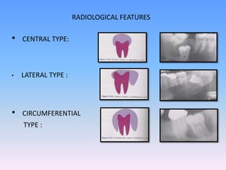
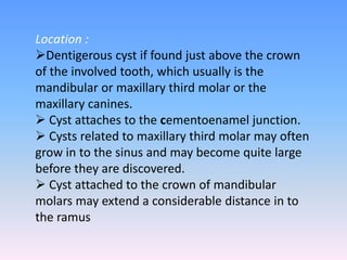
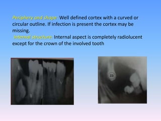
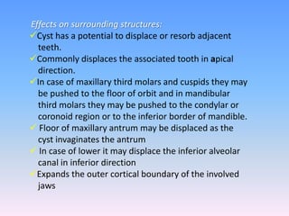
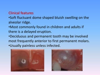
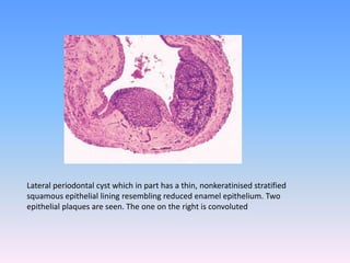
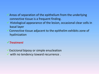
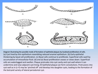
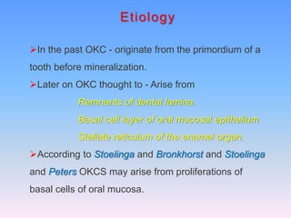
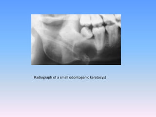
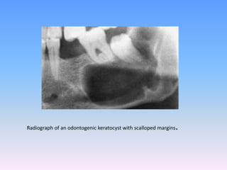
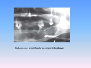
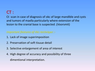
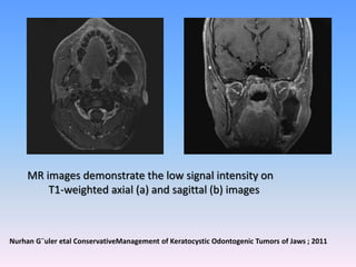
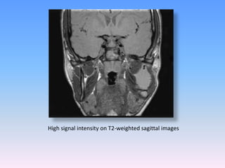
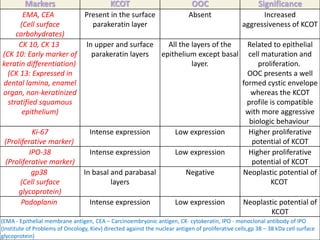
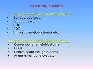
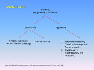
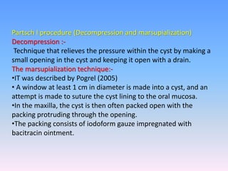
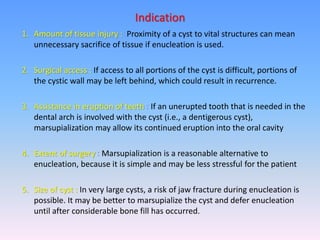
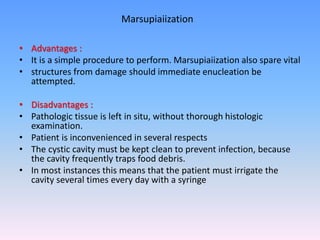
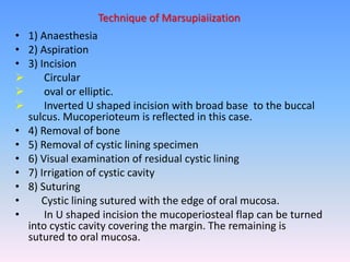
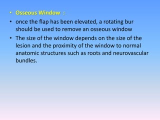
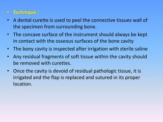
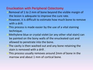
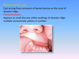
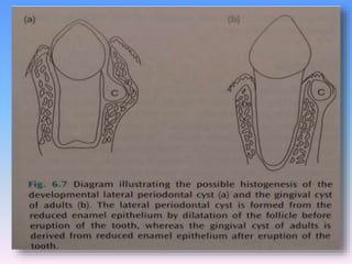
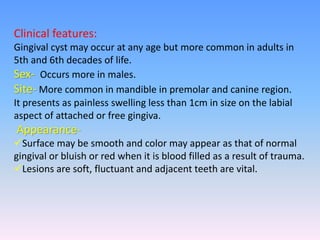
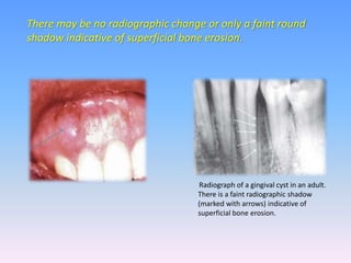
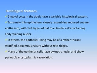
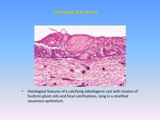
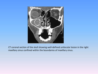
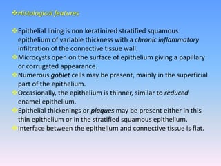
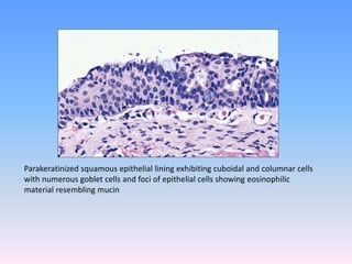
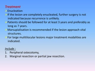
Odontogenic Cysts
SEMINAR PRESENTED BY
P.PRAVEEN
2ND YEAR PG
DEFINITION
1. Kramer (1974) has defined a cyst as ‘a pathological
cavity having fluid, semifluid or gaseous contents and
which is not created by the accumulation of pus ;
frequently, but not always, is lined by epithelium.
2. By Killey and Key 1966 described as epithelium lined
sac filled with fluid or semisolid material.
3. Most cysts, but not all, are lined by epithelium.
Cysts of the oral and maxillofacial tissues that are
not lined by epithelium are
 The mucous extravasation cyst of the salivary glands
 The aneurysmal bone cyst
 Solitary bone cyst
CLASSIFICATION (shear)
I Cysts of the jaws
A Epithelial-lined cysts
1 Developmental origin
(a) Odontogenic
i. Gingival cyst of infants
ii. Odontogenic keratocyst
iii. Dentigerous cyst
iv. Eruption cyst
v. Gingival cyst of adults
vi. Developmental lateral periodontal
cyst
vii. Botryoid odontogenic cyst
viii. Glandular odontogenic cyst
ix. Calcifying odontogenic cyst
(b) Non-odontogenic
i. Midpalatal raphé cyst of
infants
ii. Nasopalatine duct cyst
iii. Nasolabial cyst
2 Inflammatory origin
i. Radicular cyst, apical and
lateral
ii. Residual cyst
iii. Paradental cyst and
juvenile paradental cyst
iv. Inflammatory collateral
cyst
B Non-epithelial-lined cysts
1 Solitary bone cyst
2 Aneurysmal bone cyst
II Cysts associated with the maxillary antrum
1 Mucocele
2 Retention cyst
3 Pseudocyst
4 Postoperative maxillary cyst
III Cysts of the soft tissues of the mouth, face and neck
1) Dermoid and epidermoid cysts
2) Lymphoepithelial (branchial) cyst
3 )Thyroglossal duct cyst
4) Anterior median lingual cyst (intralingual cyst of foregutorigin)
5 )Oral cysts with gastric or intestinal epithelium (oral alimentary
tract cyst)
6) Cystic hygroma
7) Nasopharyngeal cyst
8) Thymic cyst
9) Cysts of the salivary glands: mucous extravasation cyst;
mucous retention cyst; ranula; polycystic (dysgenetic) disease of
the parotid
10) Parasitic cysts:
a. Hydatid cyst
b. Cysticercus cellulosae
c. Trichinosis
(WHO) Classification of cysts of jaws
A. EPITHELIAL-LINED CYSTS
1 Developmental Origin
(a) Odontogenic
Developmental
1. Odontogenic keratocyst
2. Dentigerous
3. Developmental lateral
periodontal cyst
4. Calcifying odontogenic cyst
2 INFLAMMATORY ORIGIN
i. Radicular cyst, apical and
lateral
ii. Residual cyst
b) Non-odontogenic
1. Midpalatal raphe cyst of
infants
2. Nasopalatine duct cyst
B. NON-EPITHELIAL-LINED
CYSTS
1. Solitary bone cyst
2. Aneurysmal bone cyst
Cyst of maxillary antrum
1. Surgical ciliated cysts of
maxilla
2. Benign mucosal cyst of the
maxillary antrum
A.SOFT TISSUE CYSTS
ODONTOGENIC
Gingival cysts
a) Adulits
b) Newborn
B.NON ODONTOGENIC
a) Anterior median lingual cyst
b) Nasolabial cyst
C.RETENTION CYST
SALIVARY GLAND CYSTS
a) Mucocele
b) Ranula
D.DEVELOPMENTAL CYST
a) Dermoid &epidermoid
b) Lymphoepithelial cyst
c) Thyroglossal cyst
d) Cystic hygroma
E.PARASITIC CYSTS
a) Hydatid cyst
b) Cysticercocis
F.HETEROTROPIC CYSTS
a) Oral cyst with gastric or
intestinal epithelium
• The dentigerous cyst is defined as a cyst that originates
by the separation of the follicle from around the crown
of an unerupted tooth
• The dentigerous cyst encloses the crown of an
unerupted tooth and is attached to the tooth at the
cementoenamel junction
• The pathogenesis of this cyst is uncertain, but
apparently it develops by accumulation of fluid
between the reduced enamel epithelium and the tooth
crown.
DENTIGEROUS CYST (FOLLICULAR CYST OR PERICORONAL CYST)
• Gross specimen of a dentigerous cyst.
• Cyst encloses the crown of the tooth and is attached to
its neck
Dentigerous cyst
Experimental studies on bone resorption
There is evidence that vital cyst tissue in culture
releases a potent bone resorbing factor that is
predominantly a mixture of prostaglandins (PGE2)
and E3.
 Data proposed by Harris indicated a lower levels
of PGE2 released by dentigerous cysts 12.2+/
9.4ng/mg than the radicular 16.6+/- 13ng/mg or
by OKCs 20 +/-11ng/mg.
PGE2 is one of the factor responsible for osteolytic
effects
 Interleukin 1 (IL1) may be produced by odontogenic
cysts and may account for raised levels of prostaglandin
and collagenase synthesis by the cyst capsules.
 IL1 released by the cysts leads to stimulation of
osteoclasts to resorb bone and the connective tissue
cells to produce prostaglandins that will be responsible
for further osteoclast activation.
It also stimulates connective tissue cells to produce
collagenase which is involved in the destruction of
bone matrix
Glycosaminoglycans, predominantly hyaluronic acid
but also appreciable amounts of heparin and
chrondrotoin sulphate are present in the fluids
and walls of dentigerous cyst.
Release of glycosaminoglycans from the walls and
their diffusion in to the cyst fluid is thought to have
an important role in expansive cyst growth by
increasing the osmolality of the cyst fluid and hence
raising the internal hydrostatic pressure of cyst
Clinical features
Develop around the crown of unerupted or supernumerary tooth.
Most commonly occur in the second and third decades
Male to female ratio of 2:1.
Examination reveals a missing teeth or tooth with hard swelling
Occasionally resulting in facial asymmetry.
Sites –
Mandibular and Maxillary third molars, Maxillary canines.
According to Gilibisco cyst is most often in decreasing order like
third molars, canines, second premolars.
Bilateral or multiple cysts can occur in association with number
of syndromes including cleidocranial dysplasia and Maroteaux
Lamy syndrome.
Expansion of cortical plates due to pressure extension may be
seen.
Clear, pale straw colour fluid Cholesterol crystals.
Total protein in excess 4 g / 100ml.
On aspiration
Radiographic features
Classically consists of a well corticated pericoronal radiolucency
which exceeds 5mm when measured from edge of crown to
periphery of lesion on radiographs
• CENTRAL TYPE:
• LATERAL TYPE :
• CIRCUMFERENTIAL
TYPE :
RADIOLOGICAL FEATURES
Location :
Dentigerous cyst if found just above the crown
of the involved tooth, which usually is the
mandibular or maxillary third molar or the
maxillary canines.
 Cyst attaches to the cementoenamel junction.
 Cysts related to maxillary third molar may often
grow in to the sinus and may become quite large
before they are discovered.
 Cyst attached to the crown of mandibular
molars may extend a considerable distance in to
the ramus
Periphery and shape: Well defined cortex with a curved or
circular outline. If infection is present the cortex may be
missing.
Internal structure: Internal aspect is completely radiolucent
except for the crown of the involved tooth
Effects on surrounding structures:
Cyst has a potential to displace or resorb adjacent
teeth.
Commonly displaces the associated tooth in apical
direction.
In case of maxillary third molars and cuspids they may
be pushed to the floor of orbit and in mandibular
third molars they may be pushed to the condylar or
coronoid region or to the inferior border of mandible.
 Floor of maxillary antrum may be displaced as the
cyst invaginates the antrum
 In case of lower it may displace the inferior alveolar
canal in inferior direction
Expands the outer cortical boundary of the involved
jaws
Axial contrast - enhanced CT image
obtained with soft-tissue window
settings
I. Black arrowheads => cystic with
enhancing soft-tissue septations
II. white arrowheads=> mural nodules
III. Arrow=> Cortical disruption at the
posterolateral aspect of the mass
Axial unenhanced CT image obtained
with bone window settings shows
I. Black arrow => unerupted wisdom
tooth centered in the cystic
expansile mass
II. white arrow=> cortical
breakthrough of the posterolateral
aspect of the mass is seen
Sagittal contrast-enhanced reformatted CT image shows :
The overall size of the mass and its cystic and solid components
I. Black arrowas => Unerupted tooth .
II. Arrowheads => Septations.
III. white arrow => Large mural nodule.
Ceylan Z. Cankurtaran, MD et al Ameloblastoma and Dentigerous Cyst Associated with Impacted Mandibular Third Molar Tooth1 ;2010
HISTOLOGICAL FEATURES
 Composed of connective tissue wall with a thin layer of
stratified squamous epithelium lining the lumen.
connective tissue wall is frequently thickened and composed
of a very loose fibrous connective tissue
Inflammatory cells commonly infiltrate the connective tissue.
Shows rushton bodies with in the lining epithelium.
Content of cystic lumen is usually thin, watery yellow and is
occasionally blood tinged.
cyst markers in dentigerous cyst
 Amelogenin is a lowmolecularweight enamel matrix protein.
Dentigerous cysts show positive expression for amelogenin
The expression of amelogenin is possibly an indicator of differentiation of epithelial
cells in the odontogenic lesions
amelogenin showing an intense well defined linear pattern
Natl J Maxillofac Surg. 2014 Jul-Dec; 5(2): 172–179
Dentigerous cyst with high expression of P63 in almost all epithelial layers.
P63 :
(Baretto et al (2002) Positive expression for PTCH gene was demonstrated in the
epithelium of the dentigerous cysts.
Calcifying odontogenic cysts
 Adenomatoid odontogenic tumors
 Cystic ameloblastoma
 Ameloblastic fibroma
Odontogenic keratocyst
 Radicular cyst of primary tooth
 Hyperplastic follicle
Differential diagnosis
TREATMENT
Small cysts are surgically removed which may include
tooth .
Large cysts may be treated by marsupialization before
removal.
Potential complications of dentigerous cyst
1. Ameloblasoma
2. Squamous cell carcinoma
3. Mucoepideroid
ERUPTION CYST
Odontogenic cyst with the histologic features of
dentigerous cyst that surrounds tooth crown that
has erupted through the bone but not soft tissue.
Occur when the teeth is impeded in its eruption
within the soft tissue.
Eruption cyst represents less than 1% of
odontogenic cysts (Shear 1992).
Clinical features
•Soft fluctuant dome shaped bluish swelling on the
alveolar ridge.
•Most commonly found in children and adults if
there is a delayed eruption.
•Deciduous and permanent tooth may be involved
most frequently anterior to first permanent molars.
•Usually painless unless infected.
Radiographic features
Show soft tissue shadow since it is confined with in it and there
is usually no bony involvement.
Treatment
No treatment is necessary as the cyst often ruptures by itself.
Surgical exposure of tooth crown may lead to eruption process
LATERAL PERIODONTAL CYST
Intra osseous cyst which occurs on the root surface of a vital
teeth.
Condition is unicystic but may appear as a cluster of small cysts, a
condition referred to as “Botryoid odontogenic cyst”.
 It is now widely accepted that the term lateral periodontal cyst
should be confined to cysts in the lateral periodontal position in
which an inflammatory etiology and a diagnosis of gingival cyst
of the adult and collateral keratocyst have been excluded on
clinical and histochemical grounds.
(Shear and Pindborg ,1975: Wysocki et al 1980,Cohen et al 1984: Altini and Shear ,1992)
Variant of lateral periodontal cyst is Botyroid odontogenic cyst
which was described by Weather and Waldron in 1973 for the
multilocular radiographic appearance of lateral periodontal cyst
Clinical features
Sex: More common in males.
Age: occurs particularly between the 5th to 7th decades.
with an average of 54 years.
Clinically it presents no signs or symptoms but occasionally a
small swelling of the gingiva or alveolar mucosa.
Asymptomatic and are less than 1 cm in diameter.
Detected during radiographic examination.
Radiographic features
Location 50-75% of lateral periodontal cysts develop in
mandible,mostly in the region of lateral incisor to premolar.
 Occasionally in maxilla they develop in cuspid and lateral incisor
region.
Periphery and shape
•Well defined radiolucency with a corticated border.
• Round or oval in shape.
•Rarely large cyst may have irregular outline.
• If the cyst becomes secondarily infected it mimics lateral
periodontal abscess.
i. Radiograph of a lateral periodontal cyst lying between the mandibular
premolar teeth.
ii. The margins are well corticated, indicative of slow enlargement.
Internal structure :
Totally radiolucent.
Botyroid variety may show multilocular appearance.
Effects on surrounding structure :
Small cyst may efface the lamina dura of adjacent teeth
Large cyst may displace the adjacent teeth.
Differential diagnosis :
Lateral radicular cyst
Mental foramen
Small OKCS
Lateral periodontal abcess
• The lateral periodontal cysts are lined by a thin, non-
keratinising layer of squamous or cuboidal epithelium usually
ranging from 1 to 5 cell layers wide, which resembled the
reduced enamel epithelium .
• The epithelial cells were sometimes separated by intercellular
fluid. Their nuclei were small and pyknotic .
• Small epithelial nests may be seen in connective tissue wall,
which may show signs of mild inflammation .
HISTOLOGICAL FEATURES
Lateral periodontal cyst which in part has a thin, nonkeratinised stratified
squamous epithelial lining resembling reduced enamel epithelium. Two
epithelial plaques are seen. The one on the right is convoluted
Areas of separation of the epithelium from the underlying
connective tissue is a frequent finding.
 Histological appearance of the lesion, occasional clear cells in
basal layer
Connective tissue adjacent to the epithelim exhibits zone of
hyalinization
Treatment
Excicional biposy or simple enucleation
 with no tendency toward recurrence .
Diagram illustrating the possible mode of formation of epithelial plaques by localised proliferation of cells.
(a) Cyst lined by thin epithelium resembling reduced enamel epithelium. (b) Early epithelial
thickening by basal cell proliferation. (c) Basal cells continue to proliferate. Superficial cells swell by
accumulation of intracellular fluid. (d) and (e) Basal proliferation ceases or slows down. Superficial
cells are waterlogged and swollen. Plaque protrudes into cyst cavity and cyst wall where it can
undermine and raise adjacent cyst lining. (f) Epithelial plaque can form convolutions. Protrusions into
cyst wall as in (c–f) may be ‘pinched off’ and develop into daughter cysts, leading to the formation of
the botryoid variety of lateral periodontal cyst.
ODONTOCYSTGENIC KERATOCYST
There had been a great deal of interest in the odontogenic
keratocyst since it became apparent that it may grow to large
size before it manifests clinically and that unlike other jaw cysts
It has a particular tendency to recur following surgical treatment
The term ‘odontogenic keratocyst’ was introduced by Philipsen
(1956).
Shear used the term ‘keratocystoma’ (2003).
It is termed as ‘keratinising cystic odontogenic tumor’ by Richart
and Philispen (2004).
In 2005 Philipsen proposed the term “keratocystic odontogenic
tumor”.
Etiology
In the past OKC - originate from the primordium of a
tooth before mineralization.
Later on OKC thought to - Arise from
Remnants of dental lamina.
Basal cell layer of oral mucosal epithelium
Stellate reticulum of the enamel organ.
According to Stoelinga and Bronkhorst and Stoelinga
and Peters OKCS may arise from proliferations of
basal cells of oral mucosa.
Evidence of genetic factors in the etiology of sporadic keratocysts
There is a gp 38 altered gene expression in keratocysts
Increased expression of P53 protein in keratocysts noted.
Tumor suppressor genes are expressed more strongly in
OKCS than in other cysts.
JOPM 2009( 38) 99-103
Clinical features
Occur in wide range Age distribution is bimodal with
a peak in the second and third decades of life followed
by another peak in the fifth decade of life or later.
Sex: Males are commonly affected than females.
Symptoms: OKCS usually have no symptoms .
Although mild pain and swelling may occur.
Discharge may be present.
Parasthesia of lower lip or teeth
Some are unaware of the lesion until they develop pathologic
fractures
Patients are remarkably free of symptoms until they reached
a large size
 Involving the maxillary sinus and the entire ascending ramus,
including the condylar and coronoid processes.
Occurs because the okc tends to extend in the medullary
cavity and clinically observable expansion of the bone occurs
late.
Voorsmit described the occurrence of large okc involving the
maxillary sinus that led to displacment and destruction of the
floor of thr orbit and proptosis of the eyeballs.
Patride and towers reported a case extended from maxilla
and eventually involved the base of the skull behaving rather
like a low grade squamous cell carcinoma
Parakeratinised oks have a substantially high recurrence
than orthokeratinised okc
Dayan et al have described the occurrence of a lesion
entirely with in the gingiva, had the clinical features of a
gingival cyst of adults but the histological characteristics of a
typical okc .
They have suggested the term peripheral odontoenic
keratocyst
Gorlin and Goltz syndrome
Syndrome was first described by Binkley and Johnson in 1951.
Also called as :-
-Nevoid-basal cell carcinoma syndrome
-Bifid rib syndrome.
 Gorlin and Goltz found relationship between this syndrome
and multiple odontogenic cyst.
Hereditary autosomal dominant trait with high penetrance and
variable expressively.
Caused by mutation in patched(PCTH), tumor suppressor gene
that has mapped to chromosome 9q 22.3
NBCCSyndrome composed of
Multiple odontogenic keratocysts.
 Bifid ribs.
 Frontal bossing.
Multiple nevoid basal cell carcinomas
Therefore in any case of multiple unerupted
teeth, a panaromic X ray must be taken to rule
out this syndrome.
Frontal bossing Bifid rib
Site:
 OKCS develop more in mandible than maxilla.
 In mandible majority of cases of develop in ramus and third
molar area and then anterior mandible.
 In maxilla, the most common area is third molar area followed
by cuspid area.
About half of all okcs occur at the angle of the mandible
extending for varying distances into the ascending ramus and
forward in to the body.
Woogler et al reported a high frequency in the mandibular
molar ramus area (60%) of cysts unassociated with the
syndrome than those with (44%).
Where as more syndrome (21%)than non syndrome
cysts(11%)occurred in the maxillary molar region.
POSIBLE REASONS FOR RECURRENCES :
Occurrence of satellite cysts which may be retained during
an enucleation procedures
If enucleation procedures are incomplete
New cysts arising from retained satellite microcysts or
retained mural cell islands
Okc linings are very thin and fragile particularly when the
cysts are large and are there fore more difficult to enucleate
than cyst with thick walls
Portions of the lining may be left behind and constitute the
origin of a recurrence(Kramer1963)
Enucleation in one piece may be more difficult with cysts that
have scalloped margins and this may explain the higher
recurrence rate than with those with a smoother contour
multilocular, multicystic KCOT of the right mandible not associated with a missing
tooth. The complexity of the lesion contributes to difficulty in total removal.
Voorsmit et al 1981 belived that a reccurrent okc may develop in
three different ways -
1. By incomplete removal of original cyst lining
2. By the retention of daughter cysts
3. From micro cysts or epithelial islands in the wall of the
original cyst
Radiographic features
Location:
They may appear radiologically as small round and ovoid radiolucent areas.
They may be well demarkated with distinct sclerotic margins .
Many of these are unilocular radiolucencies with a smooth periphery.
Some of the uniloular lesions have scalloped margins these may be
misinterprited as multilocular radiolucencies.
The multilocular variety is particularly liable to be misdiagnosed as
ameloblastoma .
The unilocular and multilocular lesions may involve the body and ascending
ramus of the mandible extensively.
Displacement of the inferior alveolar canal and resorption of the cortical plates
 Downward displacement of the inferior alveolar canal and resorption of the
lingual cortical plate of the mandible may be seen as will as perforation of
bone(smith and shear)
They may occur in the periapical region of the vital standing teeth
giving the appearance of a radicular cyst .
They may impede the eruption of a related teeth resulting in a
dentigerous appearance radiologically
“Main “ (1970 )has referred the variety of okc that embraces an
adjacent unerupted tooth as envelopmental .
Those that formed in place of normal tooth series called as
replacement variety
Those in the ascending ramus are away from the teeth as
extraneous
Those adjacent to the roots of teeth as collateral
Radiograph of a small odontogenic keratocyst
Radiograph of an odontogenic keratocyst with scalloped margins.
Radiograph of a multilocular odontogenic keratocyst.
CT :
Ct scan in case of diagnosis of okc of large mandible and cysts
and tumors of maxlla particularly where extension of the
lesion to the cranial base is suspected .(Voorsmit)
Important features of this technique :-
1. Lack of image superimposition
2. Preservation of soft tisuue detail
3. Selective enlargement of area of interest
4. High degree of accuracy and possibility of three
dimentional interpretation.
Odontogenic keratocyst (axial CT scan). Note marked expansion of the maxilla
posteriorly by a cystic mass, however, the sinus itself is compressed anteriorly
(arrows point to expanding posterior maxilla).
CT axial (a) and sagittal (b) images demonstrate the lingual and buccal cortical expansion
and erosion
Dentomaxillofacial Radiology (2011) 40, 133–140 ’ 2011 The British Institute of Radiology
3D CT frontal (a) sagittal (b) images demonstrate the lingual, buccal cortical and basis
of mandibular erosion with a multilocular bony defect like soup bubble appearance.
MRI:
The MRI finding of KCOTs is described as uniformly thin walls
with weak enhancement and fluids of heterogenous signal
intensity.
The contents of the cysts frequently showed intermediate or
high T1- weighted signal intensity or intermediate T2-weighted
signal intensity.
In multilocular and large lesions
T-1 weighted MR image showed the lesion with thick, strongly
enhanced walls of uniform thickness
And heterogeneous fluid contents in T-2 weighted MR image.
MR images demonstrate the low signal intensity on
T1-weighted axial (a) and sagittal (b) images
Nurhan G¨uler etal ConservativeManagement of Keratocystic Odontogenic Tumors of Jaws ; 2011
High signal intensity on T2-weighted sagittal images
ENLARGEMENT
Rate of growth:
In a number of studies pointed that inflammatory exudates
had a negligible role in the enlargement of OKCs.
As OKCS are intended to extend along the cancellous
componenent of the mandible without producing much
expansion of cortical plates, they frequently reached a large size
before they were diagnosed.
Although Browne was of opinion that these cysts grew more
rapidly than other jaw cysts.
Tollers view was that they grew at a similar rate to other
epithelial cysts of jaw.
Toller suggested that majority of OKCS take about 6 years to recur
to a clinically significant size of more than 1 cm diameter but with
a wide time range, varying from 1 to 25 years.
Forssell estimated that the rate of growth of OKCS varied from 2-
14 mm a year, with a average of about 7mm and the rate was slow
in patients over 50 years.
Role of osmolality in growth of the cysts
Toller considered the part played the osmolality of the cyst fluid
in the enlargement of OKCs.
He showed that there was statistically significant difference
between the mean osmolality of the OKCs compared with the
mean serum osmolality .
He suggested that osmotic differences between sera and cyst
fluids were not directly related to proteins in cyst fluids and may
be the result of the liberation of the products of cell lysis which
may not be proteins.
Main on the other hand, felt that mural growth in the form of
epithelial proliferation was the essential process involved in the
enlargement of OKCs
Role of inflammatory exudates in growth of the cysts
Inflammatory exudate has a negligible role in OKC enlargement.
Its cavity fluid contains low quantities of soluble protein,
composed predominantly of albumin and only relatively small
quantities of immunoglobulins.
Role of glycosaminoglycans in growth of the cysts
Smith et al reported on the presence and role of
glycosaminoglycans in odontogenic cysts, including OKCs.
Heparin sulphate showed a higher frequency and abundance in
the OKCs than the other cysts.
Dirty, creamy white
viscoid suspension
Para keratinized squames.
Total protein less than
4 g /100ml. Mostly albumin
On aspiration
• HISTOLOGICAL FEATURES :
• The epithelial lining is composed of a uniform layer of
stratified squamous epithelium,usually six to eight cells in
thickness.
• The epithelium and connective tissue interface is usually flat,
and rete ridge formation is inconspicuous.
• The basal cell layer has columnar / cuboidal cells with
reversely polarized nuclei, imparting a “picket fence” or
“tombstone” appearance.
• The luminal surface shows flattened parakeratotic epithelial
cells, which exhibit a wavy or corrugated appearance.
• Small satellite cysts, cords, or islands of odontogenic
epithelium may be seen within the fibrous wall .
• Epithelial lining is 6 to 8 cells thick, with a hyperchromatic and
palisaded basal cell layer. Note the corrugated parakeratotic surface.
OKC
TUMOR MARKERS IN KCOT
KCOT was previously grouped under odontogenic cystic lesions
with two histological Considering the biological behavior and
genetic abnormalities.
WHO working group 2005 grouped parakeratinized OKC as a
benign neoplasm and orthokeratinised variant as a separate
entity orthokeratinised odontogenic cyst (OOC).
 KCOT is an important neoplasm because of its high recurrence
rate and aggressive behaviour.
Oral Maxillofacial Surg Clin N Am 25 (2013) 21–30
Markers KCOT OOC Significance
EMA, CEA
(Cell surface
carbohydrates)
Present in the surface
parakeratin layer
Absent Increased
aggressiveness of KCOT
CK 10, CK 13
(CK 10: Early marker of
keratin differentiation)
(CK 13: Expressed in
dental lamina, enamel
organ, non-keratinized
stratified squamous
epithelium)
In upper and surface
parakeratin layers
All the layers of the
epithelium except basal
layer.
Related to epithelial
cell maturation and
proliferation.
OOC presents a well
formed cystic envelope
whereas the KCOT
profile is compatible
with more aggressive
biologic behaviour
Ki-67
(Proliferative marker)
Intense expression Low expression Higher proliferative
potential of KCOT
IPO-38
(Proliferative marker)
Intense expression Low expression Higher proliferative
potential of KCOT
gp38
(Cell surface
glycoprotein)
In basal and parabasal
layers
Negative Neoplastic potential of
KCOT
Podoplanin Intense expression Low expression Neoplastic potential of
KCOT
(EMA - Epithelial membrane antigen, CEA – Carcinoembryonic antigen, CK- cytokeratin, IPO - monoclonal antibody of IPO
(Institute of Problems of Oncology, Kiev) directed against the nuclear antigen of proliferative cells,gp 38 – 38 kDa cell surface
glycoprotein)
P63 is highly expressed as brown nuclei in OKC
throughout the epithelial lining except parakeratinized
layer
POKC. Positivity to calretinin of the intermediate and parabasal
layers, with negativity of the basal layers. Rare positivity of the
stromal cells
 Matrix metalloproteinase (MMP) 2 and 9 shows positive expression for kcot.
DIFFERENTIAL DIAGNOSIS
• In case of unilocular Radiolucencies –
• Dentigerous cyst.
• Eruption cyst.
• COC.
• AOT.
• Unicystic ameloblastoma etc.
In case of multilocular Radiolucencies –
• Conventional ameloblastoma
• CEOT
• Central giant cell granuloma,
• Aneurysmal bone cyst etc.
Treatments
are generally classified as
Conservative Aggressive
Simple enucleation,
with or without curettage
Marsupialization 1. Peripheral ostectomy .
2. Chemical Curettage with
Carnoy’s solution .
3. Cryotherapy .
4. Electrocautery and
resection .
Walid Ahmed Abdullah Surgical treatment of keratocystic odontogenic tumour: A review article 2011
MANAGEMENT :
Partsch I procedure (Decompression and marsupialization)
Decompression :-
Technique that relieves the pressure within the cyst by making a
small opening in the cyst and keeping it open with a drain.
The marsupialization technique:-
•IT was described by Pogrel (2005)
• A window at least 1 cm in diameter is made into a cyst, and an
attempt is made to suture the cyst lining to the oral mucosa.
•In the maxilla, the cyst is then often packed open with the
packing protruding through the opening.
•The packing consists of iodoform gauze impregnated with
bacitracin ointment.
1. Amount of tissue injury : Proximity of a cyst to vital structures can mean
unnecessary sacrifice of tissue if enucleation is used.
2. Surgical access : If access to all portions of the cyst is difficult, portions of
the cystic wall may be left behind, which could result in recurrence.
3. Assistance in eruption of teeth : If an unerupted tooth that is needed in the
dental arch is involved with the cyst (i.e., a dentigerous cyst),
marsupialization may allow its continued eruption into the oral cavity
4. Extent of surgery : Marsupialization is a reasonable alternative to
enucleation, because it is simple and may be less stressful for the patient
5. Size of cyst : In very large cysts, a risk of jaw fracture during enucleation is
possible. It may be better to marsupialize the cyst and defer enucleation
until after considerable bone fill has occurred.
Indication
• Advantages :
• It is a simple procedure to perform. Marsupiaiization also spare vital
• structures from damage should immediate enucleation be
attempted.
• Disadvantages :
• Pathologic tissue is left in situ, without thorough histologic
examination.
• Patient is inconvenienced in several respects
• The cystic cavity must be kept clean to prevent infection, because
the cavity frequently traps food debris.
• In most instances this means that the patient must irrigate the
cavity several times every day with a syringe
Marsupiaiization
• 1) Anaesthesia
• 2) Aspiration
• 3) Incision
 Circular
 oval or elliptic.
 Inverted U shaped incision with broad base to the buccal
sulcus. Mucoperioteum is reflected in this case.
• 4) Removal of bone
• 5) Removal of cystic lining specimen
• 6) Visual examination of residual cystic lining
• 7) Irrigation of cystic cavity
• 8) Suturing
• Cystic lining sutured with the edge of oral mucosa.
• In U shaped incision the mucoperiosteal flap can be turned
into cystic cavity covering the margin. The remaining is
sutured to oral mucosa.
Technique of Marsupiaiization
• 9) Packing-- Prevents food contamination & covers
wound margins.
• Done with ribbon gauze soaked with WHITEHEAD
VARNISH.
• COMPOSTION:
• Benzoin -10g
• Iodoform - 10g
• Storax -7.5g
• Balsam of Tolu - 5g
• Solvent ether to 100ml
• Pack changed for every 2 days.
• 10) Maintenance of cystic cavity
• Instruct the patient to clean and irrigate the cavity
regularly with oral antiseptic rinse with a disposable
syringe.
• 11) Use of plug
• Prevents contamination. Preserves patency of cyst orifice.
• Plug should be stable, retentive and safe design.
• Should be made of resilient material ( avoid irritation) like
acrylic.
• 12) Healing
• Cavity may or may not obliterate totally. Depression
remains in the alveolar process.
(A) A large, multilocular KCOT of the mandible on initial presentation.
(B) The same lesion 9 months later after biopsy, to establish the diagnosis
insertion of 2 drainage tubes (seen on the radiograph) for decompression.
The patient irrigated the drains twice daily with normal saline.
The drains were removed after 1 year
Waldron’s method(1941)
Two stage technique
Combination of two standard technique
First marsupialization
Second enucleation,when the cavity becomes smaller
Modifications of marsupialization
Indications
When bone has covered the adjacent vital structures
Adequate bone fill has strengthened the jaw to prevent fracture during
enucleation
Pt. finds difficult to clean cavity
For detection of any occult pathologic condition
Advantages
Development of a thickened cystic lining which makes enucleation easier
Spares adjacent vital structures
Combined approach reduces morbidity
Accelerated healing process
Allows histopathological examination of residual tissue
Disadvantages
Patient has to undergo secondary surgery and possible complications
Cyst that have destroyed a large portion of of the maxilla and have
ancroached on the antrum or nasal cavity
Technique
1. Anaesthesia
2. Incision – gingival curvilinear incision taken along the involving teeth
3. Two releasing incision are made at 45°angle and extending in to buccal
sulcus
4. Mucoperiosteal flap is raised
5. Removal of bone(usually in large cysts ,an opening already exist)
6. This stage a window is made by removing a portion of cystic lining like
partsch I technique
7. Second unroofing is performed by removing antral lining presents
between the cavities
Marsupialization by opening into nose or antrum
This allows the cyst cavity to become lined with normal ciliated and
mucous secreting epithelium regenrating from the respiratory mucosa
other than a squamous epithelium
Additionally intranasal antrostomy may be performed .
Cavity packed with a ribbon gauze soaked withtincture of benzoin or
antibiotic ointment
Partsch II procedure (enucleation and primary closure)
• Enucleation is the process by which the total removal of a
cystic lesion is achieved.
• By definition, it means a shelling- out of the entire cystic
lesion without rupture.
• Enucleation of cysts should be performed with care, in an
attempt to remove the cyst in one piece without
fragmentation, which reduces the chances of recurrence by
increasing the likelihood of total removal.
• However, maintenance of the cystic architecture is not always
possible, and rupture of the cystic contents may occur during
manipulation.
1. Enucleation
Indications :
• Enucleation is the treatment of choice
Advantages :
• pathologic examination of the entire cyst can be undertaken
• the initial excisional biopsy (i.e., enucleation) has also
appropriately treated the lesion.
• The patient does not have to care for a marsupial cavity with
constant irrigations.
Disadvantages
• Normal tissue may be jeopardized
• Fracture of the jaw
• Devitalization of associated teeth
• TECHNIQUE :
• Aspiration Biopsy of Radiolucent Lesions
• Mucoperiosteal Flaps
• Osseous Window
• Removal of Specimen
• Aspiration Biopsy of Radiolucent Lesions :
• Any radiolucent lesion should be aspirated before surgical
exploration.
• This provides the surgeon with valuable diagnostic
information regarding the nature of the lesion
Mucoperiosteal Flaps :
• Several varieties of mucoperiosteal flaps are available; the
choice depends chiefly on the size and location of the lesion.
• Access may necessitate extension of the mucoperiosteal flap.
The location of the lesion dictates where the flap incisions are
to be made.
• the flap design should provide 4 to 5 mm of sound bone
around the anticipated surgical margins
• mucoperiosteal flaps for biopsies in or on the jaws should be
full thickness and incised through mucosa, submucosa, and
periosteum
• Osseous Window :
• once the flap has been elevated, a rotating bur
should be used to remove an osseous window
• The size of the window depends on the size of the
lesion and the proximity of the window to normal
anatomic structures such as roots and neurovascular
bundles.
• Technique :
• A dental curette is used to peel the connective tissues wall of
the specimen from surrounding bone.
• The concave surface of the instrument should always be kept
in contact with the osseous surfaces of the bone cavity
• The bony cavity is inspected after irrigation with sterile saline
• Any residual fragments of soft tissue within the cavity should
be removed with curettes.
• Once the cavity is devoid of residual pathologic tissue, it is
irrigated and the flap is replaced and sutured in its proper
location.
ENUCLEATION OF CYST
Enucleation with Peripheral Ostectomy
Removal of 1 to 2 mm of bone beyond the visible margin of
the lesion is adequate to improve the cure rate.
However, it is difficult to estimate how much bone to remove
with a drill.
This process is made easier by the use of a vital staining
technique.
Methylene blue or crystal violet (or any other vital stain) can
be painted on the bony walls of the enucleated cyst and
allowed to penetrate into the bone.
The cavity is then washed out and any bone retaining the
stain is removed with a drill .
This process usually removes around 2mm of bone in the
marrow and about 1 mm of cortical bone.
(A)The cavity remaining after a cyst has been enucleated, and
stained with methylene blue.
(B) The same cavity after removing the methylene blue with a
peripheral ostectomy using a pineapple-type bur.
Oral Maxillofacial Surg Clin N Am 25 (2013) 21–30
Enucleation and treatment of the bony defect with Carnoy solution
As a result of the difficulty of enucleating the thin, friable wall of
the KCOT as one piece, and due to the small satellite cysts,
therefore, treatment should aim to eliminate the possible vital cells
left behind in the defect.
 For this reason a mild, not deeply penetrating, cauterizing agent is
used such as Carnoy’s solution consists (Morgan et al., 2005)
 3 ml of chloroform
 6 ml of absolute ethanol
 1 ml of glacial acetic acid
 1 g of ferric chloride
 This should be enough to do cauterization of the remaining cells.
In case the cyst has penetrated through the lingual or buccal cortex
(A) A KCOT of the left mandible enucleated.
(B) The cavity subsequently treated with Carnoy solution.
Note the brown appearance of the treated bone, which is often removed with
a pineapple bur
PROCEDURE
Painting sides of cavity with carnoy solution leave it
in space for 5 min
Wash out the cavity
Brown denatured bone removed from the walls of
the cavity
This technique involves removal of 1 to 2 mm of
bone
Disadvantages
It is nurotoxic when IAN &lingual nerve come in
contact with the solution for more than 2 min they
become fixed .
Nerve should be protected by covering with bone
wax .
Voorsmit et al. (1981) reported a decreased recurrence rate
following treatment with enucleation and Carnoy’s solution
(2.5%) compared with enucleation alone (13.5%).
According to (Blanas et al., 2000) enculation of KCOT followed
with application of Carnoy’s solution appears to be the least
invasive procedure with the lowest recurrence rate.
 And they reported that adding Carnoy’s solution to the cyst
cavity for 3 min after enucleation results in a recurrence rate
comparable to that of resection without unnecessarily
aggressive surgery.
Enucleation and liquid nitrogen cryotherapy
The ideal treatment for the KCOT would be enucleation or
curettage followed by treatment of the cavity with an agent that
would kill the epithelial remnants or satellite cysts.
The osseous framework should be left intact to allow for
osteoconduction.
KCOT <1.5 cm lesions are treated with this technique
Liquid nitrogen has the ability to devitalize bone in situ while
leaving the inorganic framework untouched.
 As a result of this, cryotherapy has been used for a number of
locally aggressive jaw lesions including :-
KCOT
Ameloblastoma
Ossifying fibroma
Cell death with cryosurgery occurs by direct damage from
intracellular and extracellular ice crystal formation plus osmotic and
electrolyte disturbances.
Schmidt and Pogrel (2001) the standardized technique is as follows
Enucleation of the cyst.
The surrounding tissues are then protected with sterile wooden
tongue blades and gauze
The cavity is sprayed with liquid nitrogen twice for 1 min
(5-min interval)
Bone graft can inserted
Mucosa is closed with tight sutures
Walid Ahmed Abdullah ;Surgical treatment of keratocystic odontogenic tumour:28 January 2011
technique of filling the cavity with KY jelly and placing a liquid nitrogen probe in
it and freezing the whole cavity.
(B) The cryoprobe has been removed, showing the frozen KY jelly and
surrounding bony walls of the cyst cavity.
The advantages
1. The bone matrix is left in place to act as a clean scaffold for
new bone formation
2. A bone graft can be placed immediately to accelerate healing
and minimize the risk of a pathologic fracture
3. Decrease of bleeding and scarring.
Disadvantages
1. Difficulty in controlling the amount of liquid nitrogen applied
to the cavity.
2. The resultant necrosis and swelling can be unpredictable
(Pogrel, 1993; Salmassy and Pogrel, 1995)
3. When the liquid nitrogen cryotherapy is given around the
inferior alveolar nerve, it is affected and patients will suffer
paraesthesia or anaesthesia.
4. Using this cryotherapy technique seems to be associated with
a recurrence rate of around 10%
Block resection, with or without preservation of the continuity of
the jaw
Segmental resection (surgical removal of a segment of the
mandible or maxilla without maintaining the continuity of the
bone)
Marginal resection (surgical removal of a lesion intact, with a rim
of uninvolved bone maintaining the continuity of the bone).
That results in considerable morbidity, particularly because
reconstructive measures are necessary to restore jaw function
and aesthetics.
Blanas et al. (2000) reported that resection was found to have
the lowest recurrence rate (0%) but the highest morbidity rate,
while enculation with application of Carnoy’s solution can result in
a recurrence rate comparable to that of resection without
unnecessarily aggressive surgery.
GINGIVAL CYST OF INFANTS (DENTAL LAMINA CYST)
o Gingival cysts are small, almost multiple white nodules found
on the alveolar ridges of newborn and infants up to about 3
months of age.
o Derived from remnants of dental lamina and resolves without
treatment
o FROMM classified oral embryological inclusion cysts as –
-Epistein pearls
-Bohns nodules
-Dental lamina cysts.
Epistein pearls
• keratin filled nodules found along the
midpalatine raphe
• Derived from entrapped epithelial remants
along the line of fusion.
Bohns nodules
• Cysts arising from remanants of mucous glands
in the palate away from the midline.
• Most numerous at the junction of hard and soft
palate.
Dental lamina cyst
Cyst arising from remnants of dental lamina on the crest of
alveolar ridge.
Clinical features–
Appears as small discrete white swellings of alveolar ridge,
multiple occassionaly solitary in number.
Histological features
•Cysts with a thin epithelial lining which lacks rete
processes.
•Lumen is filled with degenerated keratin.
Treatment
• No treatment is required.
• Cysts are superficial and with in weeks they will ruptures and spill
their content in to the oral or pharyngeal environment.
GINGIVAL CYST OF ADULT
Uncommon cyst which may be developmental or acquired in
origin.
It occurs on free or attached gingiva.
Pathogenesis
May arise from odontogenic epithelial cell rests. Or by traumatic
implantation of surface epithelium. or by cystic degeneration of
deep projections of surface epithelium (Ritchey and Orban,
1953).
Very rarely, they may be derived from glandular elements
(Traeger, 1961).
Most favoured theory of origin is from odontogenic epithelial
cell rests derived from the dental lamina, although Shafer et al.
(1983) felt that cysts arising from traumatic implantation of
surface epithelium may occur.
Wysocki et al. (1980), Theory postulates that the lateral
periodontal cyst develops from reduced enamel epithelium
before eruption of the tooth and the gingival cyst of adults from
junctional epithelium (reduced enamel epithelium) after eruption
of the tooth.
Clinical features:
Gingival cyst may occur at any age but more common in adults in
5th and 6th decades of life.
Sex- Occurs more in males.
Site- More common in mandible in premolar and canine region.
It presents as painless swelling less than 1cm in size on the labial
aspect of attached or free gingiva.
Appearance-
Surface may be smooth and color may appear as that of normal
gingival or bluish or red when it is blood filled as a result of trauma.
Lesions are soft, fluctuant and adjacent teeth are vital.
There may be no radiographic change or only a faint round
shadow indicative of superficial bone erosion.
Radiograph of a gingival cyst in an adult.
There is a faint radiographic shadow
(marked with arrows) indicative of
superficial bone erosion.
Histological features
Gingival cysts in the adult have a variable histological pattern.
Extremely thin epithelium, closely resembling reduced enamel
epithelium, with 1–3 layers of flat to cuboidal cells containing
arkly staining nuclei.
In others, the epithelial lining may be of a rather thicker,
stratified, squamous nature without rete ridges.
Many of the epithelial cells have pyknotic nuclei and show
perinuclear cytoplasmic vacuolation.
The epithelial lining of a gingival cyst of the adult (G)
lying contiguous to the junctional epithelium (J) of an
adjacent tooth.
CALCIFYING ODONTOGENIC CYST
(CALCIFYING KERATINIZING ODONTOGENIC CYST, GORLIN
CYST,CALCIFYING GHOST CELL ODONTOGENIC TUMOR)
Rare variety which was initially characterized by Gorlin and
associates.
WHO 1992 renamed as calcifying cystic odontogenic tumor.
Calcifying odontogenic cyst can be classified mainly in to two
types
Cystic lesion
Solid neoplastic lesion
classification of the odontogenic ghost cell lesions
• Group 1 : ‘Simple’ cysts Calcifying odontogenic cyst (COC)
• Group 2 : Cysts associated with odontogenic hamartomas or
benign neoplasms: calcifying cystic odontogenic tumours
(CCOT).
• Group 3 : Solid benign odontogenic neoplasms with similar
cell morphology to that in the COC, and with dentinoid
Formation
• Group 4 : Malignant odontogenic neoplasms with features
similar to those of the dentinogenic ghost cell tumour Ghost
cell odontogenic carcinoma
Clinical features
Wide age distribution that peaks at 10-19 years of age with mean
age of 36 years.
second peak incidence occurs during the seventh decade.
Aspiration yields a viscous granular yellow fluid.
Appears as slowly growing painless swelling of jaw, occasionally
the patient may complain of pain.
In some cases expanding swelling may destroy the cortical plates
Discharge may be present.
Radiographic features Location:
At least 75% of calcifying odontogenic cyst occur in bone with a
nearly equal distribution between the jaws. 75% occur anterior to
the first molar especially associated with cuspids and incisors.
Periphery and shape
well defined and corticated with a curved cyst like shape to ill
defined and irregular.
Internal structure
completely radiolucent or it may show evidence of small foci of
calcified material that appears as white fleckes or small smooth
pebbles, or it may show larger solid amorphous masses.
 Radiograph of a calcifying odontogenic cyst of the maxilla.
 There is a well-demarcated margin and calcifications suggestive of tooth material.
Axial CT image shows unilocular radiolucencies with a well-defined border in the
right mandible canine to molar area and buccolingual bony expansion.
Radio-opaque materials are located at the periphery.
Coronal CT image shows unilocular radiolucencies with a well-defined border in
the right mandible canine to molar area and buccolingual bony expansion.
Radio-opaque materials are located at the periphery.
CT FINDINGS
The British Journal of Radiology, 85 (2012), 548–554
Effects on surrounding structures
•20-50% case of cyst is associated with tooth ( commonly cuspid)
and impedes its eruption.
•Displacement and resorption of roots may occur.
•Perforation of cortical plates may occur with enlarging lesions.
• Lining is usually thin about 6 – 8 cell thick, may be thickened in
other areas.
• Lining shows characteristic odontogenic features with reversely
polarized basal cell layer.
• TYPICALLY – GHOST CELLS may be seen in thicker areas of
lining.
• Ghost cells are enlarged, ballooned, ovoid, eosinophilic cells
with well defined cell boundaries.
• Some times many cells may fuse.
• They represent abnormal keratinization and frequently calcify.
• Tubular dentinoid and even complex odontome may be found
in connective tissue wall close to epithelial lining.
Histological features
• Histological features of a calcifying odontogenic cyst with clusters of
fusiform ghost cells and focal calcifications, lying in a stratified
squamous epithelium.
Histological features
Immuno histochemistry
Kusuma et al (2005)enamelysin was detected in a portion of
the ghost cells in cocs tested
Yoshida et al confirmed the presence of amelogenin protein
in the cytoplasm of the ghost cells and also few in the
epithelial lining
Cytokeratin 19 protein was expressed in the epithelail lining
cells ,while ghost cells are devoid of staining
Bcl-2 protein was expressed in the lining of epithelial cells but
ghost cells in only few
The epithelial lining cells showed only sporadic ki-67 positive
reactions in nuclei
Fregnani et al (2003) have shown that CK 8,4,19,AE1/AE3 and
34βE12 expressed in the suprabasal cells.
CK14 and AE1/AE3 cytokeratins expressed in the basal cells of
the epithelial lining.
Ghost cell expressed only AE1/AE3 and 34βE12 .
Bcl-2 expressed in the basal and supra basal cells but negative
in ghost cells .
Proliferating cell nuclear antigen (PCNA) and KI-67 expression
was higher in the proliferative than in the non proliferative
lining epithelium.
Treatment
Conservative surgical approach. Depending up on the
site and size of the lesion and the presence if any other
odontogenic elements (odontome, ameloblastoma like
epithelium,ameloblastic fibroma) simple enuleation or
more extensive excision may be required.
DIFFERENTIAL DIAGNOSIS
1. Dentigerous cyst
2. Adenomatoid odontogenictumor
3. Ameloblastic fibroodontoma
4. Calcifying epithelial odontogenic tumor
GLANDULAR ODONTOGENIC CYST (SIALO-ODONTOGENIC CYST,
MUCOEPIDERMOID ODONTOGENIC CYST)
 Sialo odontogenic cyst was reported by Gardner.
 Mucoepidermoid odontogenic cyst” because of presence of
secretary elements and stratified squamous epithelium.
 Intrabony and multilocular radiographically with a cystic spaces
lined by nonkeratinized stratified squamous epithelium similar to
reduced enamel epithelium.
Clinical features
Frequency: The glandular odontogenic cyst is a rare lesion.
It accounts about 0.2 % of cyst.
Age: wide range of age between 10-90 years with peak in sixth
decades.
Sex- more common in females.
Site- More common in mandible than maxilla and more
commonly occurs in anterior mandible.
•Patient may present with painless swelling of jaws or face.
•Growth is slowly progressive and locally aggressive.
Radiographic features
Well defined multilocular or unilocular radiolucency
Root resorption and displacement of adjacent teeth may be seen.
Expansion and thinning of cortical plates with perforation may be
seen.
unilocular, well defined, radiolucent lesion in the left mandibular horizontal and
ascending ramus. The third molar is impacted and displaced towards the lower
border
CT coronal section of the skull showing well-defined unilocular lesion in the right
maxillary sinus confined within the boundaries of maxillary sinus.
Histological features
Epithelial lining is non keratinized stratified squamous
epithelium of variable thickness with a chronic inflammatory
infiltration of the connective tissue wall.
Microcysts open on the surface of epithelium giving a papillary
or corrugated appearance.
Numerous goblet cells may be present, mainly in the superficial
part of the epithelium.
Occasionally, the epithelium is thinner, similar to reduced
enamel epithelium.
Epithelial thickenings or plaques may be present either in this
thin epithelium or in the stratified squamous epithelium.
Interface between the epithelium and connective tissue is flat.
Parakeratinized squamous epithelial lining exhibiting cuboidal and columnar cells
with numerous goblet cells and foci of epithelial cells showing eosinophilic
material resembling mucin
Treatment
Enucleation
If the lesion are completely enucleated, further surgery is not
indicated because recurrence is unlikely.
Patients should be followed for at least 3 years and preferably as
long as 7 years.
Marsupialisation is recommended if the lesion approach vital
structures.
For large mulitilocular lesions major treatment modalities are
indicated.
Include :
1. Peripheral ostecotomy,
2. Marginal resection or partial jaw resection.
INFLAMMATORY CYSTS
Comprise a group of lesions that arise as a result of epithelial
proliferation due to inflammatory causes
Types of inflammatory cysts :-
I. Residual cyst
II. Radicular cyst
III. Inflammatory collateral cyst, Pardental cyst, Mandibular
infected cyst
RADICULAR CYST (PERIAPICAL CYST, APICAL PERIODONTAL CYST)
Most common of all odontogenic .
Accounts 70% of cysts.
Classified as an inflammatory cyst because it is thought that
inflammatory products initiate the growth of epithelial
components
Epithelial lining of radicular cysts may synthesise cytokines that
are known to be important in bone resorption.
Clinical features
Radicular cyst is the most common type of cyst in the jaws.
Age-Incidence is greater in third and sixth decades
Sex-More common in males than females
Site-About 60% occurs in maxilla, 40% occurs in mandible.
More common in maxillary anterior region
Arise from non vital tooth ( tooth that have lost vitality
due to deep caries or deep restoration or previous history
of trauma).
Most cysts are symptomless and are discovered when
periapical radiographs are taken for non vital tooth.
Patients may complain of swelling of jaws, slowly
enlarging swellings.
If it becomes secondarily infected pain may present.
On palpation swelling may feel bony hard if cortex is intact.
May demonstrate a crackling sound as the cortical plates
becomes thinned.
Swelling is rubbery and fluctuant if the outer cortex is lost.
Radiographic features Location:
Epicenter is located approximately at the apex of non vital
tooth, Occasionally it appears on the mesial or distal surface of
tooth root, at a opening of accessory canal or infrequently in
deep periodontal pockets.
About 60% found in maxilla around incisors and canines.
They also form in relation to non vital deciduous molars.
Periphery and shape
well defined with a cortical border
If secondary infection is present, the inflammatory reaction of
surrounding bone may results in loss of cortex or alteration of
the cortex in to a more sclerotic border
Outline of radicular cyst is usually curved or circular.
Internal structure
 In most cases the internal structure of cyst is radiolucent.
 Occasionally, dystrophic calcifications may develop in long
standing cysts
Effects on surrounding structures
Large cysts cause displacement and resorption of roots of
adjacent teeth.
Resorption pattern may be curved outline.
cyst may invaginate the maxillary antrum
Outer cortical plates may be expanded in a smooth curved or
circular shape.
Displacement of mandibular canal in an inferior direction may
be present.
HISTOLOGICAL FEATURES
Quiescent epithelium lining a mature, long-standing
radicular cyst (H & E).
Mucous cells in the surface layer of the stratified
squamous epithelial lining of a radicular cyst (H & E).
• Lined partly / completely by non keratinized epithelium of
varying thickness.
• Epithelium usually shows arcading around the connective
tissue.
• The connective tissue wall shows inflammatory infiltrate
mainly in the form of lymphocytes and plasma cells.
• Hyaline / Rushton bodies are found in epithelium and rarely
in connective tissue wall.
• These are curved or linear structure with eosinophilic
staining properties
• Different types of dystrophic calcification are also seen in
connective tissue wall.
• Mucus cell metaplasia as well as respiratory cells may be
seen in the epithelial lining.
HISTOLOGICAL FEATURES
Treatment
Root canal filling ( removal of necrotic pulp; the inflammatory
stimuli).
 Extraction of the involved non-vital tooth & curettage of apical
zone.
Root canal filling in association with apicoectomy(direct curretage
of the lesion).
 Surgery ( epicoectomy & curretage ) is performed for lesions that
are persistent,Indicating presence of a cyst or inadequate root canal
treatment.
Enucleation&Marsupialization.
DIFFERENTIAL DIAGNOSIS
Periapical granuloma
Periapical scar
Periapical cemental dysplasia
Surgical defect
Mandibular infected buccal cyst
Traumatic bone cyst
Residual cyst
 Cyst that remains after incomplete removal of original cyst.
 Shear have stated that the term residual cyst is frequently
applied to an apical periodontal cyst which remains after or
develops subsequent to extraction of an infected tooth.
 Shafer and associates also stated that the term can be applied
to any cyst of the jaw that remains following surgery
Clinical features
 Asymptomatic and often discovered on radiographic
examination of edentulous area.
 Some expansion of jaw may be present.
 Pain is present in case of secondary infections.
 Cysts are usually less than 1cm in size.
Age- Highest incidence over 20 years of age with an average age
of being 52years
Site-Alveolar process and body of jaw bone in edentulous areas.
Maxilla is more commonly involved than mandible.
Sex- Male predominance in the ratio 3;2.
Radiographic features
Location:
They occur in both jaws .Epicenter is positioned in a periapical
location. In mandible the epicenter is above the inferior alveolar
canal.
Periphery and shape
Residual cyst has a cortical margin unless it becomes secondarily
infected. It is oval or circular in shape.
Internal structure
• Internal aspect is radiolucent.
• Dystrophic calcifications may be present in long standing cases.
Effects on surrounding structure
• Causes displacement and resorption of adjacent teeth. Cortical
plates may be expanded.
• In some cases cyst may invaginate the maxillary antrum or
depress the mandibular canal.
Differential diagnosis :
Odontogenic keratocyst
Stafnes developmental cyst
Compared to OKC residual cyst has greater potential for
expansion.
The epicenter of stafnes cyst is located below the mandibular
canal.
Treatment
Surgical removal or marsupialization or both if the cyst is large.
PARADENTAL CYST (Buccal bifurcation cyst (bbc) Mandibular
infected cyst. Inflammatory collateral dental cyst)
Both paradental and collateral cyst have same characters.
Paradental cyst is of inflammatory origin and that it arises from
odontogenic epithelium.
Craig suggestes that either the cell rests of malassez or the
reduced enamel epithelium may provide the cells of origin.
Clinical Features
Frequency – It represents 3.7% of odontogenic cysts.
Age – BBC most common in second decade.
Sex – more common in males than females.
Site and clinical presentation
Over 60% of all para dental cyst involve the mandibular third
molar &there is usually a history of recurrent or persistent
pericoronitis.
Lesions are most often located in a buccal or distobuccal
location and cover the root surface usually involving the
bifurcation.
The tooth is allways vital.
There may be lack or delay in eruption of a mandibular first or
second molar.
On clinical examination the molar may be missing or the lingual
cusp tip may be abnormally protruding through the mucosa,
higher than the position of buccal cusps.
The first molar is involved more frequently than second molar.
A hard swelling may be present buccal to involved molar
Radiographic Features
 If paradental cyst associated with third molars there is usually a
distal as well as buccal radiolucency.
In all types of para dental cyst the periodontal ligament space is
not widened.
Location –
Mandibular first molar is the most common location of BBC
followed by the second molar.
Cyst occasionally is bilateral.
It is always located in the buccal furacation of affected molar.
Periphery and Shape:
 In some cases the periphery is not readily apparent, and the
lesion may be superimposed over the image of the roots of the
molar.
In other cases the lesion has a circular shape with a well
defined cortical border.
Internal structure : Radiolucent.
Effects on surrounding structure
Most striking character is the tipping of the involved
molar so that the root .
Tips are pushed into the lingual cortical plate of
mandible. Occlusal surface is tipped towards the buccal
aspect of mandible.
Large cyst may displace or resorb the adjacent teeth.
Periosteal bone formation is seen on the buccal cortex
adjacent to the Involved teeh.
.
Treatment
 BBC is usually removed by conservative curettage.
Involved molar should not be removed.
 BBC do not recur
Aneurysmal Bone Cyst
• Uncommon cyst, found mostly in long bones and spine.
• CLINICAL FEATURES: -
1. Age : First 3 decades.
2. Sex : Mainly females.
3. Site : molar regions of mandible & maxilla.
• Signs & symptoms:
•Hard, rapidly growing swelling which can cause malocclusion.
•If lesion perforates cortical plates, can cause “egg shell
crackling”.
• Controversy whether lesion arises de novo or from a
vascular disturbance in the form of sudden venous
occlusion or development of an AV shunt occurring
secondarily in a pre existing lesion like central giant
cell granuloma, Osteosarcoma etc.
• Due to the malformation, change in hemodynamic
forces occurs which can lead to ABC.
PATHOGENESIS
• Classically seen as a unilocular, ovoid / fusiform lucency which
balloons the cortical plates.
• Teeth displacement and root resorption also observed.
• Lesions are usually unilocular but longer-standing lesions may
show a ‘soap-bubble’ appearance and may become
progressively calcified
RADIOLOGICAL FEATURES
Radiograph of an aneurysmal bone cyst involving the angle and
ascending ramus of the mandible. There is a ballooning expansion
of the cortex.
• It consist of many capillaries and blood-filled spaces of varying
size lined by flat spindle cells and separated by delicate loose-
textured fibrous tissue
• Most lesions contain small multinucleate cells and scattered
trabeculae of osteoid and woven bone.
• In some of the solid areas, sheets of vascular tissue,
containing large numbers of multinucleate giant cells,
fibroblasts, haemorrhage and haemosiderin, look very much
like giant cell granuloma of the jaws
• The diagnosis is made primarily on the basis of the clinical and
radiological features because histologically such solid lesions
may be indistinguishable from giant cell granuloma.
Histological features
Aneurysmal bone cyst in which the solid areas have
histological features identical to those of the central
giant cell granuloma of the jaws (H & E).
Aneurysmal bone cyst of the mandible. The solid
areas show the features of cemento-ossifying
fibroma and a portion of one of the many cystic
spaces is present at the top of the
photomicrograph
(H & E).
Conventional ameloblastoma
CEOT
Central giant cell granuloma
DIFFERENTIAL DIAGNOSIS
 Also called as Hemorrhagic bone cyst, or Traumatic
bone cyst.
 Commonly seen in mandible, rare in maxilla.
 Identical to solitary bone cyst of humerus in children
and adolescents.
Solitary Bone Cyst
• Age : Young individuals
• Sex : Equal
• Site : Body and symphysismenti of mandible.
CLINICAL FEATURES
 None of the theories are certain about exact cause.
 First theory – cyst may follow trauma to bone which causes
intra medullary hemorrhage which fails to organize. This clot
subsequently liquefies - CYST.
 Recent theory osteogenic cells fail to differentiate locally and
thus instead of bone, the undifferentiated cells form synovial
tissue.
PATHOGENESIS
Asymptomatic.
Rarely, swelling and pain may be seen.
Half of all patients give a history of trauma to the
area.
Signs & symptoms
• Appears as a lucency with
irregular but well defined
edges and slight cortication.
• On occlusal view the
radiolucency is seen to
extend along cancellous
bone.
RADIOLOGICAL FEATURES
Radiograph of a solitary bone cyst involving an
extensive area in the right body of the mandible. This
example has a well-defined margin with cortication.
Interradicular scalloping is a prominent feature.
• Lumen not lined by any
epithelium (Pseudo cyst).
• Wall shows loose fibro
vascular connective tissue.
• Hemorrhage and
hemosiderin pigment
usually present.
• Multinucleated giant cells
scattered within the
connective tissue.
• Adjacent bone shows
osteoclastic resorption on
inner surface.
HISTOLOGICAL FEATURES
A solitary bone cyst of the jaw. The lining is
composed of loose vascular fibrous tissue with
osteoclastic activity on the surface of the
adjacent bone (H & E).
Mucocele
It is a common lesion of the oral mucosa that results from an
alteration of minor salivary glands due to a mucous
accumulation.
 Mucocele involves mucin accumulation causing limited
swelling
 Two types of mucocele can appear - extravasation and
retention.
Extravasation mucocele results from a broken salivary glands
duct and the consequent spillage into the soft tissues around
this gland.
Retention mucocele appears due to a decrease or absence of
glandular secretion produced by blockage of the salivary gland
ducts
 When located on the floor of the mouth these lesions are
called ranulas because the inflammation resembles the cheeks
of a frog .
Pathogenesis
Yamasoba et al. highlight two crucial etiological factors in
mucoceles: traumatism and obstruction of salivary gland ducts.
Mucoceles can appear by an extravasation or a retention
mechanism.
Extravasation mucoceles are caused by a leaking of fluid from
surrounding tissue ducts or acini. This type of mucocele is
commonly found on the minor salivary glands.
Physical trauma can cause a leakage of salivary secretion into
surrounding submucosal tissue.
Inflammation becomes obvious due to stagnant mucous
resulting from extravasation
Clinical features
Frequency:
Mucocele of the mouth are very common
Age : peak frequency in third decade
Sex:
•Equal gender frequency
•Retention cysts are some are found some what more frequently in
women than in men
Site:
The great majority are found in the lower lip,Very few occurred in the
upper lip
Retention cysts occur in the floor of the mouth followed by
Buccal mucosa ,
Lower lip
Palate tongue
Upper lip
Extravasation mucocele have been reported in the anterior ventral aspect
of the tongue associated with glands of baldin and nuhn
There is no clinical difference between extravasation and
retention mucoceles. Mucoceles present a bluish, soft and
transparent cystic swelling which frequently resolves
spontaneously. The blue colour is caused by vascular congestion
and tissular cyanosis of the tissue above and the accumulation of
fluid below .
Mucoceles of the minor salivary glands are rarely larger than
1.5 cm in diameter and are always superficial.
Mucoceles found in deeper areas are usually larger.
Mucoceles can cause a convex swelling depending on the size
and location, as well as difficulties in speaking or chewing .
HISTOLIOCAL FEATURES
Retention mucoceles
• Generally well defined with an epithelial wall covered with a
row of cuboidal or flat cells produced from the excretory duct of
the salivary glands .
• Compared to extravasation mucoceles, retention mucoceles
show no inflammatory reaction and are true cysts with an
epithelial covering
Extravasation mucoceles
• These are pseudocysts without defined walls.
• The extravasated mucous is surrounded by a layer of
inflammatory cells and then by a reactive granulation tissue
made up of fibroblasts caused by an immune reaction.
• Even though there is no epithelial covering around the mucosa,
this is well encapsulated by the granulation tissue
TREATMENT
Small mucoceles may require no surgical treatment
Small mucoceles can be removed completely with the
marginal glandular tissue .
In the case of larger mucoceles, marsupialization would
avoid damage to vital structures
Parasitic cysts
Hydated cyst
It occurs in hydatid disease or echinococcosis
Caused by the larvae of E.granulosus .
The majority of hydatid cysts are seen in the liver and lungs .
Hydatidosis commonly appear as cystic lesions and these
characteristically grow slowly (1—2 cm per year) .
The location, the size, and the pressure caused by the
enlarging cyst, define the symptoms
CT and MRI are the main facilities of diagnostic imaging.
Corona MRI revealed a cystic mass located on the right submandibular region.
•Intermediate non nucleated layer with Germinative layer
•forming brood capsules on its inner aspect
•The scolies are formed in these brood capsules
HISTOLOGICAL FEATURES
Treatment
• Since there is no effective medical treatment .
• Surgical removal without causing any spillage of the hydatid
cyst’s contents is still the most effective medical treatment .
• If it is not performed, the lesions are very likely to transform
into an untreatable multiple hydatosis or anaphylactoid
reaction may occur
Cysticercus cellulose (pork tape warm)
The adult worm may be ingested in inadequately heated or
frozen pork.
This lives attached to the small intestine .
They penetrate the intestinal mucosa and are then distributed
through the blood vessels and lymphatics.
Where they develop into cysticerci.
Clinical features
Age - range 3 to 70 years
Sex - Male : female (1:1)
Site- Most common site is tongue followed by
buccal mucosa and lips
• Asymptomatic swellings covered by normal appearing mucosa
• When cut they contain clear watery fluid and a colied white
structure apparantly attached to the inner aspect of the cyst
Histological features
Dense fibrous outer capsule derived from host tissue
Dense inflammatory infiltration seen
Foci of dystrophic calcifications are present in the capsule
A delicate double layered membrane consisting of an outer
hyaline layer and inner cellular layer
This membrane contains larval form of T. solium
cysticercus cellulose removed from tonguecontaining t.solium larva form in double layerd membrane
Treatment
Drug therapy is the treatment of choice.
High doses of praziquantel (50 mg/kg per day for 15-30 days)
Albendazole (10-15 mg/kg per day for 8 days)
cysticercosis also is treated by surgical excision of the cysts .
References
1. Mervyn Shear and Paul M.Speight Cysts of Oral and
Maxillofacial Regions –fourth edition
2. Laskin- 2nd volume
3. Ceylan Z. Cankurtaran, MD et al Ameloblastoma and
Dentigerous Cyst Associated with Impacted Mandibular
Third Molar Tooth ;2010
4. Natl J Maxillofac Surg. 2014 Jul-Dec; 5(2): 172–179
5. (Shear and Pindborg ,1975: Wysocki et al 1980,Cohen et al
1984: Altini and Shear ,1992)
6. Dentomaxillofacial Radiology (2011) 40, 133–140 ’ 2011 The
British Institute of Radiology
7. Walid Ahmed Abdullah Surgical treatment of keratocystic
odontogenic tumour: A review article 2011
8. Oral Maxillofacial Surg Clin N Am 25 (2013) 21–30
9. The British Journal of Radiology, 85 (2012), 548–554
10.Oral Maxillofacial Surg Clin N Am 25 (2013) 21–30
THANK YOU

odontogenic cysts

  • 1.
    Odontogenic Cysts SEMINAR PRESENTEDBY P.PRAVEEN 2ND YEAR PG
  • 2.
    DEFINITION 1. Kramer (1974)has defined a cyst as ‘a pathological cavity having fluid, semifluid or gaseous contents and which is not created by the accumulation of pus ; frequently, but not always, is lined by epithelium. 2. By Killey and Key 1966 described as epithelium lined sac filled with fluid or semisolid material.
  • 3.
    3. Most cysts,but not all, are lined by epithelium. Cysts of the oral and maxillofacial tissues that are not lined by epithelium are  The mucous extravasation cyst of the salivary glands  The aneurysmal bone cyst  Solitary bone cyst
  • 4.
    CLASSIFICATION (shear) I Cystsof the jaws A Epithelial-lined cysts 1 Developmental origin (a) Odontogenic i. Gingival cyst of infants ii. Odontogenic keratocyst iii. Dentigerous cyst iv. Eruption cyst v. Gingival cyst of adults vi. Developmental lateral periodontal cyst vii. Botryoid odontogenic cyst viii. Glandular odontogenic cyst ix. Calcifying odontogenic cyst (b) Non-odontogenic i. Midpalatal raphé cyst of infants ii. Nasopalatine duct cyst iii. Nasolabial cyst 2 Inflammatory origin i. Radicular cyst, apical and lateral ii. Residual cyst iii. Paradental cyst and juvenile paradental cyst iv. Inflammatory collateral cyst
  • 5.
    B Non-epithelial-lined cysts 1Solitary bone cyst 2 Aneurysmal bone cyst II Cysts associated with the maxillary antrum 1 Mucocele 2 Retention cyst 3 Pseudocyst 4 Postoperative maxillary cyst
  • 6.
    III Cysts ofthe soft tissues of the mouth, face and neck 1) Dermoid and epidermoid cysts 2) Lymphoepithelial (branchial) cyst 3 )Thyroglossal duct cyst 4) Anterior median lingual cyst (intralingual cyst of foregutorigin) 5 )Oral cysts with gastric or intestinal epithelium (oral alimentary tract cyst) 6) Cystic hygroma 7) Nasopharyngeal cyst 8) Thymic cyst 9) Cysts of the salivary glands: mucous extravasation cyst; mucous retention cyst; ranula; polycystic (dysgenetic) disease of the parotid 10) Parasitic cysts: a. Hydatid cyst b. Cysticercus cellulosae c. Trichinosis
  • 7.
    (WHO) Classification ofcysts of jaws A. EPITHELIAL-LINED CYSTS 1 Developmental Origin (a) Odontogenic Developmental 1. Odontogenic keratocyst 2. Dentigerous 3. Developmental lateral periodontal cyst 4. Calcifying odontogenic cyst 2 INFLAMMATORY ORIGIN i. Radicular cyst, apical and lateral ii. Residual cyst b) Non-odontogenic 1. Midpalatal raphe cyst of infants 2. Nasopalatine duct cyst B. NON-EPITHELIAL-LINED CYSTS 1. Solitary bone cyst 2. Aneurysmal bone cyst Cyst of maxillary antrum 1. Surgical ciliated cysts of maxilla 2. Benign mucosal cyst of the maxillary antrum
  • 8.
    A.SOFT TISSUE CYSTS ODONTOGENIC Gingivalcysts a) Adulits b) Newborn B.NON ODONTOGENIC a) Anterior median lingual cyst b) Nasolabial cyst C.RETENTION CYST SALIVARY GLAND CYSTS a) Mucocele b) Ranula D.DEVELOPMENTAL CYST a) Dermoid &epidermoid b) Lymphoepithelial cyst c) Thyroglossal cyst d) Cystic hygroma E.PARASITIC CYSTS a) Hydatid cyst b) Cysticercocis F.HETEROTROPIC CYSTS a) Oral cyst with gastric or intestinal epithelium
  • 9.
    • The dentigerouscyst is defined as a cyst that originates by the separation of the follicle from around the crown of an unerupted tooth • The dentigerous cyst encloses the crown of an unerupted tooth and is attached to the tooth at the cementoenamel junction • The pathogenesis of this cyst is uncertain, but apparently it develops by accumulation of fluid between the reduced enamel epithelium and the tooth crown. DENTIGEROUS CYST (FOLLICULAR CYST OR PERICORONAL CYST)
  • 10.
    • Gross specimenof a dentigerous cyst. • Cyst encloses the crown of the tooth and is attached to its neck Dentigerous cyst
  • 11.
    Experimental studies onbone resorption There is evidence that vital cyst tissue in culture releases a potent bone resorbing factor that is predominantly a mixture of prostaglandins (PGE2) and E3.  Data proposed by Harris indicated a lower levels of PGE2 released by dentigerous cysts 12.2+/ 9.4ng/mg than the radicular 16.6+/- 13ng/mg or by OKCs 20 +/-11ng/mg. PGE2 is one of the factor responsible for osteolytic effects
  • 12.
     Interleukin 1(IL1) may be produced by odontogenic cysts and may account for raised levels of prostaglandin and collagenase synthesis by the cyst capsules.  IL1 released by the cysts leads to stimulation of osteoclasts to resorb bone and the connective tissue cells to produce prostaglandins that will be responsible for further osteoclast activation. It also stimulates connective tissue cells to produce collagenase which is involved in the destruction of bone matrix
  • 13.
    Glycosaminoglycans, predominantly hyaluronicacid but also appreciable amounts of heparin and chrondrotoin sulphate are present in the fluids and walls of dentigerous cyst. Release of glycosaminoglycans from the walls and their diffusion in to the cyst fluid is thought to have an important role in expansive cyst growth by increasing the osmolality of the cyst fluid and hence raising the internal hydrostatic pressure of cyst
  • 14.
    Clinical features Develop aroundthe crown of unerupted or supernumerary tooth. Most commonly occur in the second and third decades Male to female ratio of 2:1. Examination reveals a missing teeth or tooth with hard swelling Occasionally resulting in facial asymmetry.
  • 15.
    Sites – Mandibular andMaxillary third molars, Maxillary canines. According to Gilibisco cyst is most often in decreasing order like third molars, canines, second premolars. Bilateral or multiple cysts can occur in association with number of syndromes including cleidocranial dysplasia and Maroteaux Lamy syndrome. Expansion of cortical plates due to pressure extension may be seen.
  • 16.
    Clear, pale strawcolour fluid Cholesterol crystals. Total protein in excess 4 g / 100ml. On aspiration
  • 17.
    Radiographic features Classically consistsof a well corticated pericoronal radiolucency which exceeds 5mm when measured from edge of crown to periphery of lesion on radiographs
  • 18.
    • CENTRAL TYPE: •LATERAL TYPE : • CIRCUMFERENTIAL TYPE : RADIOLOGICAL FEATURES
  • 19.
    Location : Dentigerous cystif found just above the crown of the involved tooth, which usually is the mandibular or maxillary third molar or the maxillary canines.  Cyst attaches to the cementoenamel junction.  Cysts related to maxillary third molar may often grow in to the sinus and may become quite large before they are discovered.  Cyst attached to the crown of mandibular molars may extend a considerable distance in to the ramus
  • 20.
    Periphery and shape:Well defined cortex with a curved or circular outline. If infection is present the cortex may be missing. Internal structure: Internal aspect is completely radiolucent except for the crown of the involved tooth
  • 21.
    Effects on surroundingstructures: Cyst has a potential to displace or resorb adjacent teeth. Commonly displaces the associated tooth in apical direction. In case of maxillary third molars and cuspids they may be pushed to the floor of orbit and in mandibular third molars they may be pushed to the condylar or coronoid region or to the inferior border of mandible.  Floor of maxillary antrum may be displaced as the cyst invaginates the antrum  In case of lower it may displace the inferior alveolar canal in inferior direction Expands the outer cortical boundary of the involved jaws
  • 23.
    Axial contrast -enhanced CT image obtained with soft-tissue window settings I. Black arrowheads => cystic with enhancing soft-tissue septations II. white arrowheads=> mural nodules III. Arrow=> Cortical disruption at the posterolateral aspect of the mass Axial unenhanced CT image obtained with bone window settings shows I. Black arrow => unerupted wisdom tooth centered in the cystic expansile mass II. white arrow=> cortical breakthrough of the posterolateral aspect of the mass is seen
  • 24.
    Sagittal contrast-enhanced reformattedCT image shows : The overall size of the mass and its cystic and solid components I. Black arrowas => Unerupted tooth . II. Arrowheads => Septations. III. white arrow => Large mural nodule. Ceylan Z. Cankurtaran, MD et al Ameloblastoma and Dentigerous Cyst Associated with Impacted Mandibular Third Molar Tooth1 ;2010
  • 25.
    HISTOLOGICAL FEATURES  Composedof connective tissue wall with a thin layer of stratified squamous epithelium lining the lumen. connective tissue wall is frequently thickened and composed of a very loose fibrous connective tissue Inflammatory cells commonly infiltrate the connective tissue. Shows rushton bodies with in the lining epithelium. Content of cystic lumen is usually thin, watery yellow and is occasionally blood tinged.
  • 26.
    cyst markers indentigerous cyst  Amelogenin is a lowmolecularweight enamel matrix protein. Dentigerous cysts show positive expression for amelogenin The expression of amelogenin is possibly an indicator of differentiation of epithelial cells in the odontogenic lesions amelogenin showing an intense well defined linear pattern Natl J Maxillofac Surg. 2014 Jul-Dec; 5(2): 172–179
  • 27.
    Dentigerous cyst withhigh expression of P63 in almost all epithelial layers. P63 : (Baretto et al (2002) Positive expression for PTCH gene was demonstrated in the epithelium of the dentigerous cysts.
  • 28.
    Calcifying odontogenic cysts Adenomatoid odontogenic tumors  Cystic ameloblastoma  Ameloblastic fibroma Odontogenic keratocyst  Radicular cyst of primary tooth  Hyperplastic follicle Differential diagnosis
  • 29.
    TREATMENT Small cysts aresurgically removed which may include tooth . Large cysts may be treated by marsupialization before removal. Potential complications of dentigerous cyst 1. Ameloblasoma 2. Squamous cell carcinoma 3. Mucoepideroid
  • 30.
    ERUPTION CYST Odontogenic cystwith the histologic features of dentigerous cyst that surrounds tooth crown that has erupted through the bone but not soft tissue. Occur when the teeth is impeded in its eruption within the soft tissue. Eruption cyst represents less than 1% of odontogenic cysts (Shear 1992).
  • 31.
    Clinical features •Soft fluctuantdome shaped bluish swelling on the alveolar ridge. •Most commonly found in children and adults if there is a delayed eruption. •Deciduous and permanent tooth may be involved most frequently anterior to first permanent molars. •Usually painless unless infected.
  • 32.
    Radiographic features Show softtissue shadow since it is confined with in it and there is usually no bony involvement. Treatment No treatment is necessary as the cyst often ruptures by itself. Surgical exposure of tooth crown may lead to eruption process
  • 33.
    LATERAL PERIODONTAL CYST Intraosseous cyst which occurs on the root surface of a vital teeth. Condition is unicystic but may appear as a cluster of small cysts, a condition referred to as “Botryoid odontogenic cyst”.  It is now widely accepted that the term lateral periodontal cyst should be confined to cysts in the lateral periodontal position in which an inflammatory etiology and a diagnosis of gingival cyst of the adult and collateral keratocyst have been excluded on clinical and histochemical grounds. (Shear and Pindborg ,1975: Wysocki et al 1980,Cohen et al 1984: Altini and Shear ,1992)
  • 34.
    Variant of lateralperiodontal cyst is Botyroid odontogenic cyst which was described by Weather and Waldron in 1973 for the multilocular radiographic appearance of lateral periodontal cyst Clinical features Sex: More common in males. Age: occurs particularly between the 5th to 7th decades. with an average of 54 years. Clinically it presents no signs or symptoms but occasionally a small swelling of the gingiva or alveolar mucosa. Asymptomatic and are less than 1 cm in diameter. Detected during radiographic examination.
  • 35.
    Radiographic features Location 50-75%of lateral periodontal cysts develop in mandible,mostly in the region of lateral incisor to premolar.  Occasionally in maxilla they develop in cuspid and lateral incisor region. Periphery and shape •Well defined radiolucency with a corticated border. • Round or oval in shape. •Rarely large cyst may have irregular outline. • If the cyst becomes secondarily infected it mimics lateral periodontal abscess.
  • 36.
    i. Radiograph ofa lateral periodontal cyst lying between the mandibular premolar teeth. ii. The margins are well corticated, indicative of slow enlargement.
  • 37.
    Internal structure : Totallyradiolucent. Botyroid variety may show multilocular appearance. Effects on surrounding structure : Small cyst may efface the lamina dura of adjacent teeth Large cyst may displace the adjacent teeth. Differential diagnosis : Lateral radicular cyst Mental foramen Small OKCS Lateral periodontal abcess
  • 38.
    • The lateralperiodontal cysts are lined by a thin, non- keratinising layer of squamous or cuboidal epithelium usually ranging from 1 to 5 cell layers wide, which resembled the reduced enamel epithelium . • The epithelial cells were sometimes separated by intercellular fluid. Their nuclei were small and pyknotic . • Small epithelial nests may be seen in connective tissue wall, which may show signs of mild inflammation . HISTOLOGICAL FEATURES
  • 39.
    Lateral periodontal cystwhich in part has a thin, nonkeratinised stratified squamous epithelial lining resembling reduced enamel epithelium. Two epithelial plaques are seen. The one on the right is convoluted
  • 40.
    Areas of separationof the epithelium from the underlying connective tissue is a frequent finding.  Histological appearance of the lesion, occasional clear cells in basal layer Connective tissue adjacent to the epithelim exhibits zone of hyalinization Treatment Excicional biposy or simple enucleation  with no tendency toward recurrence .
  • 41.
    Diagram illustrating thepossible mode of formation of epithelial plaques by localised proliferation of cells. (a) Cyst lined by thin epithelium resembling reduced enamel epithelium. (b) Early epithelial thickening by basal cell proliferation. (c) Basal cells continue to proliferate. Superficial cells swell by accumulation of intracellular fluid. (d) and (e) Basal proliferation ceases or slows down. Superficial cells are waterlogged and swollen. Plaque protrudes into cyst cavity and cyst wall where it can undermine and raise adjacent cyst lining. (f) Epithelial plaque can form convolutions. Protrusions into cyst wall as in (c–f) may be ‘pinched off’ and develop into daughter cysts, leading to the formation of the botryoid variety of lateral periodontal cyst.
  • 42.
    ODONTOCYSTGENIC KERATOCYST There hadbeen a great deal of interest in the odontogenic keratocyst since it became apparent that it may grow to large size before it manifests clinically and that unlike other jaw cysts It has a particular tendency to recur following surgical treatment The term ‘odontogenic keratocyst’ was introduced by Philipsen (1956). Shear used the term ‘keratocystoma’ (2003). It is termed as ‘keratinising cystic odontogenic tumor’ by Richart and Philispen (2004). In 2005 Philipsen proposed the term “keratocystic odontogenic tumor”.
  • 43.
    Etiology In the pastOKC - originate from the primordium of a tooth before mineralization. Later on OKC thought to - Arise from Remnants of dental lamina. Basal cell layer of oral mucosal epithelium Stellate reticulum of the enamel organ. According to Stoelinga and Bronkhorst and Stoelinga and Peters OKCS may arise from proliferations of basal cells of oral mucosa.
  • 44.
    Evidence of geneticfactors in the etiology of sporadic keratocysts There is a gp 38 altered gene expression in keratocysts Increased expression of P53 protein in keratocysts noted. Tumor suppressor genes are expressed more strongly in OKCS than in other cysts. JOPM 2009( 38) 99-103
  • 45.
    Clinical features Occur inwide range Age distribution is bimodal with a peak in the second and third decades of life followed by another peak in the fifth decade of life or later. Sex: Males are commonly affected than females. Symptoms: OKCS usually have no symptoms . Although mild pain and swelling may occur. Discharge may be present.
  • 46.
    Parasthesia of lowerlip or teeth Some are unaware of the lesion until they develop pathologic fractures Patients are remarkably free of symptoms until they reached a large size  Involving the maxillary sinus and the entire ascending ramus, including the condylar and coronoid processes. Occurs because the okc tends to extend in the medullary cavity and clinically observable expansion of the bone occurs late.
  • 47.
    Voorsmit described theoccurrence of large okc involving the maxillary sinus that led to displacment and destruction of the floor of thr orbit and proptosis of the eyeballs. Patride and towers reported a case extended from maxilla and eventually involved the base of the skull behaving rather like a low grade squamous cell carcinoma Parakeratinised oks have a substantially high recurrence than orthokeratinised okc Dayan et al have described the occurrence of a lesion entirely with in the gingiva, had the clinical features of a gingival cyst of adults but the histological characteristics of a typical okc . They have suggested the term peripheral odontoenic keratocyst
  • 48.
    Gorlin and Goltzsyndrome Syndrome was first described by Binkley and Johnson in 1951. Also called as :- -Nevoid-basal cell carcinoma syndrome -Bifid rib syndrome.  Gorlin and Goltz found relationship between this syndrome and multiple odontogenic cyst. Hereditary autosomal dominant trait with high penetrance and variable expressively. Caused by mutation in patched(PCTH), tumor suppressor gene that has mapped to chromosome 9q 22.3
  • 49.
    NBCCSyndrome composed of Multipleodontogenic keratocysts.  Bifid ribs.  Frontal bossing. Multiple nevoid basal cell carcinomas Therefore in any case of multiple unerupted teeth, a panaromic X ray must be taken to rule out this syndrome.
  • 50.
  • 51.
    Site:  OKCS developmore in mandible than maxilla.  In mandible majority of cases of develop in ramus and third molar area and then anterior mandible.  In maxilla, the most common area is third molar area followed by cuspid area. About half of all okcs occur at the angle of the mandible extending for varying distances into the ascending ramus and forward in to the body.
  • 52.
    Woogler et alreported a high frequency in the mandibular molar ramus area (60%) of cysts unassociated with the syndrome than those with (44%). Where as more syndrome (21%)than non syndrome cysts(11%)occurred in the maxillary molar region.
  • 53.
    POSIBLE REASONS FORRECURRENCES : Occurrence of satellite cysts which may be retained during an enucleation procedures If enucleation procedures are incomplete New cysts arising from retained satellite microcysts or retained mural cell islands Okc linings are very thin and fragile particularly when the cysts are large and are there fore more difficult to enucleate than cyst with thick walls Portions of the lining may be left behind and constitute the origin of a recurrence(Kramer1963) Enucleation in one piece may be more difficult with cysts that have scalloped margins and this may explain the higher recurrence rate than with those with a smoother contour
  • 54.
    multilocular, multicystic KCOTof the right mandible not associated with a missing tooth. The complexity of the lesion contributes to difficulty in total removal.
  • 55.
    Voorsmit et al1981 belived that a reccurrent okc may develop in three different ways - 1. By incomplete removal of original cyst lining 2. By the retention of daughter cysts 3. From micro cysts or epithelial islands in the wall of the original cyst
  • 56.
    Radiographic features Location: They mayappear radiologically as small round and ovoid radiolucent areas. They may be well demarkated with distinct sclerotic margins . Many of these are unilocular radiolucencies with a smooth periphery. Some of the uniloular lesions have scalloped margins these may be misinterprited as multilocular radiolucencies. The multilocular variety is particularly liable to be misdiagnosed as ameloblastoma . The unilocular and multilocular lesions may involve the body and ascending ramus of the mandible extensively. Displacement of the inferior alveolar canal and resorption of the cortical plates  Downward displacement of the inferior alveolar canal and resorption of the lingual cortical plate of the mandible may be seen as will as perforation of bone(smith and shear)
  • 57.
    They may occurin the periapical region of the vital standing teeth giving the appearance of a radicular cyst . They may impede the eruption of a related teeth resulting in a dentigerous appearance radiologically “Main “ (1970 )has referred the variety of okc that embraces an adjacent unerupted tooth as envelopmental . Those that formed in place of normal tooth series called as replacement variety Those in the ascending ramus are away from the teeth as extraneous Those adjacent to the roots of teeth as collateral
  • 58.
    Radiograph of asmall odontogenic keratocyst
  • 59.
    Radiograph of anodontogenic keratocyst with scalloped margins.
  • 60.
    Radiograph of amultilocular odontogenic keratocyst.
  • 61.
    CT : Ct scanin case of diagnosis of okc of large mandible and cysts and tumors of maxlla particularly where extension of the lesion to the cranial base is suspected .(Voorsmit) Important features of this technique :- 1. Lack of image superimposition 2. Preservation of soft tisuue detail 3. Selective enlargement of area of interest 4. High degree of accuracy and possibility of three dimentional interpretation.
  • 62.
    Odontogenic keratocyst (axialCT scan). Note marked expansion of the maxilla posteriorly by a cystic mass, however, the sinus itself is compressed anteriorly (arrows point to expanding posterior maxilla).
  • 63.
    CT axial (a)and sagittal (b) images demonstrate the lingual and buccal cortical expansion and erosion Dentomaxillofacial Radiology (2011) 40, 133–140 ’ 2011 The British Institute of Radiology
  • 64.
    3D CT frontal(a) sagittal (b) images demonstrate the lingual, buccal cortical and basis of mandibular erosion with a multilocular bony defect like soup bubble appearance.
  • 65.
    MRI: The MRI findingof KCOTs is described as uniformly thin walls with weak enhancement and fluids of heterogenous signal intensity. The contents of the cysts frequently showed intermediate or high T1- weighted signal intensity or intermediate T2-weighted signal intensity. In multilocular and large lesions T-1 weighted MR image showed the lesion with thick, strongly enhanced walls of uniform thickness And heterogeneous fluid contents in T-2 weighted MR image.
  • 66.
    MR images demonstratethe low signal intensity on T1-weighted axial (a) and sagittal (b) images Nurhan G¨uler etal ConservativeManagement of Keratocystic Odontogenic Tumors of Jaws ; 2011
  • 67.
    High signal intensityon T2-weighted sagittal images
  • 68.
    ENLARGEMENT Rate of growth: Ina number of studies pointed that inflammatory exudates had a negligible role in the enlargement of OKCs. As OKCS are intended to extend along the cancellous componenent of the mandible without producing much expansion of cortical plates, they frequently reached a large size before they were diagnosed. Although Browne was of opinion that these cysts grew more rapidly than other jaw cysts. Tollers view was that they grew at a similar rate to other epithelial cysts of jaw.
  • 69.
    Toller suggested thatmajority of OKCS take about 6 years to recur to a clinically significant size of more than 1 cm diameter but with a wide time range, varying from 1 to 25 years. Forssell estimated that the rate of growth of OKCS varied from 2- 14 mm a year, with a average of about 7mm and the rate was slow in patients over 50 years.
  • 70.
    Role of osmolalityin growth of the cysts Toller considered the part played the osmolality of the cyst fluid in the enlargement of OKCs. He showed that there was statistically significant difference between the mean osmolality of the OKCs compared with the mean serum osmolality . He suggested that osmotic differences between sera and cyst fluids were not directly related to proteins in cyst fluids and may be the result of the liberation of the products of cell lysis which may not be proteins. Main on the other hand, felt that mural growth in the form of epithelial proliferation was the essential process involved in the enlargement of OKCs
  • 71.
    Role of inflammatoryexudates in growth of the cysts Inflammatory exudate has a negligible role in OKC enlargement. Its cavity fluid contains low quantities of soluble protein, composed predominantly of albumin and only relatively small quantities of immunoglobulins. Role of glycosaminoglycans in growth of the cysts Smith et al reported on the presence and role of glycosaminoglycans in odontogenic cysts, including OKCs. Heparin sulphate showed a higher frequency and abundance in the OKCs than the other cysts.
  • 72.
    Dirty, creamy white viscoidsuspension Para keratinized squames. Total protein less than 4 g /100ml. Mostly albumin On aspiration
  • 73.
    • HISTOLOGICAL FEATURES: • The epithelial lining is composed of a uniform layer of stratified squamous epithelium,usually six to eight cells in thickness. • The epithelium and connective tissue interface is usually flat, and rete ridge formation is inconspicuous. • The basal cell layer has columnar / cuboidal cells with reversely polarized nuclei, imparting a “picket fence” or “tombstone” appearance. • The luminal surface shows flattened parakeratotic epithelial cells, which exhibit a wavy or corrugated appearance. • Small satellite cysts, cords, or islands of odontogenic epithelium may be seen within the fibrous wall .
  • 74.
    • Epithelial liningis 6 to 8 cells thick, with a hyperchromatic and palisaded basal cell layer. Note the corrugated parakeratotic surface. OKC
  • 75.
    TUMOR MARKERS INKCOT KCOT was previously grouped under odontogenic cystic lesions with two histological Considering the biological behavior and genetic abnormalities. WHO working group 2005 grouped parakeratinized OKC as a benign neoplasm and orthokeratinised variant as a separate entity orthokeratinised odontogenic cyst (OOC).  KCOT is an important neoplasm because of its high recurrence rate and aggressive behaviour. Oral Maxillofacial Surg Clin N Am 25 (2013) 21–30
  • 76.
    Markers KCOT OOCSignificance EMA, CEA (Cell surface carbohydrates) Present in the surface parakeratin layer Absent Increased aggressiveness of KCOT CK 10, CK 13 (CK 10: Early marker of keratin differentiation) (CK 13: Expressed in dental lamina, enamel organ, non-keratinized stratified squamous epithelium) In upper and surface parakeratin layers All the layers of the epithelium except basal layer. Related to epithelial cell maturation and proliferation. OOC presents a well formed cystic envelope whereas the KCOT profile is compatible with more aggressive biologic behaviour Ki-67 (Proliferative marker) Intense expression Low expression Higher proliferative potential of KCOT IPO-38 (Proliferative marker) Intense expression Low expression Higher proliferative potential of KCOT gp38 (Cell surface glycoprotein) In basal and parabasal layers Negative Neoplastic potential of KCOT Podoplanin Intense expression Low expression Neoplastic potential of KCOT (EMA - Epithelial membrane antigen, CEA – Carcinoembryonic antigen, CK- cytokeratin, IPO - monoclonal antibody of IPO (Institute of Problems of Oncology, Kiev) directed against the nuclear antigen of proliferative cells,gp 38 – 38 kDa cell surface glycoprotein)
  • 77.
    P63 is highlyexpressed as brown nuclei in OKC throughout the epithelial lining except parakeratinized layer POKC. Positivity to calretinin of the intermediate and parabasal layers, with negativity of the basal layers. Rare positivity of the stromal cells  Matrix metalloproteinase (MMP) 2 and 9 shows positive expression for kcot.
  • 78.
    DIFFERENTIAL DIAGNOSIS • Incase of unilocular Radiolucencies – • Dentigerous cyst. • Eruption cyst. • COC. • AOT. • Unicystic ameloblastoma etc. In case of multilocular Radiolucencies – • Conventional ameloblastoma • CEOT • Central giant cell granuloma, • Aneurysmal bone cyst etc.
  • 79.
    Treatments are generally classifiedas Conservative Aggressive Simple enucleation, with or without curettage Marsupialization 1. Peripheral ostectomy . 2. Chemical Curettage with Carnoy’s solution . 3. Cryotherapy . 4. Electrocautery and resection . Walid Ahmed Abdullah Surgical treatment of keratocystic odontogenic tumour: A review article 2011 MANAGEMENT :
  • 80.
    Partsch I procedure(Decompression and marsupialization) Decompression :- Technique that relieves the pressure within the cyst by making a small opening in the cyst and keeping it open with a drain. The marsupialization technique:- •IT was described by Pogrel (2005) • A window at least 1 cm in diameter is made into a cyst, and an attempt is made to suture the cyst lining to the oral mucosa. •In the maxilla, the cyst is then often packed open with the packing protruding through the opening. •The packing consists of iodoform gauze impregnated with bacitracin ointment.
  • 81.
    1. Amount oftissue injury : Proximity of a cyst to vital structures can mean unnecessary sacrifice of tissue if enucleation is used. 2. Surgical access : If access to all portions of the cyst is difficult, portions of the cystic wall may be left behind, which could result in recurrence. 3. Assistance in eruption of teeth : If an unerupted tooth that is needed in the dental arch is involved with the cyst (i.e., a dentigerous cyst), marsupialization may allow its continued eruption into the oral cavity 4. Extent of surgery : Marsupialization is a reasonable alternative to enucleation, because it is simple and may be less stressful for the patient 5. Size of cyst : In very large cysts, a risk of jaw fracture during enucleation is possible. It may be better to marsupialize the cyst and defer enucleation until after considerable bone fill has occurred. Indication
  • 82.
    • Advantages : •It is a simple procedure to perform. Marsupiaiization also spare vital • structures from damage should immediate enucleation be attempted. • Disadvantages : • Pathologic tissue is left in situ, without thorough histologic examination. • Patient is inconvenienced in several respects • The cystic cavity must be kept clean to prevent infection, because the cavity frequently traps food debris. • In most instances this means that the patient must irrigate the cavity several times every day with a syringe Marsupiaiization
  • 83.
    • 1) Anaesthesia •2) Aspiration • 3) Incision  Circular  oval or elliptic.  Inverted U shaped incision with broad base to the buccal sulcus. Mucoperioteum is reflected in this case. • 4) Removal of bone • 5) Removal of cystic lining specimen • 6) Visual examination of residual cystic lining • 7) Irrigation of cystic cavity • 8) Suturing • Cystic lining sutured with the edge of oral mucosa. • In U shaped incision the mucoperiosteal flap can be turned into cystic cavity covering the margin. The remaining is sutured to oral mucosa. Technique of Marsupiaiization
  • 84.
    • 9) Packing--Prevents food contamination & covers wound margins. • Done with ribbon gauze soaked with WHITEHEAD VARNISH. • COMPOSTION: • Benzoin -10g • Iodoform - 10g • Storax -7.5g • Balsam of Tolu - 5g • Solvent ether to 100ml • Pack changed for every 2 days. • 10) Maintenance of cystic cavity • Instruct the patient to clean and irrigate the cavity regularly with oral antiseptic rinse with a disposable syringe.
  • 85.
    • 11) Useof plug • Prevents contamination. Preserves patency of cyst orifice. • Plug should be stable, retentive and safe design. • Should be made of resilient material ( avoid irritation) like acrylic. • 12) Healing • Cavity may or may not obliterate totally. Depression remains in the alveolar process.
  • 86.
    (A) A large,multilocular KCOT of the mandible on initial presentation. (B) The same lesion 9 months later after biopsy, to establish the diagnosis insertion of 2 drainage tubes (seen on the radiograph) for decompression. The patient irrigated the drains twice daily with normal saline. The drains were removed after 1 year
  • 88.
    Waldron’s method(1941) Two stagetechnique Combination of two standard technique First marsupialization Second enucleation,when the cavity becomes smaller Modifications of marsupialization
  • 89.
    Indications When bone hascovered the adjacent vital structures Adequate bone fill has strengthened the jaw to prevent fracture during enucleation Pt. finds difficult to clean cavity For detection of any occult pathologic condition Advantages Development of a thickened cystic lining which makes enucleation easier Spares adjacent vital structures Combined approach reduces morbidity Accelerated healing process Allows histopathological examination of residual tissue Disadvantages Patient has to undergo secondary surgery and possible complications
  • 90.
    Cyst that havedestroyed a large portion of of the maxilla and have ancroached on the antrum or nasal cavity Technique 1. Anaesthesia 2. Incision – gingival curvilinear incision taken along the involving teeth 3. Two releasing incision are made at 45°angle and extending in to buccal sulcus 4. Mucoperiosteal flap is raised 5. Removal of bone(usually in large cysts ,an opening already exist) 6. This stage a window is made by removing a portion of cystic lining like partsch I technique 7. Second unroofing is performed by removing antral lining presents between the cavities Marsupialization by opening into nose or antrum
  • 91.
    This allows thecyst cavity to become lined with normal ciliated and mucous secreting epithelium regenrating from the respiratory mucosa other than a squamous epithelium Additionally intranasal antrostomy may be performed . Cavity packed with a ribbon gauze soaked withtincture of benzoin or antibiotic ointment
  • 92.
    Partsch II procedure(enucleation and primary closure) • Enucleation is the process by which the total removal of a cystic lesion is achieved. • By definition, it means a shelling- out of the entire cystic lesion without rupture. • Enucleation of cysts should be performed with care, in an attempt to remove the cyst in one piece without fragmentation, which reduces the chances of recurrence by increasing the likelihood of total removal. • However, maintenance of the cystic architecture is not always possible, and rupture of the cystic contents may occur during manipulation. 1. Enucleation
  • 93.
    Indications : • Enucleationis the treatment of choice Advantages : • pathologic examination of the entire cyst can be undertaken • the initial excisional biopsy (i.e., enucleation) has also appropriately treated the lesion. • The patient does not have to care for a marsupial cavity with constant irrigations. Disadvantages • Normal tissue may be jeopardized • Fracture of the jaw • Devitalization of associated teeth
  • 94.
    • TECHNIQUE : •Aspiration Biopsy of Radiolucent Lesions • Mucoperiosteal Flaps • Osseous Window • Removal of Specimen
  • 95.
    • Aspiration Biopsyof Radiolucent Lesions : • Any radiolucent lesion should be aspirated before surgical exploration. • This provides the surgeon with valuable diagnostic information regarding the nature of the lesion Mucoperiosteal Flaps : • Several varieties of mucoperiosteal flaps are available; the choice depends chiefly on the size and location of the lesion. • Access may necessitate extension of the mucoperiosteal flap. The location of the lesion dictates where the flap incisions are to be made. • the flap design should provide 4 to 5 mm of sound bone around the anticipated surgical margins • mucoperiosteal flaps for biopsies in or on the jaws should be full thickness and incised through mucosa, submucosa, and periosteum
  • 96.
    • Osseous Window: • once the flap has been elevated, a rotating bur should be used to remove an osseous window • The size of the window depends on the size of the lesion and the proximity of the window to normal anatomic structures such as roots and neurovascular bundles.
  • 97.
    • Technique : •A dental curette is used to peel the connective tissues wall of the specimen from surrounding bone. • The concave surface of the instrument should always be kept in contact with the osseous surfaces of the bone cavity • The bony cavity is inspected after irrigation with sterile saline • Any residual fragments of soft tissue within the cavity should be removed with curettes. • Once the cavity is devoid of residual pathologic tissue, it is irrigated and the flap is replaced and sutured in its proper location.
  • 98.
  • 99.
    Enucleation with PeripheralOstectomy Removal of 1 to 2 mm of bone beyond the visible margin of the lesion is adequate to improve the cure rate. However, it is difficult to estimate how much bone to remove with a drill. This process is made easier by the use of a vital staining technique. Methylene blue or crystal violet (or any other vital stain) can be painted on the bony walls of the enucleated cyst and allowed to penetrate into the bone. The cavity is then washed out and any bone retaining the stain is removed with a drill . This process usually removes around 2mm of bone in the marrow and about 1 mm of cortical bone.
  • 100.
    (A)The cavity remainingafter a cyst has been enucleated, and stained with methylene blue. (B) The same cavity after removing the methylene blue with a peripheral ostectomy using a pineapple-type bur. Oral Maxillofacial Surg Clin N Am 25 (2013) 21–30
  • 101.
    Enucleation and treatmentof the bony defect with Carnoy solution As a result of the difficulty of enucleating the thin, friable wall of the KCOT as one piece, and due to the small satellite cysts, therefore, treatment should aim to eliminate the possible vital cells left behind in the defect.  For this reason a mild, not deeply penetrating, cauterizing agent is used such as Carnoy’s solution consists (Morgan et al., 2005)  3 ml of chloroform  6 ml of absolute ethanol  1 ml of glacial acetic acid  1 g of ferric chloride  This should be enough to do cauterization of the remaining cells. In case the cyst has penetrated through the lingual or buccal cortex
  • 102.
    (A) A KCOTof the left mandible enucleated. (B) The cavity subsequently treated with Carnoy solution. Note the brown appearance of the treated bone, which is often removed with a pineapple bur
  • 103.
    PROCEDURE Painting sides ofcavity with carnoy solution leave it in space for 5 min Wash out the cavity Brown denatured bone removed from the walls of the cavity This technique involves removal of 1 to 2 mm of bone Disadvantages It is nurotoxic when IAN &lingual nerve come in contact with the solution for more than 2 min they become fixed . Nerve should be protected by covering with bone wax .
  • 104.
    Voorsmit et al.(1981) reported a decreased recurrence rate following treatment with enucleation and Carnoy’s solution (2.5%) compared with enucleation alone (13.5%). According to (Blanas et al., 2000) enculation of KCOT followed with application of Carnoy’s solution appears to be the least invasive procedure with the lowest recurrence rate.  And they reported that adding Carnoy’s solution to the cyst cavity for 3 min after enucleation results in a recurrence rate comparable to that of resection without unnecessarily aggressive surgery.
  • 105.
    Enucleation and liquidnitrogen cryotherapy The ideal treatment for the KCOT would be enucleation or curettage followed by treatment of the cavity with an agent that would kill the epithelial remnants or satellite cysts. The osseous framework should be left intact to allow for osteoconduction. KCOT <1.5 cm lesions are treated with this technique Liquid nitrogen has the ability to devitalize bone in situ while leaving the inorganic framework untouched.  As a result of this, cryotherapy has been used for a number of locally aggressive jaw lesions including :- KCOT Ameloblastoma Ossifying fibroma Cell death with cryosurgery occurs by direct damage from intracellular and extracellular ice crystal formation plus osmotic and electrolyte disturbances.
  • 106.
    Schmidt and Pogrel(2001) the standardized technique is as follows Enucleation of the cyst. The surrounding tissues are then protected with sterile wooden tongue blades and gauze The cavity is sprayed with liquid nitrogen twice for 1 min (5-min interval) Bone graft can inserted Mucosa is closed with tight sutures Walid Ahmed Abdullah ;Surgical treatment of keratocystic odontogenic tumour:28 January 2011
  • 107.
    technique of fillingthe cavity with KY jelly and placing a liquid nitrogen probe in it and freezing the whole cavity. (B) The cryoprobe has been removed, showing the frozen KY jelly and surrounding bony walls of the cyst cavity.
  • 108.
    The advantages 1. Thebone matrix is left in place to act as a clean scaffold for new bone formation 2. A bone graft can be placed immediately to accelerate healing and minimize the risk of a pathologic fracture 3. Decrease of bleeding and scarring.
  • 109.
    Disadvantages 1. Difficulty incontrolling the amount of liquid nitrogen applied to the cavity. 2. The resultant necrosis and swelling can be unpredictable (Pogrel, 1993; Salmassy and Pogrel, 1995) 3. When the liquid nitrogen cryotherapy is given around the inferior alveolar nerve, it is affected and patients will suffer paraesthesia or anaesthesia. 4. Using this cryotherapy technique seems to be associated with a recurrence rate of around 10%
  • 110.
    Block resection, withor without preservation of the continuity of the jaw Segmental resection (surgical removal of a segment of the mandible or maxilla without maintaining the continuity of the bone) Marginal resection (surgical removal of a lesion intact, with a rim of uninvolved bone maintaining the continuity of the bone). That results in considerable morbidity, particularly because reconstructive measures are necessary to restore jaw function and aesthetics. Blanas et al. (2000) reported that resection was found to have the lowest recurrence rate (0%) but the highest morbidity rate, while enculation with application of Carnoy’s solution can result in a recurrence rate comparable to that of resection without unnecessarily aggressive surgery.
  • 111.
    GINGIVAL CYST OFINFANTS (DENTAL LAMINA CYST) o Gingival cysts are small, almost multiple white nodules found on the alveolar ridges of newborn and infants up to about 3 months of age. o Derived from remnants of dental lamina and resolves without treatment o FROMM classified oral embryological inclusion cysts as – -Epistein pearls -Bohns nodules -Dental lamina cysts.
  • 112.
    Epistein pearls • keratinfilled nodules found along the midpalatine raphe • Derived from entrapped epithelial remants along the line of fusion. Bohns nodules • Cysts arising from remanants of mucous glands in the palate away from the midline. • Most numerous at the junction of hard and soft palate.
  • 113.
    Dental lamina cyst Cystarising from remnants of dental lamina on the crest of alveolar ridge. Clinical features– Appears as small discrete white swellings of alveolar ridge, multiple occassionaly solitary in number.
  • 114.
    Histological features •Cysts witha thin epithelial lining which lacks rete processes. •Lumen is filled with degenerated keratin. Treatment • No treatment is required. • Cysts are superficial and with in weeks they will ruptures and spill their content in to the oral or pharyngeal environment.
  • 115.
    GINGIVAL CYST OFADULT Uncommon cyst which may be developmental or acquired in origin. It occurs on free or attached gingiva. Pathogenesis May arise from odontogenic epithelial cell rests. Or by traumatic implantation of surface epithelium. or by cystic degeneration of deep projections of surface epithelium (Ritchey and Orban, 1953).
  • 116.
    Very rarely, theymay be derived from glandular elements (Traeger, 1961). Most favoured theory of origin is from odontogenic epithelial cell rests derived from the dental lamina, although Shafer et al. (1983) felt that cysts arising from traumatic implantation of surface epithelium may occur. Wysocki et al. (1980), Theory postulates that the lateral periodontal cyst develops from reduced enamel epithelium before eruption of the tooth and the gingival cyst of adults from junctional epithelium (reduced enamel epithelium) after eruption of the tooth.
  • 118.
    Clinical features: Gingival cystmay occur at any age but more common in adults in 5th and 6th decades of life. Sex- Occurs more in males. Site- More common in mandible in premolar and canine region. It presents as painless swelling less than 1cm in size on the labial aspect of attached or free gingiva. Appearance- Surface may be smooth and color may appear as that of normal gingival or bluish or red when it is blood filled as a result of trauma. Lesions are soft, fluctuant and adjacent teeth are vital.
  • 119.
    There may beno radiographic change or only a faint round shadow indicative of superficial bone erosion. Radiograph of a gingival cyst in an adult. There is a faint radiographic shadow (marked with arrows) indicative of superficial bone erosion.
  • 120.
    Histological features Gingival cystsin the adult have a variable histological pattern. Extremely thin epithelium, closely resembling reduced enamel epithelium, with 1–3 layers of flat to cuboidal cells containing arkly staining nuclei. In others, the epithelial lining may be of a rather thicker, stratified, squamous nature without rete ridges. Many of the epithelial cells have pyknotic nuclei and show perinuclear cytoplasmic vacuolation.
  • 121.
    The epithelial liningof a gingival cyst of the adult (G) lying contiguous to the junctional epithelium (J) of an adjacent tooth.
  • 122.
    CALCIFYING ODONTOGENIC CYST (CALCIFYINGKERATINIZING ODONTOGENIC CYST, GORLIN CYST,CALCIFYING GHOST CELL ODONTOGENIC TUMOR) Rare variety which was initially characterized by Gorlin and associates. WHO 1992 renamed as calcifying cystic odontogenic tumor. Calcifying odontogenic cyst can be classified mainly in to two types Cystic lesion Solid neoplastic lesion
  • 123.
    classification of theodontogenic ghost cell lesions • Group 1 : ‘Simple’ cysts Calcifying odontogenic cyst (COC) • Group 2 : Cysts associated with odontogenic hamartomas or benign neoplasms: calcifying cystic odontogenic tumours (CCOT). • Group 3 : Solid benign odontogenic neoplasms with similar cell morphology to that in the COC, and with dentinoid Formation • Group 4 : Malignant odontogenic neoplasms with features similar to those of the dentinogenic ghost cell tumour Ghost cell odontogenic carcinoma
  • 124.
    Clinical features Wide agedistribution that peaks at 10-19 years of age with mean age of 36 years. second peak incidence occurs during the seventh decade. Aspiration yields a viscous granular yellow fluid. Appears as slowly growing painless swelling of jaw, occasionally the patient may complain of pain. In some cases expanding swelling may destroy the cortical plates Discharge may be present.
  • 125.
    Radiographic features Location: Atleast 75% of calcifying odontogenic cyst occur in bone with a nearly equal distribution between the jaws. 75% occur anterior to the first molar especially associated with cuspids and incisors. Periphery and shape well defined and corticated with a curved cyst like shape to ill defined and irregular. Internal structure completely radiolucent or it may show evidence of small foci of calcified material that appears as white fleckes or small smooth pebbles, or it may show larger solid amorphous masses.
  • 126.
     Radiograph ofa calcifying odontogenic cyst of the maxilla.  There is a well-demarcated margin and calcifications suggestive of tooth material.
  • 127.
    Axial CT imageshows unilocular radiolucencies with a well-defined border in the right mandible canine to molar area and buccolingual bony expansion. Radio-opaque materials are located at the periphery. Coronal CT image shows unilocular radiolucencies with a well-defined border in the right mandible canine to molar area and buccolingual bony expansion. Radio-opaque materials are located at the periphery. CT FINDINGS The British Journal of Radiology, 85 (2012), 548–554
  • 128.
    Effects on surroundingstructures •20-50% case of cyst is associated with tooth ( commonly cuspid) and impedes its eruption. •Displacement and resorption of roots may occur. •Perforation of cortical plates may occur with enlarging lesions.
  • 129.
    • Lining isusually thin about 6 – 8 cell thick, may be thickened in other areas. • Lining shows characteristic odontogenic features with reversely polarized basal cell layer. • TYPICALLY – GHOST CELLS may be seen in thicker areas of lining. • Ghost cells are enlarged, ballooned, ovoid, eosinophilic cells with well defined cell boundaries. • Some times many cells may fuse. • They represent abnormal keratinization and frequently calcify. • Tubular dentinoid and even complex odontome may be found in connective tissue wall close to epithelial lining. Histological features
  • 130.
    • Histological featuresof a calcifying odontogenic cyst with clusters of fusiform ghost cells and focal calcifications, lying in a stratified squamous epithelium. Histological features
  • 131.
    Immuno histochemistry Kusuma etal (2005)enamelysin was detected in a portion of the ghost cells in cocs tested Yoshida et al confirmed the presence of amelogenin protein in the cytoplasm of the ghost cells and also few in the epithelial lining Cytokeratin 19 protein was expressed in the epithelail lining cells ,while ghost cells are devoid of staining Bcl-2 protein was expressed in the lining of epithelial cells but ghost cells in only few The epithelial lining cells showed only sporadic ki-67 positive reactions in nuclei
  • 132.
    Fregnani et al(2003) have shown that CK 8,4,19,AE1/AE3 and 34βE12 expressed in the suprabasal cells. CK14 and AE1/AE3 cytokeratins expressed in the basal cells of the epithelial lining. Ghost cell expressed only AE1/AE3 and 34βE12 . Bcl-2 expressed in the basal and supra basal cells but negative in ghost cells . Proliferating cell nuclear antigen (PCNA) and KI-67 expression was higher in the proliferative than in the non proliferative lining epithelium.
  • 133.
    Treatment Conservative surgical approach.Depending up on the site and size of the lesion and the presence if any other odontogenic elements (odontome, ameloblastoma like epithelium,ameloblastic fibroma) simple enuleation or more extensive excision may be required.
  • 134.
    DIFFERENTIAL DIAGNOSIS 1. Dentigerouscyst 2. Adenomatoid odontogenictumor 3. Ameloblastic fibroodontoma 4. Calcifying epithelial odontogenic tumor
  • 135.
    GLANDULAR ODONTOGENIC CYST(SIALO-ODONTOGENIC CYST, MUCOEPIDERMOID ODONTOGENIC CYST)  Sialo odontogenic cyst was reported by Gardner.  Mucoepidermoid odontogenic cyst” because of presence of secretary elements and stratified squamous epithelium.  Intrabony and multilocular radiographically with a cystic spaces lined by nonkeratinized stratified squamous epithelium similar to reduced enamel epithelium.
  • 136.
    Clinical features Frequency: Theglandular odontogenic cyst is a rare lesion. It accounts about 0.2 % of cyst. Age: wide range of age between 10-90 years with peak in sixth decades. Sex- more common in females. Site- More common in mandible than maxilla and more commonly occurs in anterior mandible. •Patient may present with painless swelling of jaws or face. •Growth is slowly progressive and locally aggressive.
  • 137.
    Radiographic features Well definedmultilocular or unilocular radiolucency Root resorption and displacement of adjacent teeth may be seen. Expansion and thinning of cortical plates with perforation may be seen. unilocular, well defined, radiolucent lesion in the left mandibular horizontal and ascending ramus. The third molar is impacted and displaced towards the lower border
  • 138.
    CT coronal sectionof the skull showing well-defined unilocular lesion in the right maxillary sinus confined within the boundaries of maxillary sinus.
  • 139.
    Histological features Epithelial liningis non keratinized stratified squamous epithelium of variable thickness with a chronic inflammatory infiltration of the connective tissue wall. Microcysts open on the surface of epithelium giving a papillary or corrugated appearance. Numerous goblet cells may be present, mainly in the superficial part of the epithelium. Occasionally, the epithelium is thinner, similar to reduced enamel epithelium. Epithelial thickenings or plaques may be present either in this thin epithelium or in the stratified squamous epithelium. Interface between the epithelium and connective tissue is flat.
  • 140.
    Parakeratinized squamous epitheliallining exhibiting cuboidal and columnar cells with numerous goblet cells and foci of epithelial cells showing eosinophilic material resembling mucin
  • 141.
    Treatment Enucleation If the lesionare completely enucleated, further surgery is not indicated because recurrence is unlikely. Patients should be followed for at least 3 years and preferably as long as 7 years. Marsupialisation is recommended if the lesion approach vital structures. For large mulitilocular lesions major treatment modalities are indicated. Include : 1. Peripheral ostecotomy, 2. Marginal resection or partial jaw resection.
  • 142.
    INFLAMMATORY CYSTS Comprise agroup of lesions that arise as a result of epithelial proliferation due to inflammatory causes Types of inflammatory cysts :- I. Residual cyst II. Radicular cyst III. Inflammatory collateral cyst, Pardental cyst, Mandibular infected cyst
  • 143.
    RADICULAR CYST (PERIAPICALCYST, APICAL PERIODONTAL CYST) Most common of all odontogenic . Accounts 70% of cysts. Classified as an inflammatory cyst because it is thought that inflammatory products initiate the growth of epithelial components Epithelial lining of radicular cysts may synthesise cytokines that are known to be important in bone resorption.
  • 144.
    Clinical features Radicular cystis the most common type of cyst in the jaws. Age-Incidence is greater in third and sixth decades Sex-More common in males than females Site-About 60% occurs in maxilla, 40% occurs in mandible. More common in maxillary anterior region
  • 145.
    Arise from nonvital tooth ( tooth that have lost vitality due to deep caries or deep restoration or previous history of trauma). Most cysts are symptomless and are discovered when periapical radiographs are taken for non vital tooth. Patients may complain of swelling of jaws, slowly enlarging swellings. If it becomes secondarily infected pain may present.
  • 146.
    On palpation swellingmay feel bony hard if cortex is intact. May demonstrate a crackling sound as the cortical plates becomes thinned. Swelling is rubbery and fluctuant if the outer cortex is lost.
  • 147.
    Radiographic features Location: Epicenteris located approximately at the apex of non vital tooth, Occasionally it appears on the mesial or distal surface of tooth root, at a opening of accessory canal or infrequently in deep periodontal pockets. About 60% found in maxilla around incisors and canines. They also form in relation to non vital deciduous molars.
  • 149.
    Periphery and shape welldefined with a cortical border If secondary infection is present, the inflammatory reaction of surrounding bone may results in loss of cortex or alteration of the cortex in to a more sclerotic border Outline of radicular cyst is usually curved or circular. Internal structure  In most cases the internal structure of cyst is radiolucent.  Occasionally, dystrophic calcifications may develop in long standing cysts
  • 150.
    Effects on surroundingstructures Large cysts cause displacement and resorption of roots of adjacent teeth. Resorption pattern may be curved outline. cyst may invaginate the maxillary antrum Outer cortical plates may be expanded in a smooth curved or circular shape. Displacement of mandibular canal in an inferior direction may be present.
  • 151.
    HISTOLOGICAL FEATURES Quiescent epitheliumlining a mature, long-standing radicular cyst (H & E). Mucous cells in the surface layer of the stratified squamous epithelial lining of a radicular cyst (H & E).
  • 152.
    • Lined partly/ completely by non keratinized epithelium of varying thickness. • Epithelium usually shows arcading around the connective tissue. • The connective tissue wall shows inflammatory infiltrate mainly in the form of lymphocytes and plasma cells. • Hyaline / Rushton bodies are found in epithelium and rarely in connective tissue wall. • These are curved or linear structure with eosinophilic staining properties • Different types of dystrophic calcification are also seen in connective tissue wall. • Mucus cell metaplasia as well as respiratory cells may be seen in the epithelial lining. HISTOLOGICAL FEATURES
  • 153.
    Treatment Root canal filling( removal of necrotic pulp; the inflammatory stimuli).  Extraction of the involved non-vital tooth & curettage of apical zone. Root canal filling in association with apicoectomy(direct curretage of the lesion).  Surgery ( epicoectomy & curretage ) is performed for lesions that are persistent,Indicating presence of a cyst or inadequate root canal treatment. Enucleation&Marsupialization.
  • 154.
    DIFFERENTIAL DIAGNOSIS Periapical granuloma Periapicalscar Periapical cemental dysplasia Surgical defect Mandibular infected buccal cyst Traumatic bone cyst
  • 155.
    Residual cyst  Cystthat remains after incomplete removal of original cyst.  Shear have stated that the term residual cyst is frequently applied to an apical periodontal cyst which remains after or develops subsequent to extraction of an infected tooth.  Shafer and associates also stated that the term can be applied to any cyst of the jaw that remains following surgery
  • 156.
    Clinical features  Asymptomaticand often discovered on radiographic examination of edentulous area.  Some expansion of jaw may be present.  Pain is present in case of secondary infections.  Cysts are usually less than 1cm in size. Age- Highest incidence over 20 years of age with an average age of being 52years Site-Alveolar process and body of jaw bone in edentulous areas. Maxilla is more commonly involved than mandible. Sex- Male predominance in the ratio 3;2.
  • 157.
    Radiographic features Location: They occurin both jaws .Epicenter is positioned in a periapical location. In mandible the epicenter is above the inferior alveolar canal. Periphery and shape Residual cyst has a cortical margin unless it becomes secondarily infected. It is oval or circular in shape.
  • 158.
    Internal structure • Internalaspect is radiolucent. • Dystrophic calcifications may be present in long standing cases. Effects on surrounding structure • Causes displacement and resorption of adjacent teeth. Cortical plates may be expanded. • In some cases cyst may invaginate the maxillary antrum or depress the mandibular canal.
  • 159.
    Differential diagnosis : Odontogenickeratocyst Stafnes developmental cyst Compared to OKC residual cyst has greater potential for expansion. The epicenter of stafnes cyst is located below the mandibular canal. Treatment Surgical removal or marsupialization or both if the cyst is large.
  • 160.
    PARADENTAL CYST (Buccalbifurcation cyst (bbc) Mandibular infected cyst. Inflammatory collateral dental cyst) Both paradental and collateral cyst have same characters. Paradental cyst is of inflammatory origin and that it arises from odontogenic epithelium. Craig suggestes that either the cell rests of malassez or the reduced enamel epithelium may provide the cells of origin.
  • 161.
    Clinical Features Frequency –It represents 3.7% of odontogenic cysts. Age – BBC most common in second decade. Sex – more common in males than females.
  • 162.
    Site and clinicalpresentation Over 60% of all para dental cyst involve the mandibular third molar &there is usually a history of recurrent or persistent pericoronitis. Lesions are most often located in a buccal or distobuccal location and cover the root surface usually involving the bifurcation. The tooth is allways vital. There may be lack or delay in eruption of a mandibular first or second molar.
  • 163.
    On clinical examinationthe molar may be missing or the lingual cusp tip may be abnormally protruding through the mucosa, higher than the position of buccal cusps. The first molar is involved more frequently than second molar. A hard swelling may be present buccal to involved molar
  • 164.
    Radiographic Features  Ifparadental cyst associated with third molars there is usually a distal as well as buccal radiolucency. In all types of para dental cyst the periodontal ligament space is not widened. Location – Mandibular first molar is the most common location of BBC followed by the second molar. Cyst occasionally is bilateral. It is always located in the buccal furacation of affected molar.
  • 166.
    Periphery and Shape: In some cases the periphery is not readily apparent, and the lesion may be superimposed over the image of the roots of the molar. In other cases the lesion has a circular shape with a well defined cortical border. Internal structure : Radiolucent.
  • 167.
    Effects on surroundingstructure Most striking character is the tipping of the involved molar so that the root . Tips are pushed into the lingual cortical plate of mandible. Occlusal surface is tipped towards the buccal aspect of mandible. Large cyst may displace or resorb the adjacent teeth. Periosteal bone formation is seen on the buccal cortex adjacent to the Involved teeh. .
  • 168.
    Treatment  BBC isusually removed by conservative curettage. Involved molar should not be removed.  BBC do not recur
  • 169.
    Aneurysmal Bone Cyst •Uncommon cyst, found mostly in long bones and spine. • CLINICAL FEATURES: - 1. Age : First 3 decades. 2. Sex : Mainly females. 3. Site : molar regions of mandible & maxilla. • Signs & symptoms: •Hard, rapidly growing swelling which can cause malocclusion. •If lesion perforates cortical plates, can cause “egg shell crackling”.
  • 170.
    • Controversy whetherlesion arises de novo or from a vascular disturbance in the form of sudden venous occlusion or development of an AV shunt occurring secondarily in a pre existing lesion like central giant cell granuloma, Osteosarcoma etc. • Due to the malformation, change in hemodynamic forces occurs which can lead to ABC. PATHOGENESIS
  • 171.
    • Classically seenas a unilocular, ovoid / fusiform lucency which balloons the cortical plates. • Teeth displacement and root resorption also observed. • Lesions are usually unilocular but longer-standing lesions may show a ‘soap-bubble’ appearance and may become progressively calcified RADIOLOGICAL FEATURES
  • 172.
    Radiograph of ananeurysmal bone cyst involving the angle and ascending ramus of the mandible. There is a ballooning expansion of the cortex.
  • 173.
    • It consistof many capillaries and blood-filled spaces of varying size lined by flat spindle cells and separated by delicate loose- textured fibrous tissue • Most lesions contain small multinucleate cells and scattered trabeculae of osteoid and woven bone. • In some of the solid areas, sheets of vascular tissue, containing large numbers of multinucleate giant cells, fibroblasts, haemorrhage and haemosiderin, look very much like giant cell granuloma of the jaws • The diagnosis is made primarily on the basis of the clinical and radiological features because histologically such solid lesions may be indistinguishable from giant cell granuloma. Histological features
  • 174.
    Histological features Aneurysmal bonecyst in which the solid areas have histological features identical to those of the central giant cell granuloma of the jaws (H & E). Aneurysmal bone cyst of the mandible. The solid areas show the features of cemento-ossifying fibroma and a portion of one of the many cystic spaces is present at the top of the photomicrograph (H & E).
  • 175.
    Conventional ameloblastoma CEOT Central giantcell granuloma DIFFERENTIAL DIAGNOSIS
  • 176.
     Also calledas Hemorrhagic bone cyst, or Traumatic bone cyst.  Commonly seen in mandible, rare in maxilla.  Identical to solitary bone cyst of humerus in children and adolescents. Solitary Bone Cyst
  • 177.
    • Age :Young individuals • Sex : Equal • Site : Body and symphysismenti of mandible. CLINICAL FEATURES
  • 178.
     None ofthe theories are certain about exact cause.  First theory – cyst may follow trauma to bone which causes intra medullary hemorrhage which fails to organize. This clot subsequently liquefies - CYST.  Recent theory osteogenic cells fail to differentiate locally and thus instead of bone, the undifferentiated cells form synovial tissue. PATHOGENESIS
  • 179.
    Asymptomatic. Rarely, swelling andpain may be seen. Half of all patients give a history of trauma to the area. Signs & symptoms
  • 180.
    • Appears asa lucency with irregular but well defined edges and slight cortication. • On occlusal view the radiolucency is seen to extend along cancellous bone. RADIOLOGICAL FEATURES Radiograph of a solitary bone cyst involving an extensive area in the right body of the mandible. This example has a well-defined margin with cortication. Interradicular scalloping is a prominent feature.
  • 181.
    • Lumen notlined by any epithelium (Pseudo cyst). • Wall shows loose fibro vascular connective tissue. • Hemorrhage and hemosiderin pigment usually present. • Multinucleated giant cells scattered within the connective tissue. • Adjacent bone shows osteoclastic resorption on inner surface. HISTOLOGICAL FEATURES A solitary bone cyst of the jaw. The lining is composed of loose vascular fibrous tissue with osteoclastic activity on the surface of the adjacent bone (H & E).
  • 182.
    Mucocele It is acommon lesion of the oral mucosa that results from an alteration of minor salivary glands due to a mucous accumulation.  Mucocele involves mucin accumulation causing limited swelling  Two types of mucocele can appear - extravasation and retention. Extravasation mucocele results from a broken salivary glands duct and the consequent spillage into the soft tissues around this gland. Retention mucocele appears due to a decrease or absence of glandular secretion produced by blockage of the salivary gland ducts  When located on the floor of the mouth these lesions are called ranulas because the inflammation resembles the cheeks of a frog .
  • 183.
    Pathogenesis Yamasoba et al.highlight two crucial etiological factors in mucoceles: traumatism and obstruction of salivary gland ducts. Mucoceles can appear by an extravasation or a retention mechanism. Extravasation mucoceles are caused by a leaking of fluid from surrounding tissue ducts or acini. This type of mucocele is commonly found on the minor salivary glands. Physical trauma can cause a leakage of salivary secretion into surrounding submucosal tissue. Inflammation becomes obvious due to stagnant mucous resulting from extravasation
  • 186.
    Clinical features Frequency: Mucocele ofthe mouth are very common Age : peak frequency in third decade Sex: •Equal gender frequency •Retention cysts are some are found some what more frequently in women than in men Site: The great majority are found in the lower lip,Very few occurred in the upper lip Retention cysts occur in the floor of the mouth followed by Buccal mucosa , Lower lip Palate tongue Upper lip Extravasation mucocele have been reported in the anterior ventral aspect of the tongue associated with glands of baldin and nuhn
  • 187.
    There is noclinical difference between extravasation and retention mucoceles. Mucoceles present a bluish, soft and transparent cystic swelling which frequently resolves spontaneously. The blue colour is caused by vascular congestion and tissular cyanosis of the tissue above and the accumulation of fluid below . Mucoceles of the minor salivary glands are rarely larger than 1.5 cm in diameter and are always superficial. Mucoceles found in deeper areas are usually larger. Mucoceles can cause a convex swelling depending on the size and location, as well as difficulties in speaking or chewing .
  • 188.
    HISTOLIOCAL FEATURES Retention mucoceles •Generally well defined with an epithelial wall covered with a row of cuboidal or flat cells produced from the excretory duct of the salivary glands . • Compared to extravasation mucoceles, retention mucoceles show no inflammatory reaction and are true cysts with an epithelial covering Extravasation mucoceles • These are pseudocysts without defined walls. • The extravasated mucous is surrounded by a layer of inflammatory cells and then by a reactive granulation tissue made up of fibroblasts caused by an immune reaction. • Even though there is no epithelial covering around the mucosa, this is well encapsulated by the granulation tissue
  • 189.
    TREATMENT Small mucoceles mayrequire no surgical treatment Small mucoceles can be removed completely with the marginal glandular tissue . In the case of larger mucoceles, marsupialization would avoid damage to vital structures
  • 190.
    Parasitic cysts Hydated cyst Itoccurs in hydatid disease or echinococcosis Caused by the larvae of E.granulosus . The majority of hydatid cysts are seen in the liver and lungs . Hydatidosis commonly appear as cystic lesions and these characteristically grow slowly (1—2 cm per year) . The location, the size, and the pressure caused by the enlarging cyst, define the symptoms CT and MRI are the main facilities of diagnostic imaging.
  • 191.
    Corona MRI revealeda cystic mass located on the right submandibular region.
  • 192.
    •Intermediate non nucleatedlayer with Germinative layer •forming brood capsules on its inner aspect •The scolies are formed in these brood capsules HISTOLOGICAL FEATURES
  • 193.
    Treatment • Since thereis no effective medical treatment . • Surgical removal without causing any spillage of the hydatid cyst’s contents is still the most effective medical treatment . • If it is not performed, the lesions are very likely to transform into an untreatable multiple hydatosis or anaphylactoid reaction may occur
  • 194.
    Cysticercus cellulose (porktape warm) The adult worm may be ingested in inadequately heated or frozen pork. This lives attached to the small intestine . They penetrate the intestinal mucosa and are then distributed through the blood vessels and lymphatics. Where they develop into cysticerci.
  • 195.
    Clinical features Age -range 3 to 70 years Sex - Male : female (1:1) Site- Most common site is tongue followed by buccal mucosa and lips • Asymptomatic swellings covered by normal appearing mucosa • When cut they contain clear watery fluid and a colied white structure apparantly attached to the inner aspect of the cyst
  • 196.
    Histological features Dense fibrousouter capsule derived from host tissue Dense inflammatory infiltration seen Foci of dystrophic calcifications are present in the capsule A delicate double layered membrane consisting of an outer hyaline layer and inner cellular layer This membrane contains larval form of T. solium cysticercus cellulose removed from tonguecontaining t.solium larva form in double layerd membrane
  • 197.
    Treatment Drug therapy isthe treatment of choice. High doses of praziquantel (50 mg/kg per day for 15-30 days) Albendazole (10-15 mg/kg per day for 8 days) cysticercosis also is treated by surgical excision of the cysts .
  • 198.
    References 1. Mervyn Shearand Paul M.Speight Cysts of Oral and Maxillofacial Regions –fourth edition 2. Laskin- 2nd volume 3. Ceylan Z. Cankurtaran, MD et al Ameloblastoma and Dentigerous Cyst Associated with Impacted Mandibular Third Molar Tooth ;2010 4. Natl J Maxillofac Surg. 2014 Jul-Dec; 5(2): 172–179 5. (Shear and Pindborg ,1975: Wysocki et al 1980,Cohen et al 1984: Altini and Shear ,1992) 6. Dentomaxillofacial Radiology (2011) 40, 133–140 ’ 2011 The British Institute of Radiology 7. Walid Ahmed Abdullah Surgical treatment of keratocystic odontogenic tumour: A review article 2011 8. Oral Maxillofacial Surg Clin N Am 25 (2013) 21–30 9. The British Journal of Radiology, 85 (2012), 548–554 10.Oral Maxillofacial Surg Clin N Am 25 (2013) 21–30
  • 199.

Editor's Notes

  • #16 JOINT ABNORMALITIES ,hepatomegaly,skeletal deformities,corneal clouding,hearing loss=>mucopolysaccaroidosis= accumlation of glycosaminoglycons
  • #43 o
  • #49 PROTIEN PATCHED HOMOLOG1